Tissue analogs for in vitro testing and method of use therefor

Inactive Publication Date: 2005-07-07
CHONDROS +1
View PDF3 Cites 38 Cited by
  • Summary
  • Abstract
  • Description
  • Claims
  • Application Information

AI Technical Summary

Benefits of technology

[0063]FIG. 13. Results from a test in a 96-well assay format: e

Problems solved by technology

However, these in vitro models are limited in providing sufficient numbers of chondrocytes that exhibit features of their original phenotype.
When damaged as a consequence of disease or mechanical injury, cartilage is unable to heal.
This is attributed to the limited capacity of chondrocytes to proliferate and to the absence of blood supply in the tissue.
Altered chemical composition compromises biomechanical function and eventually requires joint replacement.
In OA, the dynamic balance between the MMPs and TIMPs is disrupted resulting in increased breakdown of cartilage.
Exposure of chondrocytes to compression, cyclic strain, or fluid-induced shear results in altered proliferation rates and metabolism.
However, use of cartilage models is time-consuming and subject to great variability.
The major limitation of this appr

Method used


Image

  • Tissue analogs for in vitro testing and method of use therefor
  • Tissue analogs for in vitro testing and method of use therefor
  • Tissue analogs for in vitro testing and method of use therefor

Examples


example 1

Preparation of Cartilage Analogs

[0106] Human nasal septum chondrocytes were isolated by collagenase digestion from tissue discarded following deviated septum reconstruction. The cells were seeded at 4×10³ / cm2 in monolayer culture and propagated in HY medium (Hank's Balanced Salt Solution, HBSS+10% Fetal Calf Serum) until nearly confluent (about 2 weeks). Cells were harvested by trypsinization and seeded onto collagen microcarriers (4×10³ cells / cm2 on Cellagen® beads, ICN, Cleveland, Ohio). The cultures (P3) were incubated for 5 to 15 days at 37° C., 5% CO2. These chondrocyte-microcarrier-ECM aggregates are referred to as “cartilage analogs”.

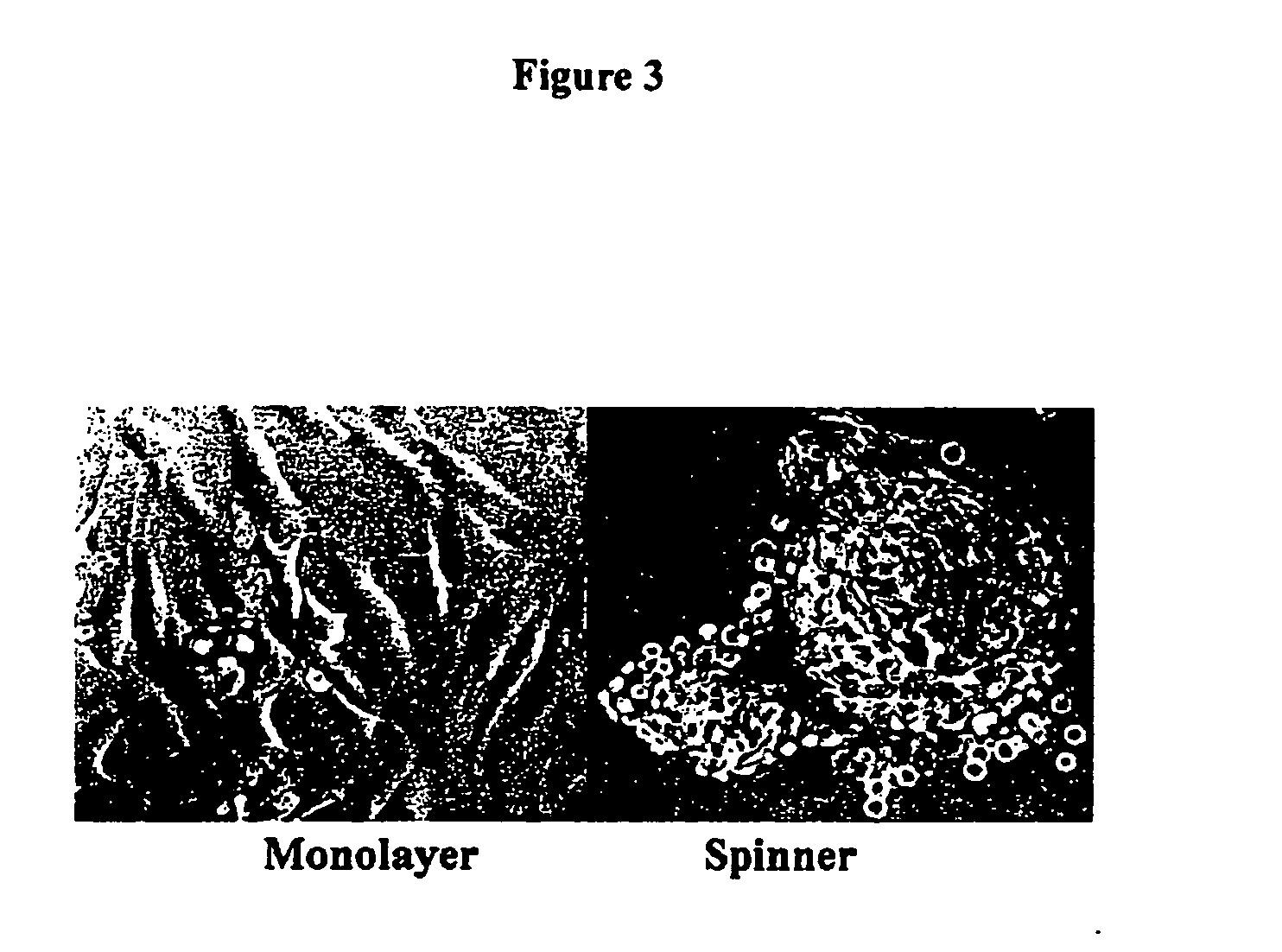
[0107] Chondrocytes directly propagated in microcarrier spinner culture (primary cultures) retain their expression of type II collagen and aggrecan [1]. In contrast, matched chondrocytes propagated in monolayer culture decrease production of type II collagen while increasing their production of type I collagen. Furthermore, dedifferentiated cho...

example 2

Preparation of Bone Analogs

[0115] Human trabecular bone-derived osteoblasts were isolated by collagenase digestion from tissue discarded following hip or knee reconstruction. The cells were seeded at 4×10³ / cm2 in monolayer culture and propagated in HY medium (Hank's Balanced Salt Solution, HBSS+10% Fetal Calf Serum) until nearly confluent (about 2 weeks). Cells were harvested by trypsinization and seeded onto collagen microcarriers (4×10³ / cm2 on Cellagen® beads, ICN, Cleveland, Ohio). The cultures (P3) were incubated for 5 to 15 days at 37° C., 5% CO2. After 15 days, osteoblasts were recovered from microcarrier cultures and were reseeded (4×10³ / cm2) onto microcarriers for 5 to 15 days (P4). These P3 or P4 osteoblast-microcarrier-ECM aggregates are referred to as “bone analogs”.

[0116] After the culture period, the supernatant was removed and saved in microcentrifuge tubes for osteocalcin determination. DNA synthesis rates were determined by labeling the cells with 1 μCi / well of 3H-...

example 3

Preparation of Mesenchyme Analogs

[0118] Human Mesenchymal Stem Cells (hMSCs) were harvested by trypsinization from 4 flasks. 4.0 million hMSCs were seeded into a spinner culture reactor containing 1000 cm2 of microcarrier (Cellagen) surface area (4 thousand cells per cm2) in 120 ml of hMSC culture medium. On days 7 and 14, 10 ml of suspension was withdrawn from the spinner culture and frozen. Cell counts from spinner cultures on days 7 and 14 were 5.3 and 12.8 thousand cells per cm2, respectively. HMSCs on some microcarrier beads were nearly confluent by day 7. RT-PCR on samples from spinner cultures from day 14 was consistent with expected hMSC profiles (positive for type I collagen and aggrecan; negative for type II collagen). Day 14 aggregates are suitable as Mesenchyme Analogs.


PUM


Abstract

A test system and method are disclosed for using tissue analogs The method includes the following steps: (1) isolating the cells to be implanted from donor tissue; (2) seeding the cells onto a particulate microcarrier bead; (3) culturing the cells on the microcarriers to achieve an expansion in the number of cells; and (4) further culturing the cell-particle aggregates to form a tissue analog. The resulting tissue analog and test system may be used for use in screening drugs for diseases and pathological conditions, for testing for toxicity of chemical agents, or for genomic or proteomic screening. The tissue analogs may also be subjected to conditions that will induce disease-like conditions such that the resulting diseased tissue analogs may be used to screen for therapeutic drugs that modify the diseased tissue analog physiology or block progression of the diseased conditions. Kits may be developed for the purpose of conducting multiple tests in conventional multi-well plate systems.

Description

RELATED APPLICATIONS [0001] This application is related to provisional application Ser. No. 60 / 367,682 filed Mar. 25, 2002 and U.S. patent application Ser. No. 09 / 825,632, filed Apr. 4, 2001.FIELD OF THE INVENTION [0002] The herein disclosed invention finds applicability in the field of cell culture, as well as in the field of cell-based in vitro testing of biocompatibility, drug screening, genomics and proteomics. BACKGROUND [0003] Cultured cells and tissue explants have been used extensively in vitro to study the biology, metabolism and function and many connective tissues including cartilage, bone, tendon, ligament, synovium, and meniscus, and many organs such as liver, kidney and pancreas. [0004] For example, cultured cartilage cells (chondrocytes) and cartilage explants have been used extensively to study cartilage biology, metabolism and function. Isolated chondrocytes have been propagated in monolayer culture and in constrained environments such as in pellet culture or in thr...

Claims


Application Information

Patent Timeline
IPC IPC(8): A01N1/02C12N5/071C12N5/077C12Q1/00G01N33/50
CPCC12N5/0655C12N5/0697C12N2501/23G01N33/5082C12N2503/04C12N2531/00C12N2501/25
Inventor FRONDOZA, CARMELITA G.FINK, DAVID J.
Owner CHONDROS
Why Eureka
Try Eureka