[0031] Co-pending
patent application PCT / IL01 / 01108, by one of the present inventors, entitled “device and method for the examination of samples in a non-vacuum environment using a
scanning electron microscope”, discloses a non-vacuum
Scanning Electron Microscope (SEM) device that enables the imaging of wet samples in a wet environment, at near-
atmospheric pressure and a wide range of temperatures. This obviates the need for extensive
sample preparation procedures, thus combining some advantages of electron
microscopy, such as
high resolution and
high contrast or spatial
signal to
noise ratio, with advantages of light microscopy, such as ease and speed of
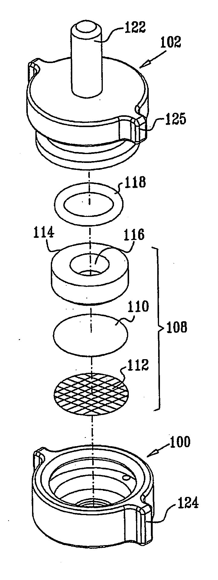
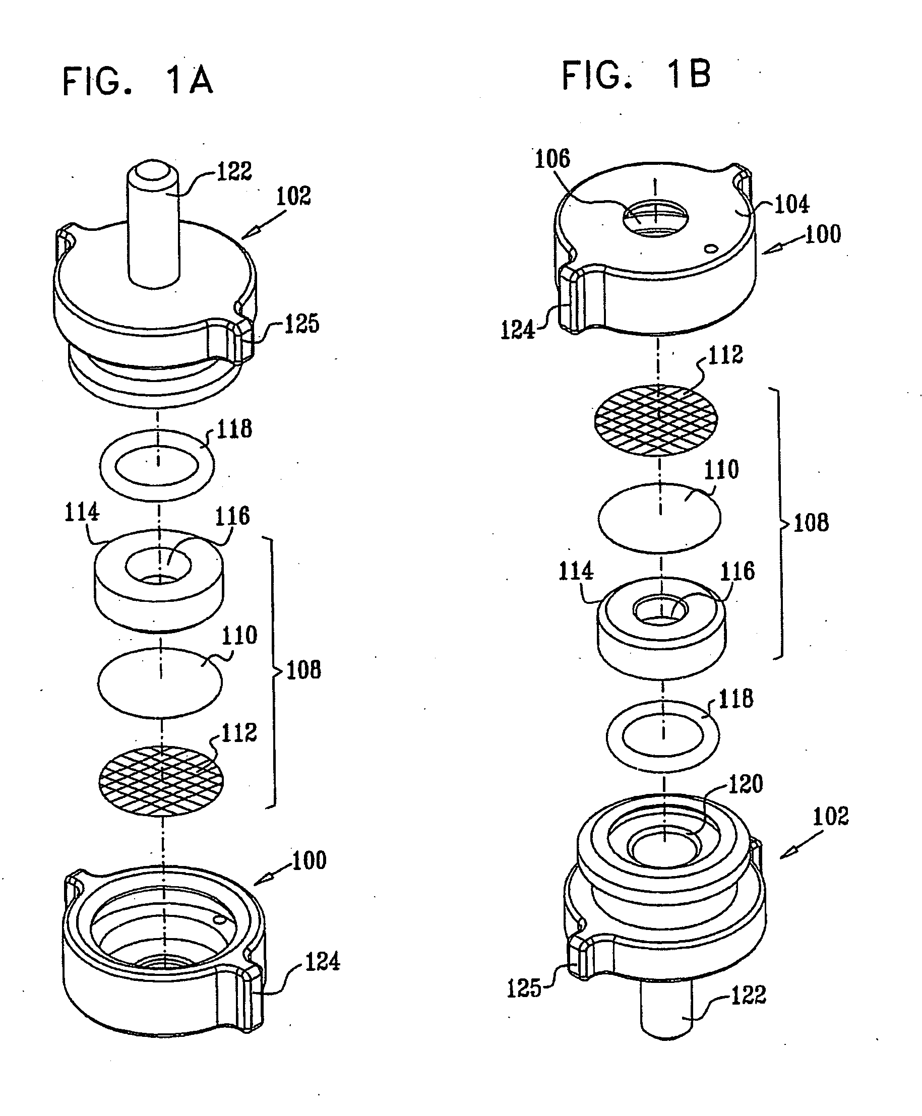
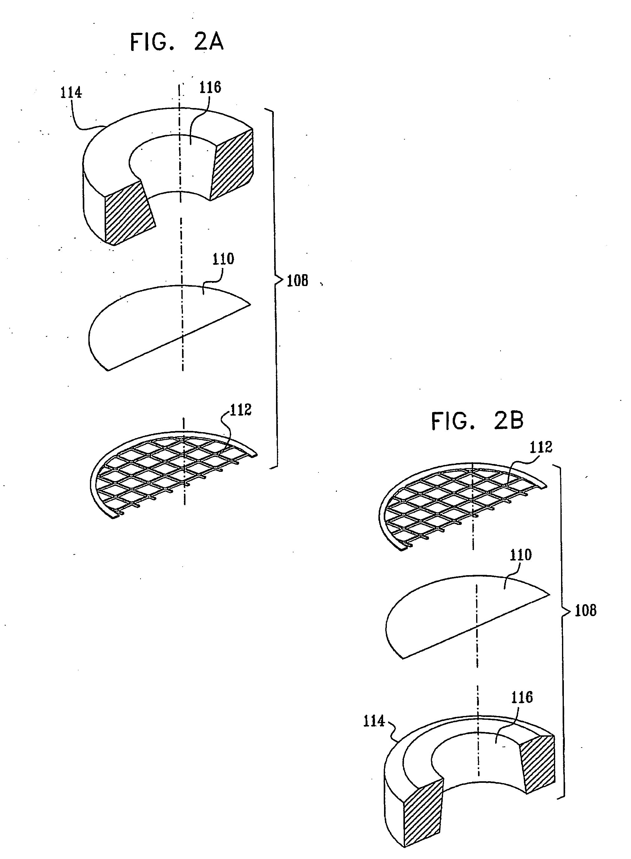
sample preparation. This is accomplished by the use of a sample container covered by a thin partition membrane that is permeable to electrons but is fluid impermeable and sufficiently strong to withstand the
pressure difference between the interior of the container, which is typically one
atmosphere, and the vacuum in the imaging region of an SEM. This type of membrane is referred to hereinbelow as a partition membrane or as an electron-permeable, fluid impermeable, membrane.
[0033] 1. Separation of the sample from the vacuum allows direct
visualization of wet samples. This immediately obviates the need for all
dehydration procedures, including water replacement and critical-point
drying. The wet state most closely resembles the
native state of the sample, preserving features that are distorted or destroyed during
dehydration. This
advantage is particularly important in the observation of tissues, where the true architecture involves both cells and
extracellular matrix. In addition, the presence of fluid in and around the sample allows efficient dissipation of electrical charge and of
excess heat. This eliminates artifacts due to sample charging, as well as
thermal damage.
[0034] 2.
Electron microscopy of biological tissues is most frequently done in two imaging
modes.
Transmission electron microscopy (TEM) utilizes electrons transmitted through the sample; the entire thickness of the sample contributes to the image. Transmission techniques impose a severe constraint on the thickness of the samples: typically, 50 nm, which can be increased to 3 μm in ultrahigh
voltage microscopes. Scanning electron microscopy uses a reflective mode, most frequently detecting
secondary electrons that image only the surface
topography of the sample. The non-vacuum SEM technique uses
backscattered electron detection in a
scanning electron microscope. The electron beam penetrates into the sample, and the backscattered electrons reveal sample features beyond the sample surface to a depth of up to a few microns. Thus, although an electron scanning / reflecting mode of imaging is employed, the image is not limited to the surface, and internal structure of the sample is revealed Furthermore, because detection is done in a reflective mode, any material
lying beyond the interaction volume has no effect on imaging. Therefore, the samples can be of a thickness far exceeding the imaged region. Typically, a tissue fragment several millimeters thick can be viewed; only the material layer of a few micrometers or less that is closest to the surface contributes to the scanned image, without interference from the bulk of the sample. The thickness of the imaged region can be modulated by varying the
acceleration voltage of the electron beam. Non-vacuum SEM thus yields “virtual sections” without the need for actual sectioning of the sample. This eliminates the need for embedding or freezing the sample, which are otherwise required to enable sectioning of the sample. Finally, the dependence of electron backscattering efficiency on the material composition of the sample (through the
atomic number Z) creates contrast even in the absence of heavy
metal staining that is characteristic of TEM imaging. Subcellular organelles can be distinguished based on differences in local concentrations of lipids, phosphates, and salts within biological samples; and a wide variety of stains and labels can be used to enhance contrast.
[0036] It is another objective of the present invention to provide means for automated electron microscopy of wet samples, and specifically of biological samples. Such
automated microscopy has been widely applied in the
semiconductor industry. The main barrier to the application of automated electron microscopy to wet samples is the need to employ
sample preparation procedures such as
drying, embedding, sectioning or
coating, which are highly complex and not amenable to
automation. The present invention provides means for
direct imaging of wet samples in a scanning
electron microscope, thus obviating the need for the aforementioned preparative procedures. The present invention thus provides means for automated electron microscopy of wet samples.
[0053] In accordance with another preferred embodiment of the present invention, the inspecting includes introducing a sample including at least the portion into a specimen
enclosure in a wet environment and scanning the sample in the specimen enclosure in the scanning
electron microscope, thereby visualizing the sample.

