Mass intensity profiling system and uses thereof
a mass intensity profiling and profiling technology, applied in the field of mass spectrometry, bioinformatics, computational molecular biology, can solve the problems of inability to accurately predict other data types, limited sample quantity and stability, and enormous challenges, and achieve the effect of rapid and efficient identification of individual biomolecules and rapid identification of sets
- Summary
- Abstract
- Description
- Claims
- Application Information
AI Technical Summary
Benefits of technology
Problems solved by technology
Method used
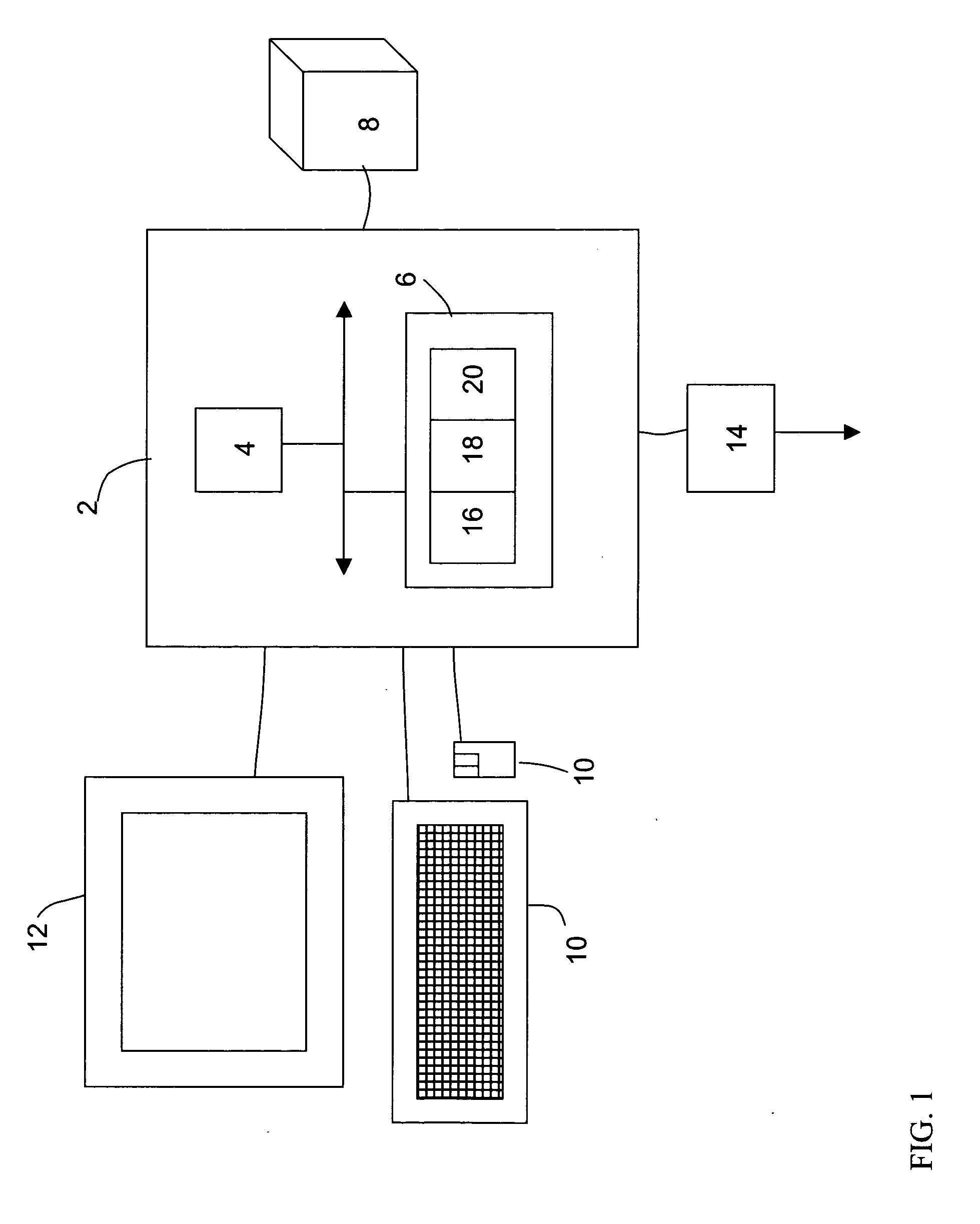
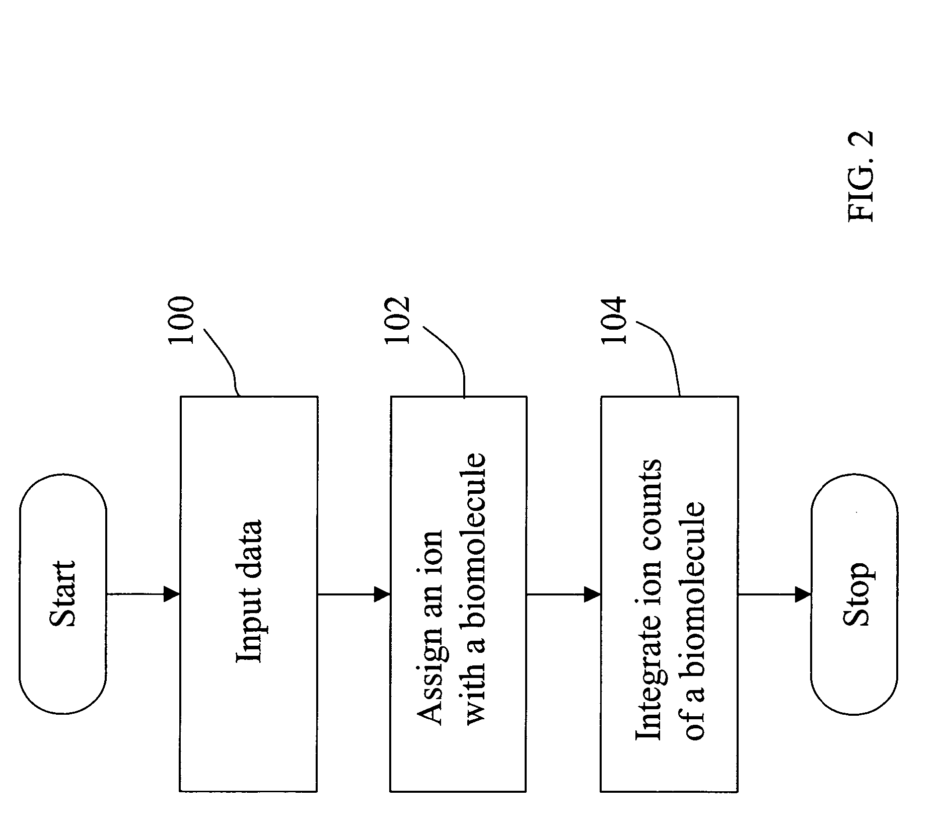
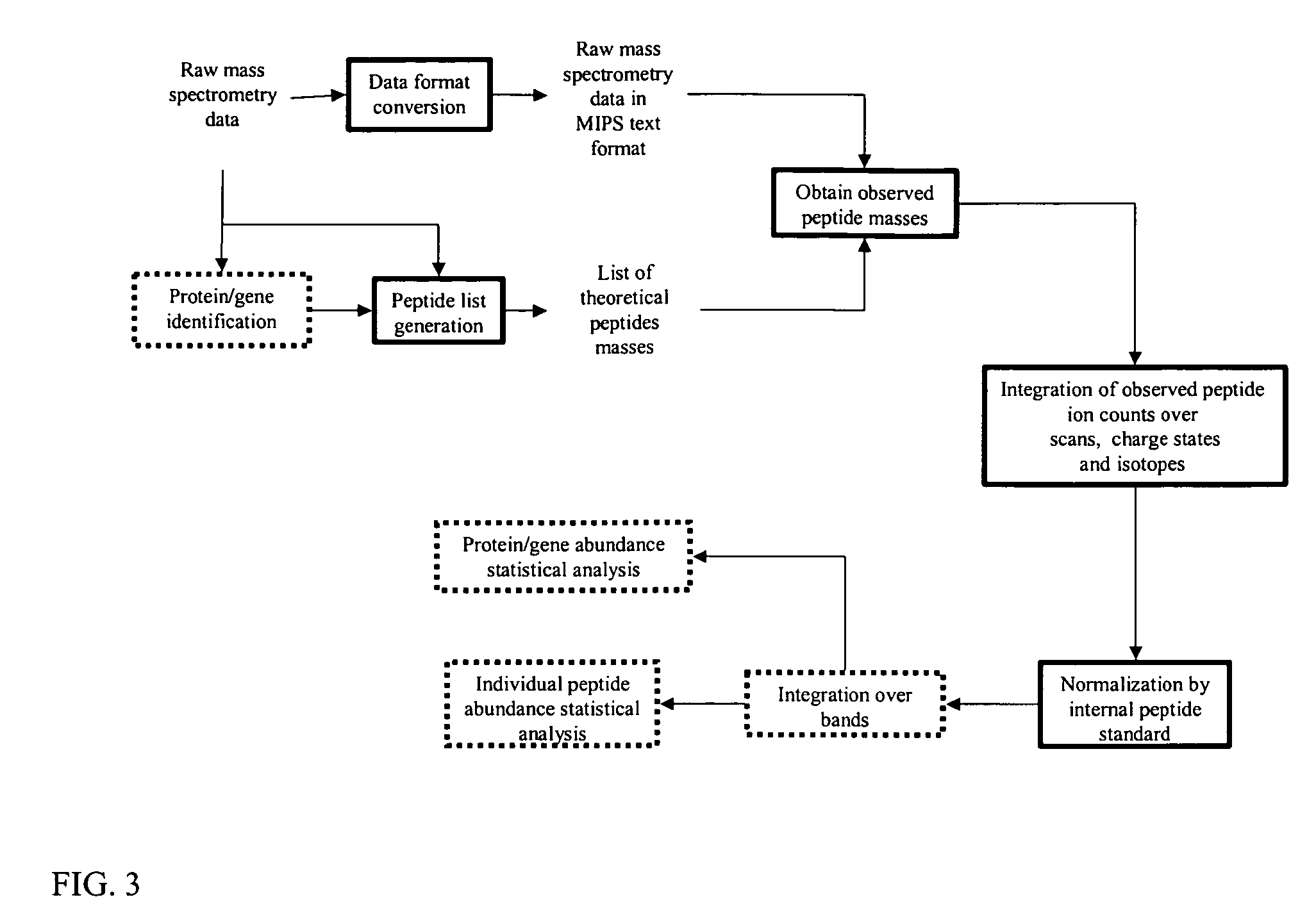
Image

Examples
example 1
Reproducibility
[0120] A total cellular lysate of U937 cells was prepared. Fifty μg of this lysate was mixed with 30 ng of bovine serum albumin (BSA). Five samples of this mixture were prepared, and each sample was separated by SDS gel electrophoresis. A sixth sample was prepared without the addition of BSA. After electrophoresis, the gel was stained, and the band containing the BSA was excised from the gel.
[0121] Tryptic digestion of the BSA band was performed according to standard methods. Three hundred and fifty fmol of Leu-enkephalin was added to each tryptic digest as an internal standard. The peptide mixture was separated using a Waters CapLC HPLC system that was coupled to a Micromass quadropole-time of flight (Q-ToF) mass spectrometer. The conditions for the separation of this mixture were as follows: a reversed phase column (1D, 10 cm×75 μm, C 18), a flow rate of 300 nL / min, and a linear gradient of 10% to 80% acetonitrile / deionized water (containing 0.2% formic acid) in 2...
example 2
[0123] U937 cells were cultured for 48 hours with or without 25 nM phorbol myristate acetate (PMA) to generate macrophages and untransformed cells (monocytes) using standard culturing techniques. Approximately 100 million cells suspended in 10 mM Tris / 200 mM sucrose, pH 7.5 homogenization buffer were placed into a cavitations chamber, pressurized with N2 at 1000 psi and kept on ice for 60 minutes. After each incubation period, samples were released to atmospheric pressure rapidly, and centrifuged at 900 g for 15 minutes at 4° C. (low speed centrifugation). The supernatant (post nuclear supernatant, PNS) was collected and either kept or immediately centrifuged at 40000 rpm for 60 minutes at 4° C. to produce the post nuclear membrane (PNM) samples.
[0124] Five samples of 50 μg of monocyte or macrophage proteins were prepared. The first sample contained only monocyte proteins, the second sample contained 75% monocyte and 25% macrophage proteins, the third sample contained 50%...
PUM
| Property | Measurement | Unit |
|---|---|---|
| flow rate | aaaaa | aaaaa |
| pH | aaaaa | aaaaa |
| mass spectrometry | aaaaa | aaaaa |
Abstract
Description
Claims
Application Information
PatSnap Eureka turns technology decisions into work you can execute. Powered by our Innovation Knowledge Graph, it runs expert workflows across engineering, life sciences, materials and intellectual property. Get your review-ready output in minutes.


