
Amorphous cell delivery vehicle treated with physical/physicochemical stimuli

a delivery vehicle and amorphous cell technology, applied in the field of functional tissue engineering, can solve the problems of cartilage not being able to regenerate in vivo, physical wear and dislocation of non-living tissues, and the inability to physically regenerate in vivo

Inactive Publication Date: 2006-02-16
THE BRIGHAM & WOMEN S HOSPITAL INC
View PDF7 Cites 11 Cited by
  • Summary
  • Abstract
  • Description
  • Claims
  • Application Information

AI Technical Summary

Benefits of technology

[0057] In one aspect the invention provides a method of treating osteoarthritis in a subject. The method according to this aspect of the invention includes the step of introducing, in a subject having osteoarthritis of a joint, an effective amount of a chondrocytic cell / matrix composition of the invention into a space defined by a surface zone cartilage overlying a site of damaged articular cartilage surface and subchondral bone beneath the site of damaged articular cartilage surface of the joint, to treat the osteoarthritis.

Problems solved by technology

These methods can have certain drawbacks, including physical wear and dislodgement of non-living tissues and availability or suitability of living tissue for certain purposes.
Cartilage is not vascularized and its ability to regenerate in vivo is very limited, particularly in adult individuals and if the piece of cartilage to be regenerated exceeds even a small volume.
However, articular cartilage often suffers degeneration due to wear, age, disease, or traumatic or overuse injuries, involving a significantly greater volume than might be naturally regenerated.
This kind of defect of the cartilage layer makes movement and loading of the affected joint painful and can lead to further complications such as inflammation, which can contribute to further damage to the cartilage layer.
A problem encountered in these methods is the fact that chondrocytes in these in vitro conditions have the tendency to de-differentiate into fibroblasts relatively rapidly.

Method used


Image

  • Amorphous cell delivery vehicle treated with physical/physicochemical stimuli
  • Amorphous cell delivery vehicle treated with physical/physicochemical stimuli
  • Amorphous cell delivery vehicle treated with physical/physicochemical stimuli

Examples


example 1

Apparatus for Cultivating Cells or Tissue

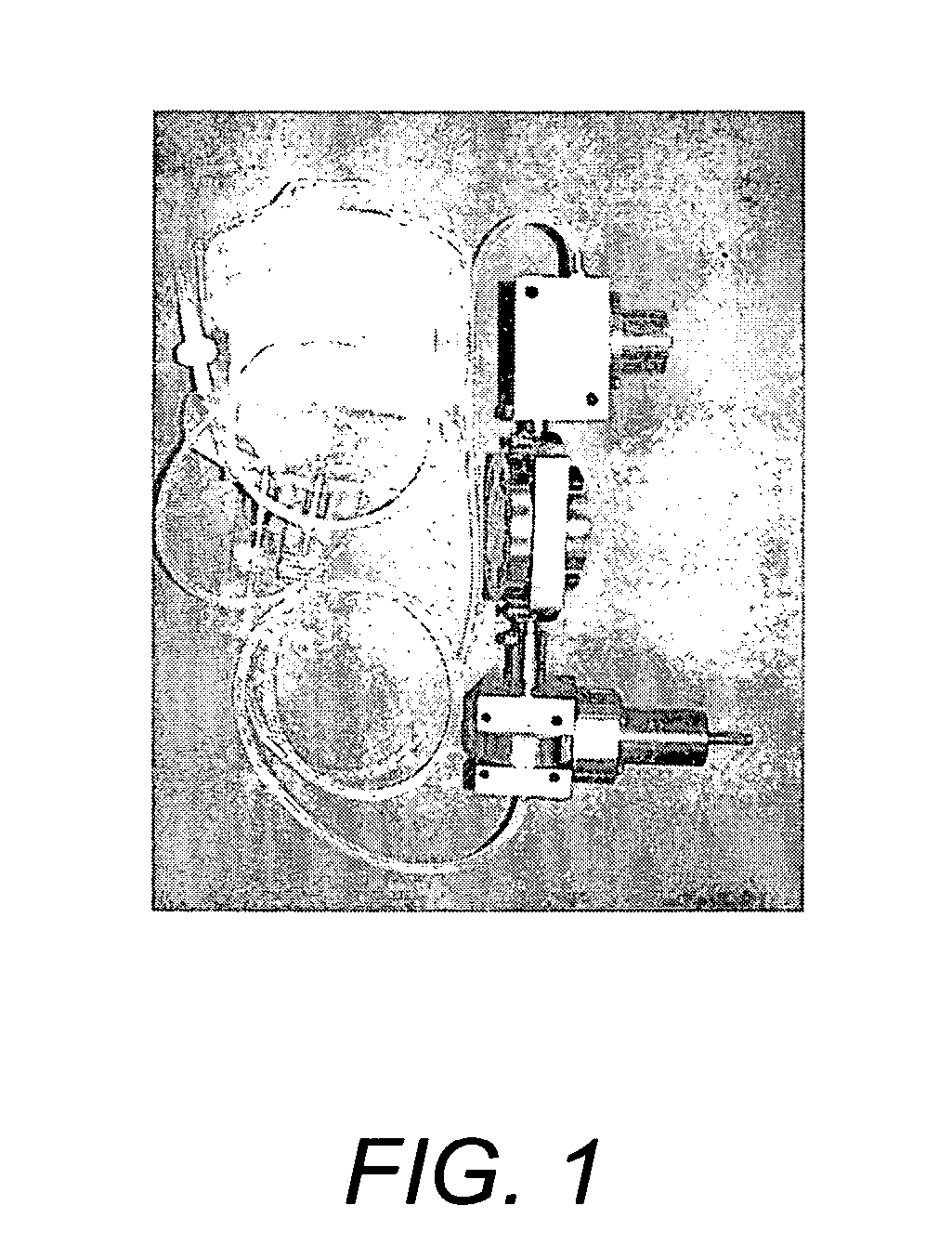
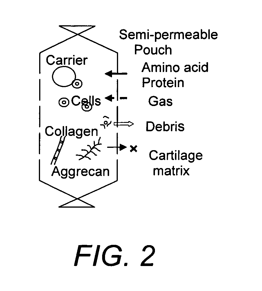
[0143] A hydrostatic pressure / perfusion culture system (bioreactor) suitable for use in the in vitro culture methods of the invention is depicted in FIG. 1 and described in U.S. Pat. No. 6,432,713, the entire contents of which are incorporated herein by reference. A schematic drawing depicting the use of a semi-permeable membrane pouch for the culture of cells in a biodegradable amorphous carrier is depicted in FIG. 2. The semi-permeable membrane pouch, containing cells and biodegradable amorphous carrier, is placed within the culture chamber, which is kept horizontal and maintained at 37° C. for a culture period of one to six weeks or more.

example 2

Evaluation of Mass Transfer of Molecular Markers with Biodegradable Polymers in a Semi-Permeable Membrane Pouch After Application of Hydrostatic Pressure In Vitro

[0144] Experiments are performed to evaluate the mass transfer of molecular markers in an amorphous cell carrier through a semi-permeable membrane at static culture conditions as well as at different magnitudes and cycles of fluid pressure. Cartilage ECM has high molecular weight and will stay within the semi-permeable membrane pouch. Degraded cell carrier debris (small molecules) and metabolic waste will be exuded into the medium phase. Under static conditions, nutrients can infiltrate the pouch according to Fick's law. In addition, the bioreactor is used to manipulate mass transfer with defined hydrostatic fluid pressure, medium flow, and controlled oxygen / carbon dioxide concentration. As a model of mass transfer, molecular weight markers are used to evaluate mass transfer under a series of experimental conditions (Table...

example 3

Effects of Hydrostatic Fluid Pressure on Mass Transfer and Degradation of Amorphous Cell Carrier in Semi-Permeable Membrane Pouch

[0146] Biodegradable amorphous polymer (hydrogel or sol / gel reversible polymer) is tested at defined cell culture conditions using a tissue culture system. A semi-permeable membrane pouch is used to hold the cell construct and extracellular matrix products of large molecular weight produced by the chondrocytes. Performance of the pouch is analyzed according to molecular weight cut-off size from 100 to 500 kDa under hydrostatic fluid pressure (HFP) ranging 0 to 5 MPa and 0 to 0.5 Hz. The test carrier is injected into the pouch and evaluated in terms of kinetics of degradation. Fluorescent molecular tracers (e.g., dextran-FITC) ranging from 100 to 500 kDa are used as markers. The fluorescence intensity is measured with a fluorometer using suitably selected wavelengths for fluorescence excitation and detection. From a preliminary study it was observed that d...


PUM

PropertyMeasurementUnit
molecular weightaaaaaaaaaa

Abstract

Provided are compositions and methods for in vitro generation and in vivo use of tissue for the repair of defective tissue, especially cartilage. Chondrocytes or other cells are cultured in vitro in a biodegradable amorphous carrier within the confines of a space bounded by a semi-permeable membrane with a molecular weight cut-off of greater than 100 kDa. The culture can be subjected to physical / physicochemical conditions that mimic in vivo conditions of the tissue in need of repair or replacement. In one embodiment the invention provides an amorphous preparation of chondrocytes and their extracellular products, suitable for injection.

Description

RELATED APPLICATION [0001] This application claims benefit under 35 U.S.C. 119(e) of U.S. Provisional Patent Application Ser. No. 60 / 592,786, filed Jul. 30, 2004, the entire content of which is incorporated herein by reference.FIELD OF THE INVENTION [0002] The invention is in the field of functional tissue engineering. More particularly, the invention in some aspects involves in vitro culture methods and products thereof, useful for regenerating tissue suitable for use in replacement or repair sites of damaged tissue. BACKGROUND OF THE INVENTION [0003] There are various methods for restoring a damaged tissue or a pathogenic part of a living body. One method is to substitute the damaged tissue or pathogenic part with materials other than a living tissue, such as plastic, metal, and / or ceramic to restore the damaged tissue or the pathogenic part. Another method is to substitute the damaged tissue or pathogenic part with parts from other individuals or other animals, or from a differen...

Claims


Application Information

Patent Timeline
Patent Type & Authority Applications(United States)
IPC IPC(8): A61K45/00C12N5/08A61K35/12A61K35/32C12N5/077
CPCC12N5/0068C12N5/0655C12N2533/80C12N2533/30C12N2533/54C12N2521/00A61P19/00A61P19/02A61P19/08C12N5/0602C12N5/00A61K35/32
Inventor MIZUNO, SHUICHI
Owner THE BRIGHAM & WOMEN S HOSPITAL INC
Learn More