
Method and apparatus for non-invasive measurement of blood analytes

Inactive Publication Date: 2006-03-23
YU DEJIN +1
View PDF30 Cites 139 Cited by
  • Summary
  • Abstract
  • Description
  • Claims
  • Application Information

AI Technical Summary

Benefits of technology

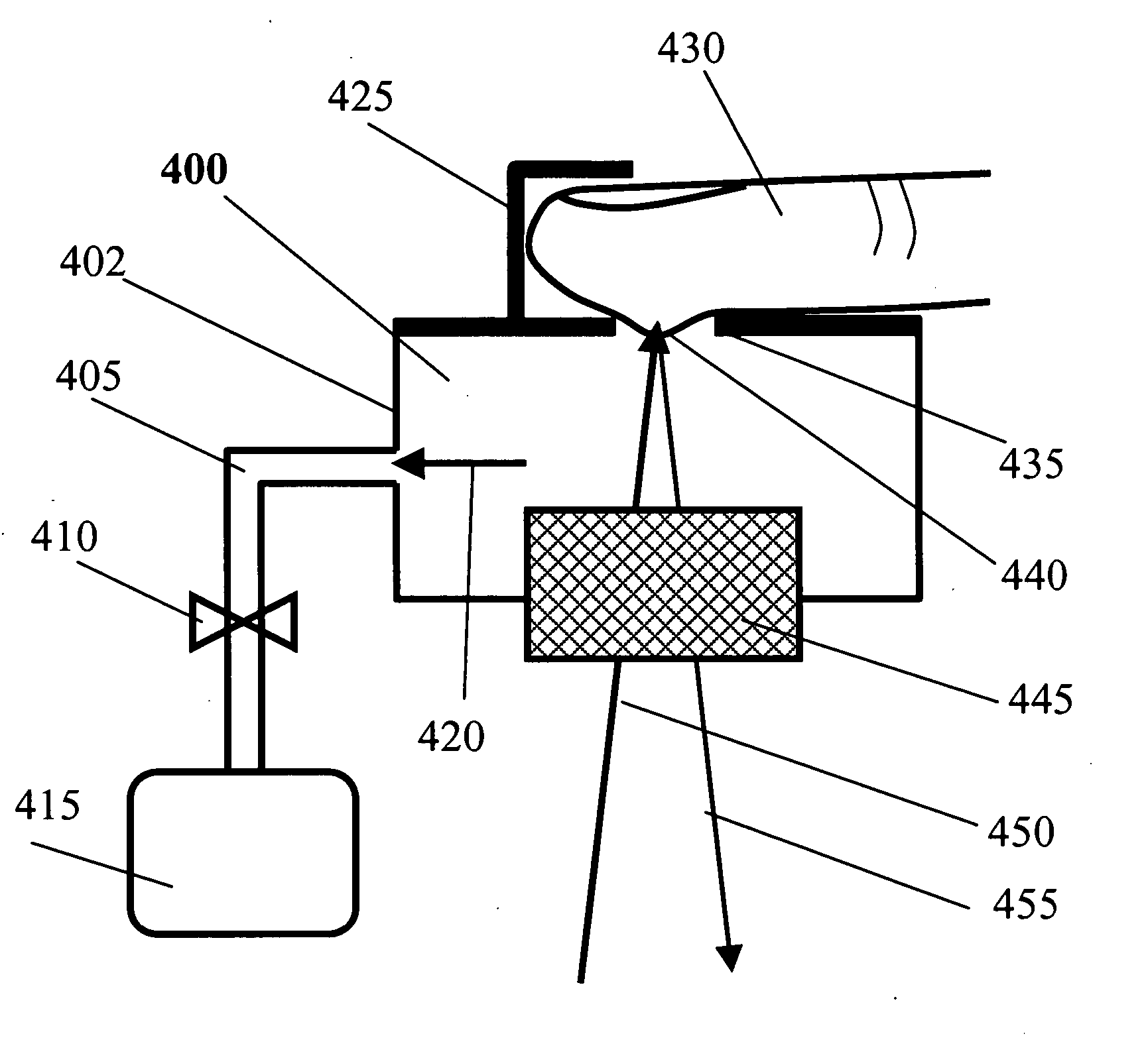
[0018] The tissue permeation unit modulates tissue and blood locally. It will increase the blood amount at the beginning of the measurement so that it intensifies the Raman scattering and increases the signal-to-noise ratio, and then gradually decrease the local blood amount with time until blood depletion. In one embodiment, the unit may be made of a vacuum chamber with a transparent window and small opening or hole, which is connected with an electrically or manually driven vacuum pump that creates a negative air pressure inside the vacuum chamber. The pressure inside the chamber can be changed. The user's fingertip is placed on the hole to form a closed chamber. Under the negative air pressure, a substantial amount of blood is “sucked” into a small area of the human finger after finger is placed on the hole. As the time is increased, the blood amount will be decreased gradually.
[0021] Other mechanical methods can be also used for varying the level of blood in the region being measured. For example, a mechanical means can be used to press the finger and then slowly release the finger. Another example could include a variable pressure tourniquet that could slow or speed up blood flow to the region being measured. For commercial use, the approach used should be relatively low cost and not discomfort the patient.
[0023] In another preferred embodiment, the blood permeation unit is so controlled that the blood amount is increased at the beginning and then is decreased until blood depletion while keeping the target tissue area stationary and eliminating the effects from heart pulse, respiration and body movement during the data acquisition. The blood depletion is eventually accomplished due to the distributed tension around contact region between the skin and chamber material. In a preferred embodiment, after reaching its maximum level, the blood amount is decreased linearly with time. The measurement starts at the moment when the blood amount is at its maximum, from which the strongest Raman scattering from the blood analytes is substantially achieved. Over time, the signal intensity attributed from the blood will decrease gradually while the signal components arising from the surrounding tissues will remain relatively unchanged due to the effect of blood permeation. The so-acquired Raman spectra can be processed in various ways. In a preferred embodiment, the spectral data obtained at a given time is subtracted by the spectrum acquired when the blood is depleted, i.e., Rin=Ri−Rn with i=1, 2, 3, . . . , n−1 where Ri is the Raman spectrum obtained at time ti and Rn is the last Raman spectrum acquired at the blood depletion. Rl is the first spectrum with the strongest Raman scattering from blood substances. The direct advantage embedded in the new series of spectra over the raw data is that the spectral contributions arising from the surrounding static tissues are removed and the resulted spectra (Rin) are dominated by the contribution from the blood.

Problems solved by technology

Currently, daily blood glucose monitoring for diabetes patients can only be done through the use of invasive techniques.
The invasive methods require drawing blood from patients, which is painful and inconvenient since the skin has to be lanced in order to collect the blood sample for measurement.
It is an unpleasant practice, but that is exactly what many diabetics have to do daily in order to measure blood glucose level to provide feedback for insulin dosing and other treatment.
Several large clinical studies have shown that tight control of blood sugar slows the progression of and development of long-term complications of diabetes, such as blindness and kidney failures.
However, many people with diabetes do not test their blood glucose levels regularly due to physical pain and high material cost, as well as the risk of infections when finger was lanced.
This is mainly because many barriers exist for the current monitoring methods.
To date, none of these approaches has been proven to be clinically feasible.
Further, while mid-infrared absorption detects fundamental tones of molecular vibration, the optical penetration depth over this wavelength range is extremely short, typically at the magnitude of order of the thickness of epidermis due to strong absorption of water.
Unfortunately, there are some fundamental issues to be addressed: 1) laser eye safety and 2) time delay between glucose in blood and aqueous humor and correlation between ocular and artery glucose levels.
These unresolved issues limit the effectiveness of this approach.
Together with other inventions based on Raman scattering, these methods experience the following problems: 1) Raman scattering is quite weak, 2) biological effects from heart pulses, respiration, and body movement, etc., degrade measurement, and 3) calibration against that portion of the optical response caused by the skin and other tissue substances is difficult.
The last issue is critical because the amounts of protein, fats, water, etc.
In different people and different skin surface conditions such as oily and turbid fingers will seriously degrade the measurement results if not properly calibrated out.

Method used


Image

  • Method and apparatus for non-invasive measurement of blood analytes
  • Method and apparatus for non-invasive measurement of blood analytes
  • Method and apparatus for non-invasive measurement of blood analytes

Examples


Embodiment Construction

[0038] The present invention provides a method and apparatus for non-invasive measurement of blood analytes with dynamic spectral calibration against the influence from skin and other tissues other than blood. The working principle is described based on Raman spectroscopy, but it can be applied to other lightwave methods including near-infrared spectroscopy, mid-infrared spectroscopy, infrared spectroscopy, reflectance spectroscopy, fluorescence spectroscopy, Fourier-transform infrared (FTIR) spectroscopy, polarization changes, scatter changes, and photo-acoustic spectroscopy.

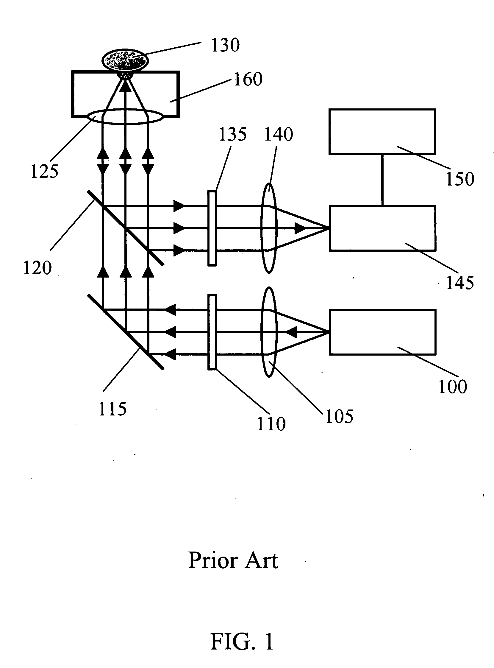
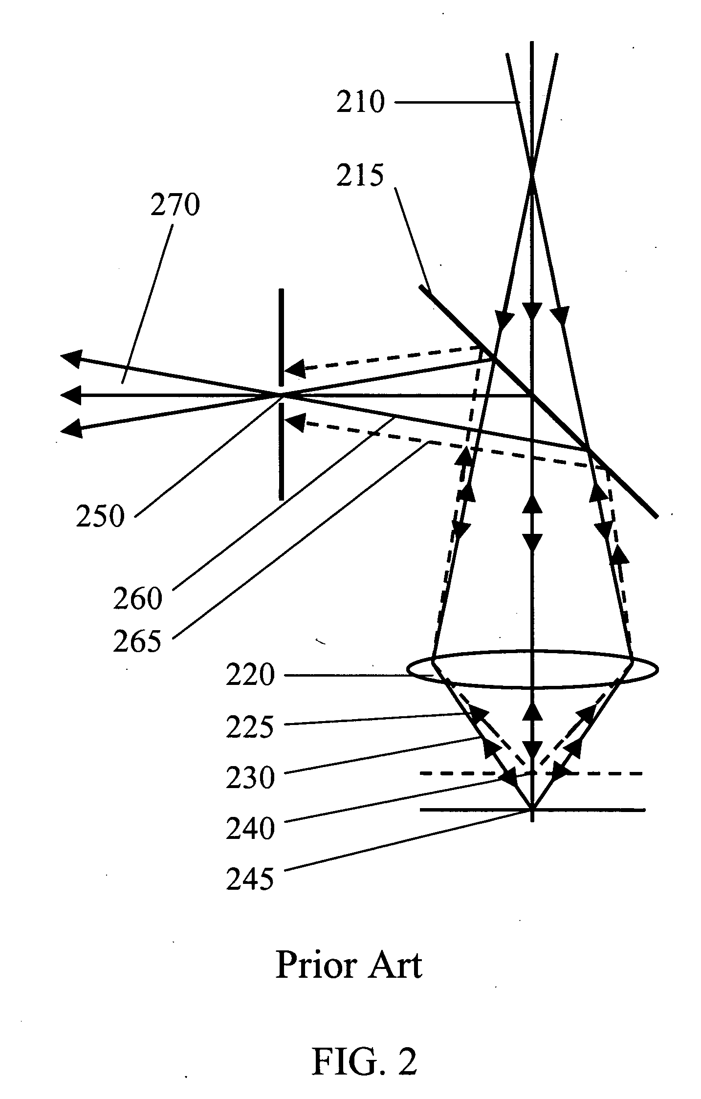
[0039] Referring now to the drawings, FIG. 1 illustrates a basic Raman configuration of the apparatus used for non-invasive measurement of blood glucose level in accordance with the prior art (U.S. Pat. No. 6,167,290). It consists of five parts: 1) excitation laser 100, 2) Raman spectrometer 145, 3) light excitation and collection unit, 4) tissue permeation unit 160, and 5) data processing unit 150. The CW exc...


PUM


Abstract

The present invention discloses a method and apparatus and method for achieving non-invasive measurement of analytes from human and animal blood through the skin using Raman lightwave technology. The apparatus includes a hydraulic tissue permeation unit, which controls the amount of blood in the laser tissue interaction region. Two or more spectra are obtained at different blood levels. These spectra are used to improve the measurements.

Description

CLAIM OF PRIORITY [0001] The present application is a continuation-in-part of U.S. patent application Ser. No. 10 / 914,761, filed Aug. 9, 2004, the disclosure of which is incorporated in this document by reference.TECHNICAL FIELD OF THE INVENTION [0002] This invention in general relates to methods and apparatus for non-invasive measurement of the concentrations of analytes within human / animal blood through the skin, and in particular, for monitoring the blood glucose levels in vivo for diabetes using light scattering technology and calibrating the effects from skin and other surrounding tissue constituents. BACKGROUND OF THE INVENTION [0003] Currently, daily blood glucose monitoring for diabetes patients can only be done through the use of invasive techniques. The invasive methods require drawing blood from patients, which is painful and inconvenient since the skin has to be lanced in order to collect the blood sample for measurement. 6-8 times a day, it is the same routine for the d...

Claims


Application Information

Patent Timeline
IPC IPC(8): A61B5/00
CPCA61B5/14532A61B5/1455G01N21/65A61B5/6838A61B5/702A61B5/6826A61B5/6834A61B5/0075
Inventor YU, DEJINYANG, WEI
Owner YU DEJIN
Learn More