
Photometric determination of coagulation time in undiluted whole blood

a technology of coagulation time and photometric measurement, which is applied in the field of system, device and method for detecting coagulation of a freeflowing liquid, can solve the problems of background art that the art also does not teach or suggest such a device, etc., to achieve easy implementation and operation, simple construction, and easy to obtain regulatory approval/clearance

Inactive Publication Date: 2006-05-25
INVERNESS MEDICAL SWITZERLAND GMBH
View PDF13 Cites 43 Cited by
  • Summary
  • Abstract
  • Description
  • Claims
  • Application Information

AI Technical Summary

Benefits of technology

[0014] The present invention overcomes these drawbacks of the background art by providing a device, system and method for photometric detection of coagulation in whole blood. The present invention is easy to implement and operate. Furthermore, the present invention has the advantage of being considered to fulfill the desired standard of using photometry for measuring blood coagulation. Also, a photometric coagulation test device for whole blood specimens according to the present invention provides medical accuracy to the home user and, at the same time, is simple to construct. The present invention can more easily obtain regulatory approval / clearance because of its adherence to the photometric technology. The present invention is also useful for testing cessation of movement in a free flowing liquid other than blood, for example due to other types of coagulation.
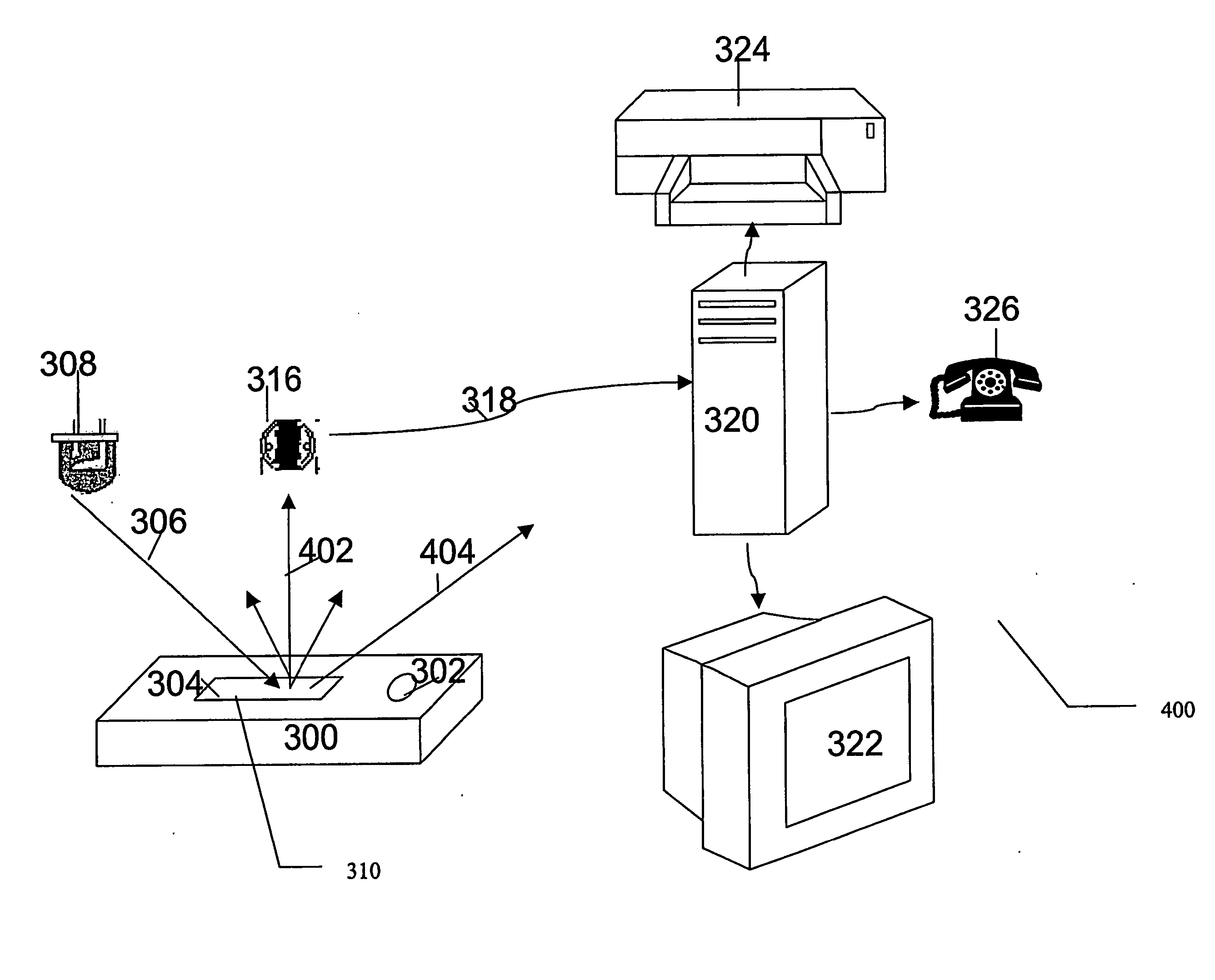
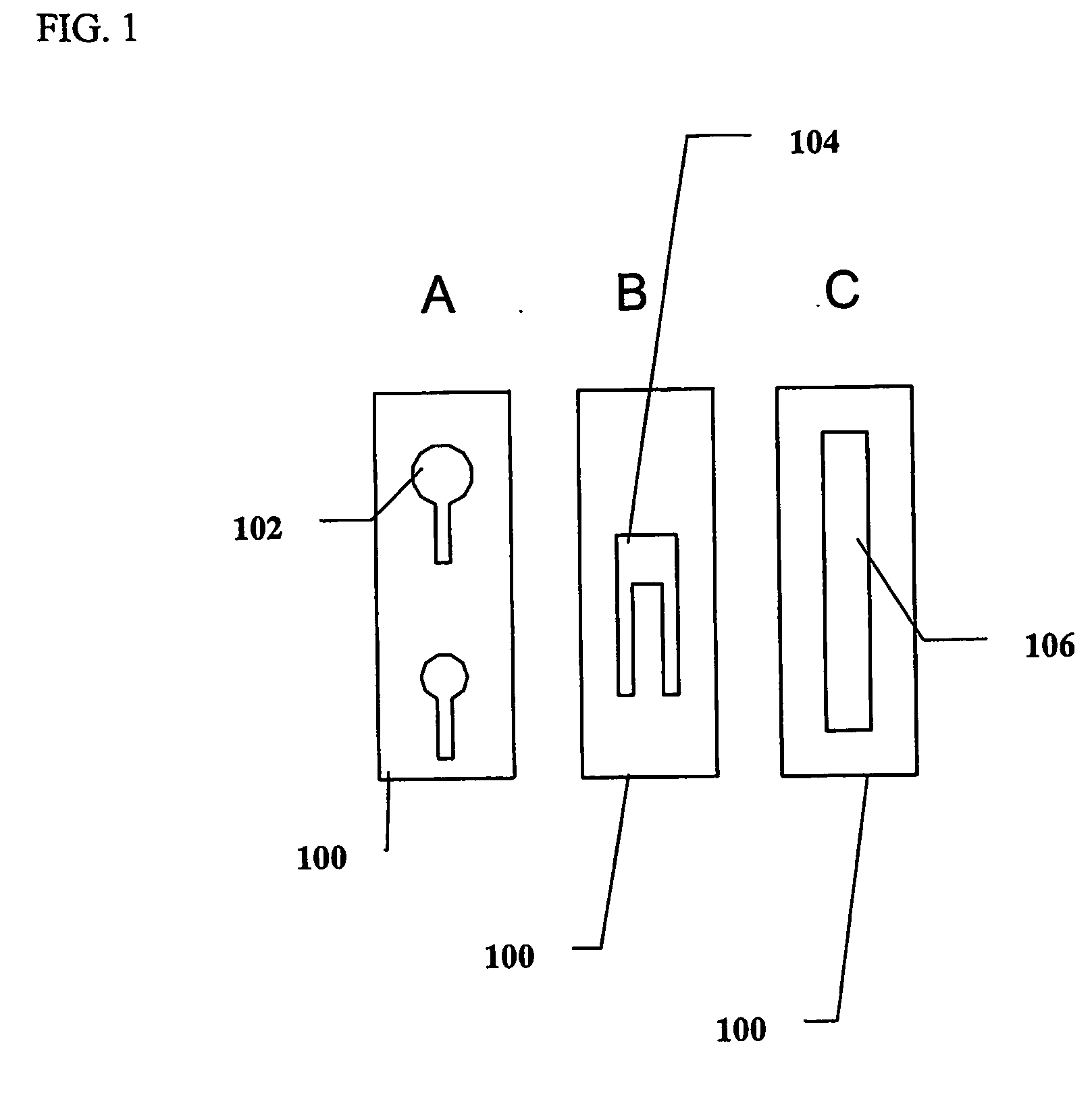
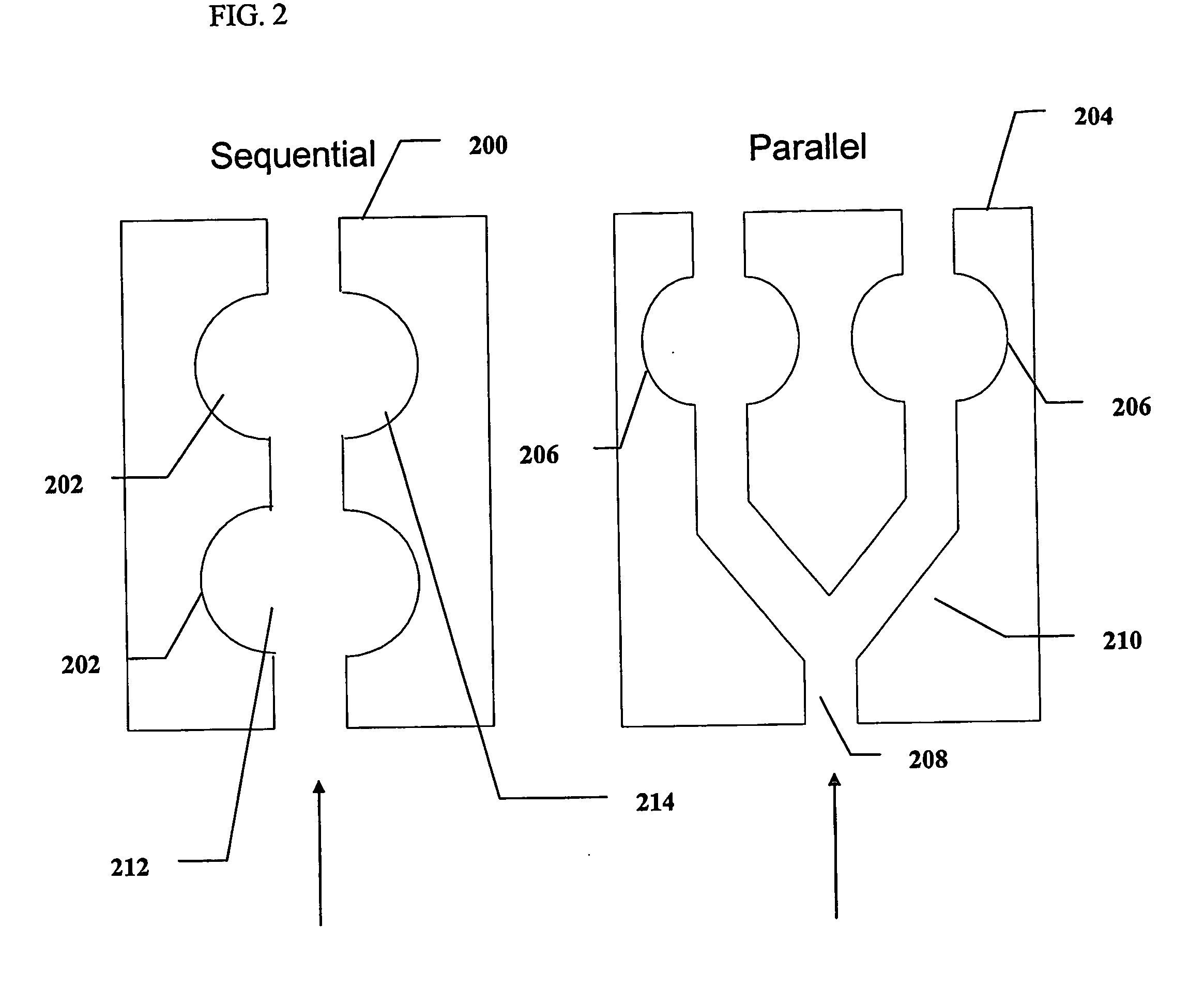
[0017] In one optional embodiment of the invention, photometry may optionally be performed inside a disposable, test-strip-like hollow device, which contains the coagulation reagents in a dry, ready-for-use form. The size of the strip can be reduced, so that the amount of blood required is sufficiently small (microliters or fractions thereof) to reduce discomfort and pain to the patient. The strip may then optionally be inserted into a small, portable meter, which may optionally and preferably include a low cost, low power light source and a basic light sensor. Alternatively, the light source and light sensitive devices may optionally be incorporated in their chip form into the disposable test strip, thus further improving the user interface and increasing the ease of operation. The time required for the determination of coagulation time may optionally be shortened by employing novel signal analysis strategies.

Problems solved by technology

The background art also does not teach or suggest such a device, system and method which is simple and easy to use.
The background art also does not teach or suggest such a photometric device, system and method which are capable of detecting coagulation of a free-flowing liquid.

Method used


Image

  • Photometric determination of coagulation time in undiluted whole blood
  • Photometric determination of coagulation time in undiluted whole blood
  • Photometric determination of coagulation time in undiluted whole blood

Examples


example 1

Preparation of Test-Strips

[0143] Thromboplastin Reagent: Dried Innovin® Reagent with calcium (Dade Behring, Inc.; Deerfield, Ill.) was resuspended in water according to the manufacturer's instructions and stored at 4-8° C. for up to 1 week.

[0144] Innovin salts solution, which is functionally equivalent to the contents of the original Innovin solution (buffer, calcium and protein), but which does not include thromboplastin or lipids, was prepared according to U.S. Pat. No. 5,625,036 and included:

1.4415 gr HEPES

0.16 gr CaCl2.2H2O

0.5844 NaCl

5.0 gr glycine

0.135 gr Bovine Serum Albumin (Bovuminar® Biotechnology Premium Grade pH 7, Serologicals Corp, Norcross, Ga.)

Reagent Grade Water to 100 mL

pH adjusted to 7.0

(The Bovine Serum Albumin is not a component of the original Innovin® reagent. It is added to the salts solution to simulate the protein load in that original reagent)

[0145] The base of the test-strips was constructed from a flat optically clear Lexan® film, 250 o...

example 2

Determination of Preferred Color of Light and LED Output for Reflectance-Scattering

[0147] A reflectance measurement test was built according to the block diagram in FIG. 4, employing red green and white LEDs (LiteON, Taipei, Taiwan) as the light source and a Texas Instruments TSL250 light-to-voltage sensor (Texas Instruments, USA) as the light measurement device. The sensor's lens was covered with a mask having a 1 mm pinhole. The background (see definitions) behind the test-strip was a matt-black vinyl film (Ritrama, USA). The voltage output of the sensor was recorded by an Extech 380281 digital multi-meter (Extech Instruments Corp., Waltham, Mass., USA) connected to a computer, running Extech's DMM data acquisition software. The angle of light incidence was 32.5° or 60°.

[0148] Innovin containing strips and empty strips (i.e control strips) were filled with whole, citrate preserved, fresh capillary blood. Following 5 minutes incubation at room temperature the reflected light valu...

example 3

Effect of Thromboplastin Coagulation Reagent on Reflectance-Scattering of Light

[0152] The temporal reflectance-scattering of light from test-strips containing Innovin (i.e reagent with thromboplastin and calcium), or Innovin salts (i.e. reagent with calcium but without thromboplastin) and control strips (i.e. no reagent) was recorded following the introduction of an 18 μL citrate preserved, fresh capillary blood specimen. The raw light measurements are depicted in FIG. 5A. The calculated ratios of each measurement to the starting measurement (taken at blood entry) are depicted in FIG. 5B. The reaction chamber of the control strip used to produce FIGS. 5A and 5B did not contain any reagent. The salts strip contains salts and proteins but no thromboplastin. The Innovin strip contains dried Innovin. FIG. 5A presents the temporal light measurements. FIG. 5B presents ratios of the light measurement in each time point to the starting light measurement for each of the different types of t...


PUM


Abstract

A device, system and method for photometric detection of coagulation in whole blood. The present invention is easy to implement and operate. Furthermore, the present invention has the advantage of being considered to fulfill the desired standard of using photometry for measuring blood coagulation. Also, a photometric coagulation test device for whole blood specimens according to the present invention provides medical accuracy to the home user and, at the same time, is simple to construct. The present invention is also useful for detecting and determining blood agglutination, for example as the results of a serological reaction with an antibody.

Description

FIELD OF THE INVENTION [0001] The present invention is of a system, device and method for detecting coagulation of a free-flowing liquid, such as blood. BACKGROUND OF THE INVENTION [0002] Anticoagulant therapy is prescribed to an increasing number of patients with a variety of cardiovascular conditions, such as venous or arterial thrombosis, embolism and cardiac valve replacement. Since the anticoagulant drug activity and efficacy is affected by the patient's lifestyle and diet, frequent monitoring of the coagulation status of the blood is required, to maintain a suitable dosage level within the therapeutic window for such drugs. For the patient, this requirement results in at least weekly travel to a clinic with painful drawing of a venous blood specimen. [0003] The classical and standard reference blood coagulation tests involve the measurement of the time required to form a clot. Clot formation is determined by two general approaches: (a) detecting a change in the mechanical (e.g...

Claims


Application Information

Patent Timeline
IPC IPC(8): G01N21/64G01N33/487G01N33/49
CPCG01N21/03G01N21/272G01N21/82G01N21/8483G01N33/4905G01N33/86G01N2021/0321G01N2021/0328G01N2021/0346G01N2021/825G01N2201/0221G01N2201/0624
Inventor FISH, FALK
Owner INVERNESS MEDICAL SWITZERLAND GMBH
Learn More