

Multilayered microcultures
- Summary
- Abstract
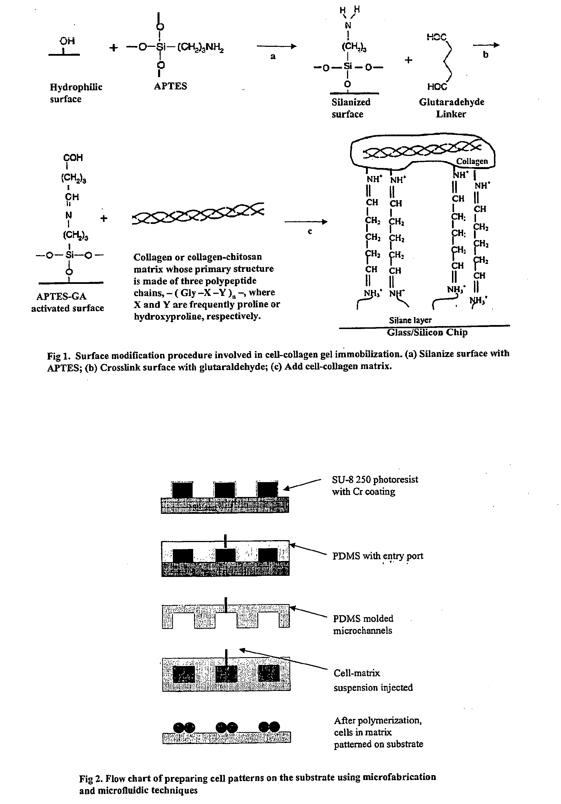
- Description
- Claims
- Application Information
AI Technical Summary
Benefits of technology
Problems solved by technology
Method used
Image

Examples
example 1
[0091] A microscale 3-layer microculturing structure for vascular tissue engineering was prepared. The multilayer system described above was used to model the vascular system, and the in-channel culture mode was used. The biomimetic system for vascular tissue engineering was investigated by separating the system into two parts: co-culture settings with a bilayer of “neo-adventitia” and “neo-media,” as well as co-culture settings with a bilayer of “neo-media” and endothelium.
[0092] Our investigation revealed the differences among the various co-culture designs. FIG. 11 shows the 3-D reconstructed confocal images for different co-culture settings. For co-cultures with an SMC-collagen layer directly attached on top of the fibroblast-collagen layer, SMC (labeled with green fluorescence) invaded into and migrated inside the fibroblast layer easily, so that the two-layered structure could not be maintained in this situation (FIG. 11a). This is a typical iin...
example 2
[0098] Structure-function relationships in neomedia and endothelial bilayers were investigated.
[0099] Adhesion molecule expression was measured by means of fluorescence microscopy applying Image-pro plus software analysis. Mean fluorescence intensities (MI) for HUVECs and SMCs in co-culture settings were compared to the MFI of unstimulated cells. After removing the microchannel stamp from the slides, the slides were washed with PBS (or HBSS) three times. Then, serum PBS (10% serum in PBS, SPBS) was added on top of the slides. After incubating for 30 minutes at room temperature with gentle shaking, the slides were washed with PBS three times, for 5 minutes every time. Then, FITC-conjugated mouse monoclonal antibody (Sigma) in SPBS (diluted 1:100) was applied to the cell patterns on the slides. Slides were again incubated for 60 minutes at room temperature with gentle shaking. Finally, slides were washed with PBS three times, and observed using fluorescence micros...
example 3
Directed Cell Migration
[0107] Flipping the position of cells in the matrix, wound healing models for cells can be established. As fibroblasts play an important role in vascular formation [Roy, 1997], they were chosen to co-culture with HUVECS. Sprout formation of ECs may be nonspecifically stimulated by nonendothelial cells possessing fibrinolytic activity; e.g., fibroblasts [Brown et al., Am J Pathol. 142(1), 273-83, 1993]. Such cells may support the migration and tubule formation of ECs by creation of a permissive matrix with formation of fibroblast-aligned channels, which might serve as guiding tracks for endothelial sprouts. Sprout formation of ECs was examined in the three types of co-culture configurations shown in FIG. 16. The “out-channel” culturing mode was used in all three co-cultures. However, HUVECs directed migration such that sprout formation was only found in the configuration shown in FIG. 16a, but not in the other two configurations. In the co-culture setting of ...
PUM
 Abstract
Description
Claims
Application Information