Biosynthetic composite for osteochondral defect repair

a biosynthetic composite and osteochondral technology, applied in the field of biosynthetic composite for osteochondral defect repair, can solve the problems of limited healing ability, premature arthritis, frequent problem of damaged cartilage in our population, etc., and achieve the effect of enhancing the healing of large osteochondral defects

Active Publication Date: 2006-08-31
MAYO FOUND FOR MEDICAL EDUCATION & RES
View PDF11 Cites 40 Cited by
  • Summary
  • Abstract
  • Description
  • Claims
  • Application Information

AI Technical Summary

Benefits of technology

[0021] As a result, a porous scaffold (e.g., tantalum) may be used instead of a bone graft as a primary structural support and interface between a periosteal explant and the subchondral bone to from a biosynthetic composite for the repair of major osteochondral defects. One advantage over other approaches is the security of graft fixation by bonding / integration to subchondral bone. Additionally, the invention is applicable with scaffolds other than tantalum, or with other cartilage-generating sources than periosteum like mesenchymal stem cells, chondrocytes, perichondrium, or a combination of them. Finally, the scaffold and cell carriers may be loaded with different matrices to carry chemical signals and / or one or more growth factors / drug release.
[0029] Therefore, the invention provides a composite for osteochondral defect repair that allows for cartilage growth from autologous periosteum chondrogenesis. Biological resurfacing of large osteochondral defects, or a complete joint is feasible using the porous scaffold / autologous periosteal composite. The use of this composite eliminates the necessity of using normal cartilage surface as a donor site and its respective associated morbidity. In one form, the strong bone integration capacity of a porous metal (e.g., tantalum) scaffold and the high grade of integration observed from periosteal chondrogenesis into the normal cartilage eliminates the lack of chondral-chondral integration observed in the autologous osteochondral graft technique.

Problems solved by technology

Damaged cartilage is a frequent problem in our population.
Unfortunately, damaged articular cartilage has a limited ability to heal and can lead to premature arthritis.
As a result, partial thickness defects that do not reach the subchondral bone stimulate only a transient induction of chondrocyte replication and matrix production in the area adjacent to the wound.
Nevertheless, when a young patient presents with a large focal osteochondral defect due to trauma or avascular necrosis, and the defect of both bone and cartilage are too large to accept any regenerative procedure, there are currently no clinically proven treatment options.
Continuous passive motion is helpful for small defects, less than 3 millimeters in diameter, and electricity has not been proven to induce cartilage healing.
Problems associated with intra-articular use of growth factors such as TGF-β include the undesirable side effects such as development of osteophytes.
The clinical outcomes of these procedures remain controversial.
For example, debridement, subchondral penetration and abrasion arthroplasty are limited to the treatment of small lesions (2) and result in a fibrous repair tissue with poor biomechanical properties.
However, the long-term viability of the hyaline-like repair tissue has not been proven.
In addition, recent results have been reported that indicate that autologous chondrocyte transplantation is no better then either autologous osteochondral cylinder transplantation or microfracture techniques.
Furthermore, progressive deterioration of the cartilage surrounding the donor site and involving weight bearing surface areas remains a main concern with this technique.
However, potential problems with the use of bone grafts include obtaining grafts of the appropriate size and shape, graft-site morbidity, and tissue integration with the surrounding tissue.
Therefore, there is no satisfactory treatment at the present time for deep osteochondral defects.

Method used


Image

  • Biosynthetic composite for osteochondral defect repair
  • Biosynthetic composite for osteochondral defect repair
  • Biosynthetic composite for osteochondral defect repair

Examples


examples

[0050] The following Examples have been presented in order to further illustrate the invention and are not intended to limit the invention in any way.

Experimental Methods

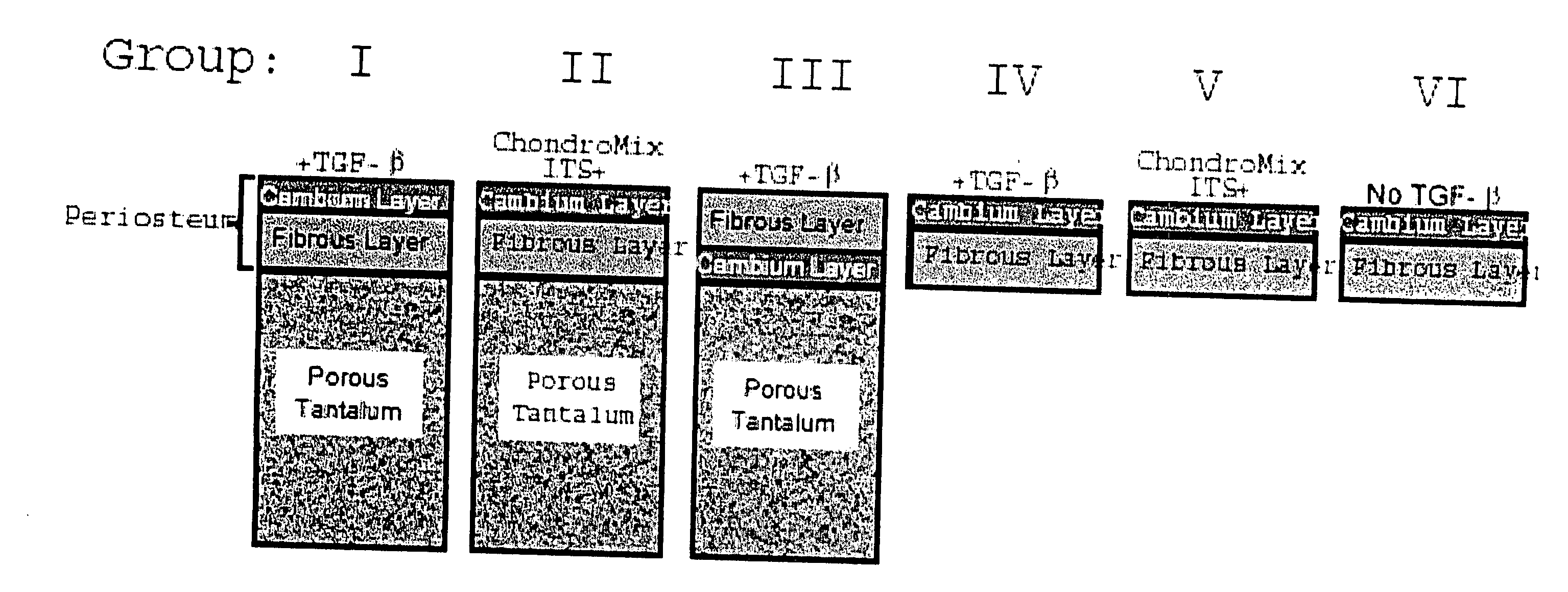
1. Periosteal Harvesting

[0051] Using a dermal punch 3.5-mm in diameter and sharp subperiosteal elevation, explants of periosteum are harvested from the medial proximal tibiae from two-month-old New Zealand white rabbits. Immediately after surgical harvesting, the periosteal explants are placed in Dulbecco's Modified Eagle Media (DMEM), with penicillin / streptomycin and 1 mM proline at 4° C. for no more than 1.5 hours prior to placement into the defect or into culture wells.
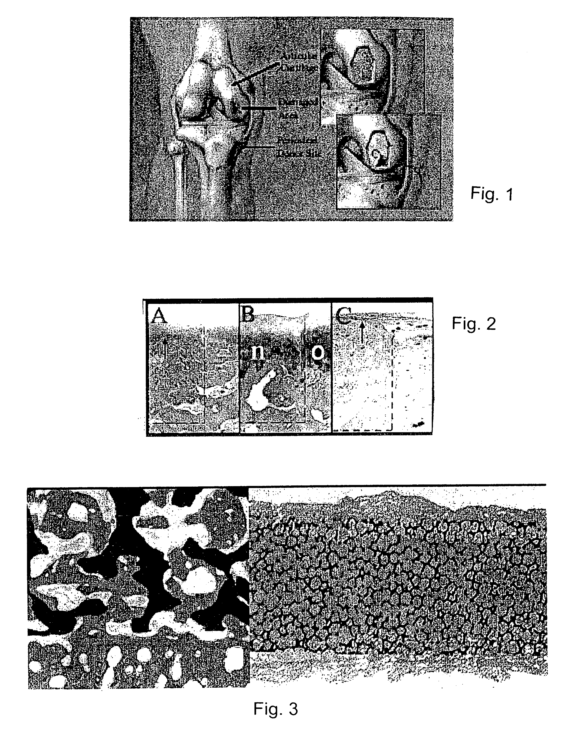
2. Histology

[0052] Samples are embedded in methyl methacrylate and sectioned using an Exakt™ System and stained with safranin O / fast green.

3. Histological Analysis

[0053] A standardized cartilage yield assay was performed after six weeks of culture (see O'Driscoll et al., “A method for automated cartilage histomorphometry.”Tissue Eng. 5:13-23...


PUM

PropertyMeasurementUnit
diameteraaaaaaaaaa
elastic modulusaaaaaaaaaa

Abstract

A composite for osteochondral defect repair includes a porous scaffold and a periosteal graft secured to a surface of the scaffold. The composite provides cartilage growth from autologous periosteum chondrogenesis. Biological resurfacing of large osteochondral defects, or a complete joint is feasible using the porous scaffold / autologous periosteal composite. The use of this composite eliminates the necessity of using normal cartilage surface as a donor site and its respective associated morbidity. In one form, the strong bone integration capacity of a porous metal (e.g., tantalum) scaffold and the high grade of integration observed from periosteal chondrogenesis into the normal cartilage eliminates the lack of chondral-chondral integration observed in the autologous osteochondral graft technique.

Description

CROSS-REFERENCES TO RELATED APPLICATIONS [0001] This application claims priority from U.S. Provisional Patent Application No. 60 / 630,622 filed Nov. 24, 2004.STATEMENT REGARDING FEDERALLY SPONSORED RESEARCH [0002] Not Applicable. BACKGROUND OF THE INVENTION [0003] 1. Field of the Invention [0004] This invention relates to an improved method and new devices for the treatment of osteochondral defects. More particularly, the invention relates to a biosynthetic composite for osteochondral defect repair. [0005] 2. Description of the Related Art [0006] Articular cartilage is the remarkably durable tissue that covers the articulating bone surfaces in our joints and permits pain-free movement by greatly reducing friction between bones and distributing stress. Damaged cartilage is a frequent problem in our population. Unfortunately, damaged articular cartilage has a limited ability to heal and can lead to premature arthritis. [0007] Adult articular cartilage contains no blood supply, neural n...

Claims


Application Information

Patent Timeline
Patent Type & Authority Applications(United States)
IPC IPC(8): A61F2/28C12N5/08
CPCA61F2/30756A61F2/468A61F2002/2817A61F2002/30764A61F2002/30766A61F2310/00131A61F2310/00365A61L27/3604A61L27/365A61L27/3654A61L27/3683A61L27/427A61L27/56
Inventor O'DRISCOLL, SHAWN W.LEWALLEN, DAVID G.MARDONES, RODRIGO
Owner MAYO FOUND FOR MEDICAL EDUCATION & RES
Why Eureka
Try Eureka