
Use of apotosis inhibiting compounds in degenerative neurological disorders

a technology of apotosis and inhibiting compounds, which is applied in the direction of applications, drug compositions, ligases, etc., can solve the problems of invariably fatal outcome, low relief, and damage that they cause, and achieve minimal immunological or inflammatory responses, reduce the need for immunosuppression, and reduce the risk

Active Publication Date: 2006-09-21
MEIRAGTX UK II LTD
View PDF11 Cites 33 Cited by
  • Summary
  • Abstract
  • Description
  • Claims
  • Application Information

AI Technical Summary

Benefits of technology

[0008] It appears that abnormal proteasome activity in neuronal cells is a contributing factor in neurodegenerative diseases such that the cells lose their ability to adequately degrade proteins, especially the mutated or misfolded proteins that may be pathological components of neurodegenerative diseases. Insofar as loss of function, or change in function, of the proteasome is a contributing factor in neuron degeneration. It has been discovered that blocking apoptotic cell death protects neurons from death following proteasome inhibition in vivo. The invention provides a method for inhibiting death of a cell of the nervous system, e.g., a neuron. Compositions comprising an inhibitor of apoptosis, e.g. an X-linked inhibitor of apoptosis protein (XIAP), which act as a potent inhibitor of caspases, e.g., caspases 9, 3 and 7, provide a therapeutic neuroprotective effect. The methods include increasing the biological activity (e.g., levels or neuroprotective effects) of a inhibitor of apoptosis protein in a region of the central nervous system.
[0011] Particularly preferred methods of delivering the vector to specific regions of the brain are those techniques that are simple, safe, and have a lower risk associated with them than lesioning, electrode implantation or cell transplantation. For example, delivery of the vector using stereotactic microinjection techniques, or delivery of the vector using specialized probes, or percutaneous delivery via disruption of the blood-brain barrier. Delivery of the vector using the method of the invention results in minimal immunological or inflammatory responses within the regions of the brain, thus eliminating the need for immunosupression. After delivery of the vector to a specific region of the brain, regional dispersion and / or diffusion of vector occurs ensuring local distribution of gene and stable gene expression.

Problems solved by technology

These diseases are debilitating and the damage that they cause is often irreversible.
Moreover, in the case of a number of these diseases, the outcome is invariably fatal.
DNA laddering precedes cell death and may be a key event leading to death.
Nonetheless, the present therapies for most, if not all, of these diseases provide very little relief.
One problem has been the relevance of current animal models to human disease.
To date, the cause of neuronal death has remained elusive.
In order to test the value of protective or curative strategies, however, the mechanism of cell death must be relevant to human disease otherwise successful experimental studies will not translate into effective human therapy.

Method used


Image

  • Use of apotosis inhibiting compounds in degenerative neurological disorders
  • Use of apotosis inhibiting compounds in degenerative neurological disorders
  • Use of apotosis inhibiting compounds in degenerative neurological disorders

Examples


example 1

Methods and Materials

(i) XIAP Expression Plasmids

[0111] A full length human XIAP cDNA was amplified from U87-MG cells and cloned into pcDNA4. To produce pdXIAP, C-terminal 48 amino acid comprising the RING domain were removed in a second round of PCR. The integrity of all constructs was verified by sequencing.

(ii) Recombinant AAV Vectors

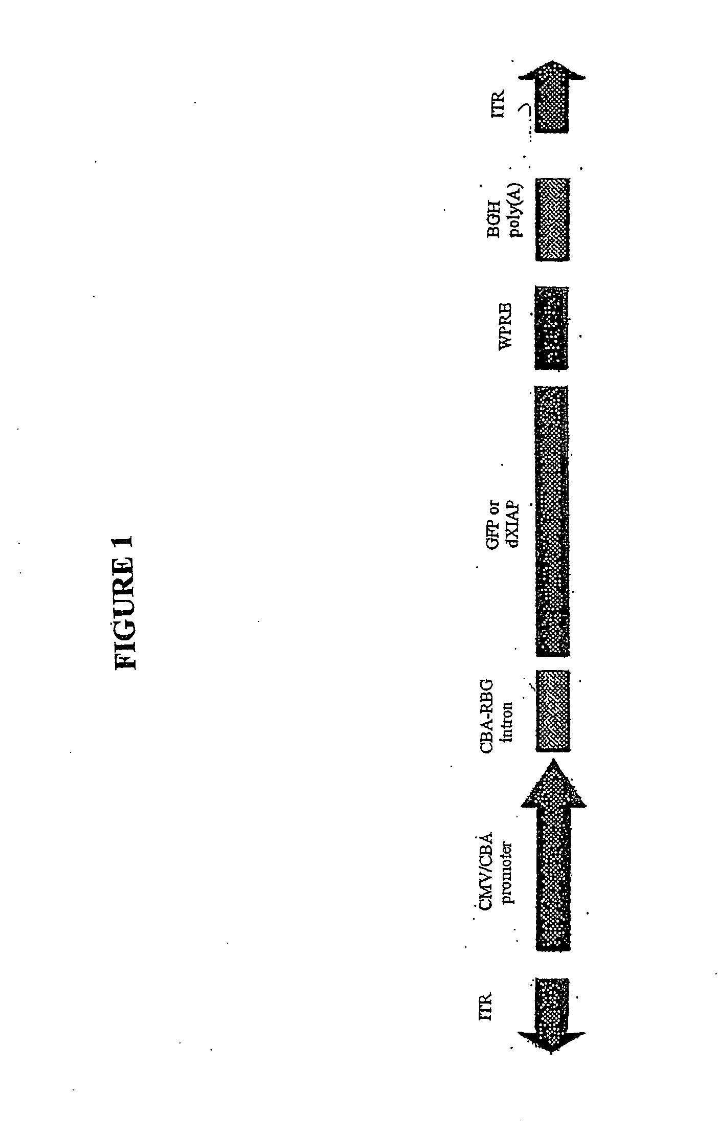
[0112] To generate AAV.dXIAP, the XIAP ORF lacking C-terminal 48 amino acid was PCR-amplified and cloned into an AAV expression plasmid (FIG. 1). It was engineered to contain the Kozak consensus translation start site. A control vector was generated by subcloning the EGFP cDNA into the same AAV backbone. Virus stocks were prepared by packaging the vector plasmids into AAV cerotype 2 particles using helper-free plasmid transfection system. The vectors were purified using heparin affinity chromatography and dialyzed against PBS. rAAV titers were determined by quantitative PCR using CMV-enhancer-specific primers and adjusted to 1012 genomic partic...

example 2

XIAP Protects SH-SY5Y Cells from 6-OHDA-Induced Apoptosis

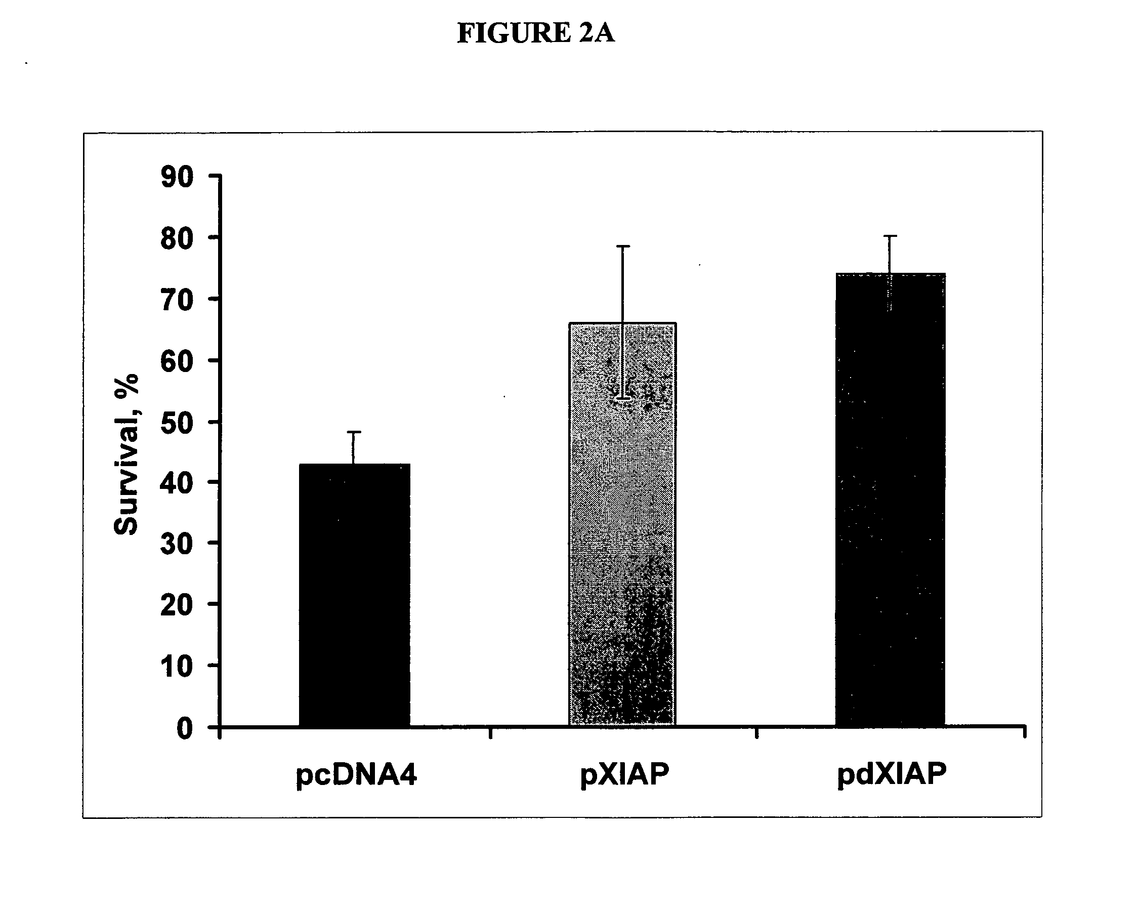
[0118] This example describes how an inhibitor of apoptosis protein, XIAP, protects from 6-OHDA-induced apoptosis. SH-SY5Y cells were transfected with indicated plasmids, treated with 70 nM 6-OHDA and stained with propidium iodide. The results from the study are shown in FIG. 2A. Note a significant reduction of cell death by either XIAP or dXIAP compared to a control. All transfections were performed in triplicates. * p<0.001.

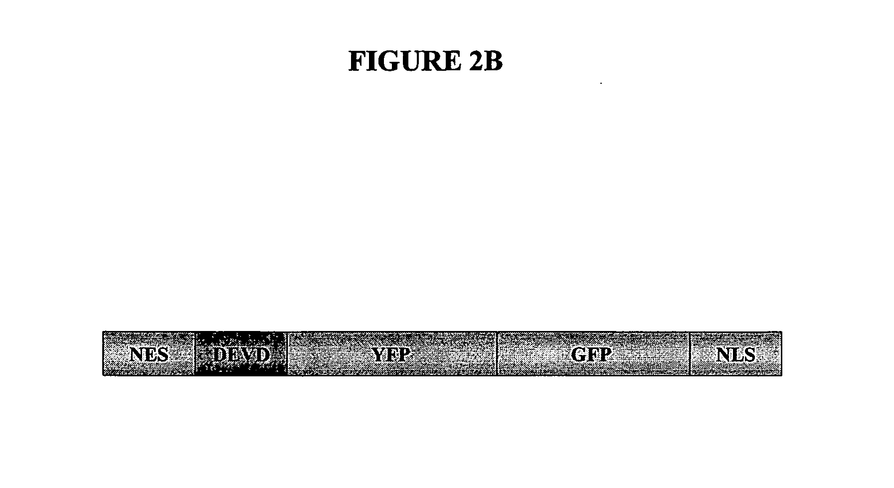
[0119] The location of apoptotic proteins was determined in an apoptosis assay using the pCaspase3-Sensor vector shown in FIG. 2B. The caspase-3 / 7 cleavage site appears in the DEVD region of the vector. The vector was transfected into cells and the results are shown in FIG. 2C. This fusion protein resides predominantly in the cytoplasm in normal cells (green). During apoptosis a nuclear exclusion signal (NES) at the N-terminus is cleaved by caspase-3 and the protein quickly translocates into the nucleus...

example 3

XIAP is protective in In Vitro Models of Familial Parkinson's and Huntington's Diseases

[0121] This example describes how an inhibitor of apoptosis protein, XIAP, is protective in in vitro models of familial Parkinson's and Huntington's diseases. pAAV.GFP and pAAV.dXIAP were cotransfected into SH-SY5Y cells with pa-syn-RFP (FIG. 3A) or pQ111-RFP encoding for RFP fusions of the murine α-synuclein and the first exon of the mouse Huntington gene containing additional polyglutamine repeats, respectively. Apoptotic cells were scored 24 h post-transfection using YO-PRO-1 vital DNA dye (FIG. 3B). Note that live cells shown in the top panel (dashed arrow) are impermeable to YO-PRO-1 while cells undergoing apoptosis (solid arrow) are stained positive. * p<0.001. Phase contrast is shown in FIG. 3C. The percentage of cells undergoing apoptosis is shown in FIG. 3D.

[0122] These results demonstrate that dXIAP is a potent inhibitor of apoptosis in several in vitro models of Parkinson's disease in...


PUM


Abstract

The invention provides methods and compositions for localized delivery of a vector comprising a therapeutic agent to a specific region of the brain associated with a neurodegenerative diseases that is characterized by an excess buildup of buildup of intracellular protein aggregates. In particular, the invention provides methods and compositions used to deliver an adeno-associated virus vector (AAV) comprising a nucleotide sequence encoding an inhibitor of apoptosis protein (IAP) to cells in the region.

Description

CROSS REFERENCE TO RELATED APPLICATIONS [0001] This application claims priority to U.S. Provisional Patent Application No. 60 / 686,588 filed on Jun. 2, 2005 and entitled “Use of Apotosis Inhibiting Compounds in Degenerative Neurological Disorders,” and to U.S. Provisional Patent Application No. 60 / 621,307 filed on Oct. 22, 2004 and entitled “Use of IAP-Proteosome Inhibiter in Degenerative Neurological Disorders.”BACKGROUND OF THE INVENTION [0002] The invention is generally in the field of methods and compositions for treating neurodegenerative diseases characterized by excess buildup of intracellular protein aggregates such as Parkinson's disease (PD), using viral and non-viral delivery systems that deliver therapeutic agents to specific regions of the brain. More specifically, using an adeno-associated viral vector to deliver a nucleotide sequence encoding an inhibitor of apoptosis protein (IAP) to specific regions of the brain associated with such neurodegenerative diseases. [0003]...

Claims


Application Information

Patent Timeline
IPC IPC(8): A61K48/00C12N15/86
CPCA61K48/00C07K14/4747C07K2319/02C12N9/93C12N15/86C12N2750/14111C12N2750/14143C12N2800/107C12N2830/15C12N2830/48A61P25/00A61P25/16A61P25/28
Inventor KAPLITT, MICHAELMOUSSATOV, SERGUEI
Owner MEIRAGTX UK II LTD
Learn More