Apparatus and methods for efficient processing of biological samples on slides

a technology of slides and apparatuses, applied in the field of apparatuses for processing biological samples on slides, can solve the problems of time and labor, inability to use most commercial ready-to-use reagents in automated systems, and high labor intensity of assays, so as to achieve the effect of faster and convenient assays

Inactive Publication Date: 2006-09-28
AMERICAN REGISTRY OF PATHOLOGY
View PDF14 Cites 9 Cited by
  • Summary
  • Abstract
  • Description
  • Claims
  • Application Information

AI Technical Summary

Benefits of technology

[0006] The present invention relates to an apparati and methods for performing assays on biological samples mounted on microscope slides. Use of the apparati and / or methods aid in making assays more rapid and convenient. One aspect of the invention is the use of reagents which are predried in the wells of the tray thereby simply necessitating the addition of water or buffer to the well without having to add the reagents at the time of assay. The well is then covered with a slide with a biological sample premounted on the slide. The different wells of a multiwell tray can be pretreated with different reagents dried in each well. Multistep assays can be performed by moving a slideholder with attached slides from one multiwell tray to the next, with each well of a multiwell tray having the desired reagents predried on it. A variation of this is to employ a multilayer coating of reagents in each well such that the first set of reagents dissolves quickly and acts upon the biological sample, the second layer then dissolves releasing the reagents for the second step, etc., thereby requiring the use of fewer trays, possibly only a single tray.

Problems solved by technology

These assays are also quite labor intensive although there are now some automated systems (e.g., the Ventana ESIHC Staining System, the Shandon Lipshaw Cadenza Automated Immunostainer; also see Brigati et al.
Most automated systems which only perform immunocytochemistry do not perform deparaffinizing, histochemistry (such as hematoxylin and eosin staining) and coverslipping steps and these consequently must be done separately by hand which is time and labor intensive.
Furthermore, most commercial ready-to-use reagents are not suitable for automated systems which are required to use specially designed reagents.
Laboratories which process large numbers of samples are likely to be willing to pay the high cost associated with buying these automated systems as well as the high cost of using the disposable accessories and reagents to perform the assays, but small to intermediate sized laboratories find it more cost effective to continue to process samples manually.
Besides being very labor intensive, there are drawbacks associated with the commonly used method of simply dropping solutions on top of the mounted sample on the microscope slide.
These features require use of extra reagents which are quite expensive.
Leaving the solutions open to the air as they sit on the slide also may lead to evaporation if the samples must incubate for a long period of time.
Evaporation leads to concentration or drying out of the reagents and high concentrations may lead to increased background levels which are clearly undesirable.
If the solutions evaporate totally the assay will fail.
Incubating samples in humidity chambers with covers may prevent evaporation problems, but water droplets which condense onto the humidity chamber cover may fall onto the slides and this will ruin the assay.

Method used


Image

  • Apparatus and methods for efficient processing of biological samples on slides
  • Apparatus and methods for efficient processing of biological samples on slides
  • Apparatus and methods for efficient processing of biological samples on slides

Examples


example 1

Immunocytochemistry

[0034] In this Example a biological sample is treated with antibodies (primary and secondary), treated for chromogen color development, and finally counterstained.

A. Proteolytic Pretreatment of Mounted Tissue Samples

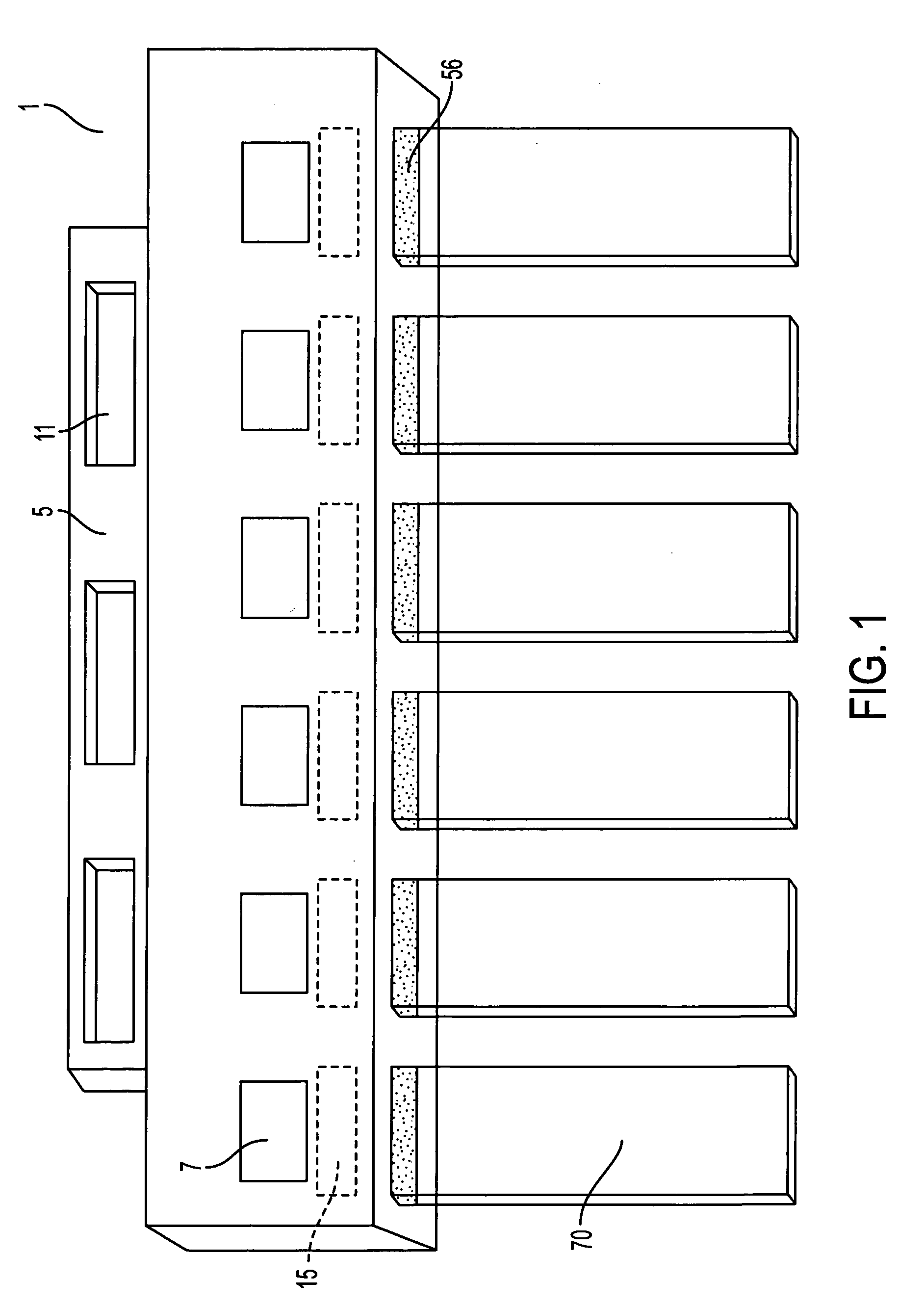
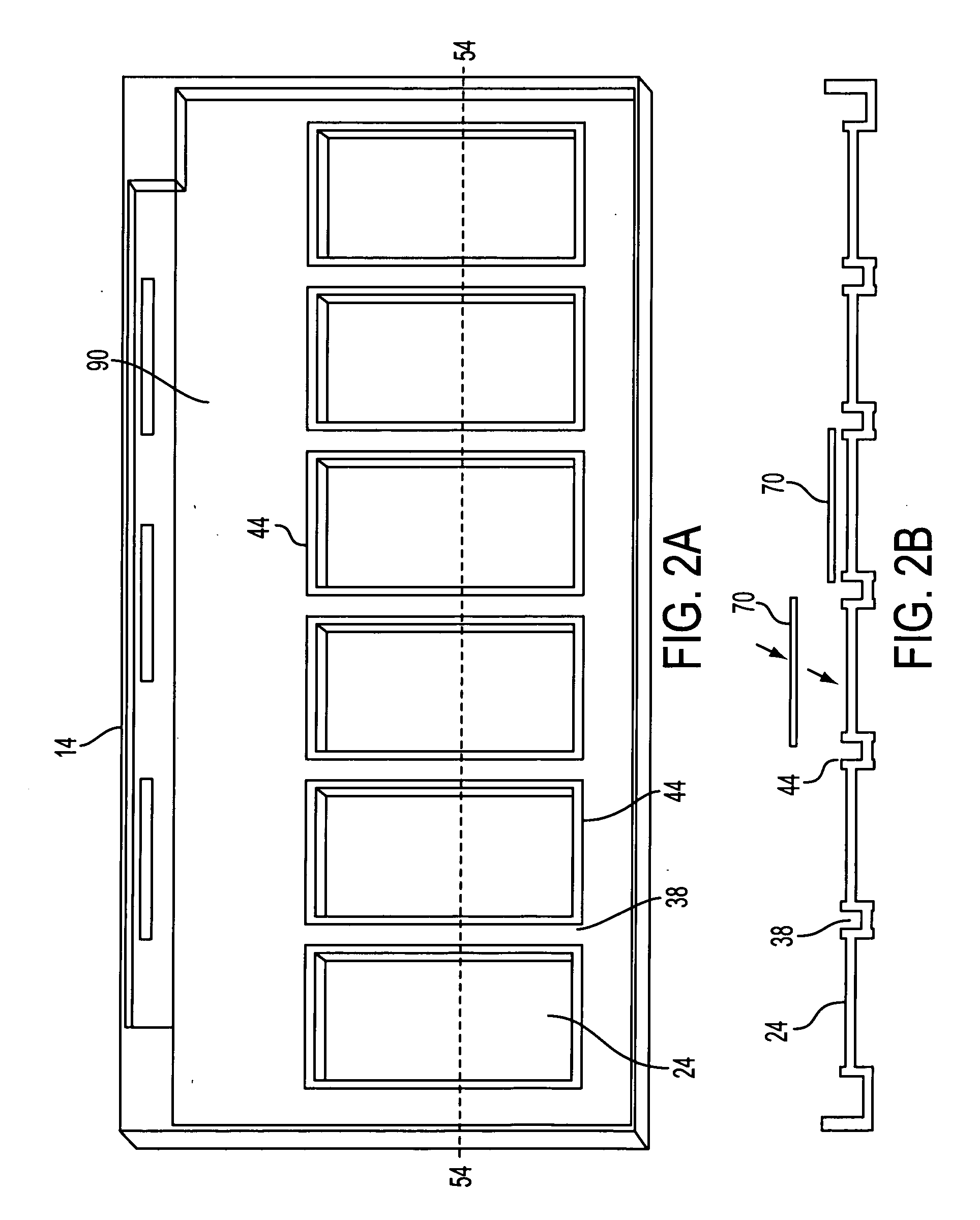
[0035] It is well known in the art that when using certain antibodies for immunocytochemical staining it is necessary to pretreat the formalin fixed tissue section with proteolytic enzymes such as 0.4% pepsin, pH 2.0. When this is necessary the following steps may be utilized. A few drops (150-200 μL) of the proteolytic digestion solution are placed on each well 24 of the 3 or 6 well tray 14. The tissue side of the slides 70 is faced down on the wells 24. The slideholder 1 with the slides 70 should be slowly laid down and placed on the wells 24 of the tray 14. No air bubbles should remain between the tissue side of the slides 70 and the solution in the wells 24 of the tray 14. The slides 70, slideholder 1 and tray 14 with solution are incubated for...

example 2

In Situ Hybridization

[0049] In this example biological samples are mounted onto slides 70, hybridized with biotin or digoxigenin labeled probes and reacted with anti-biotin or anti-digoxigenin antibody. The samples are then stained.

A. Preparation and Mounting of Tissue Sample

[0050] A tissue sample is prepared as described above but with extra measures to prevent nucleic acid degradation. A tissue sample is fixed in 10% neutral buffered formalin, processed overnight on a tissue processor, embedded in paraffin, cut into serial sections of 4-5 microns, mounted onto Probe-On-Plus Slides (#15-188-52; Fisher Scientific), and dried overnight at room temperature. The slides 70 are inserted into a slideholder 1 and are deparaffinized by placing into a staining dish. The slides 70 are treated with four changes of xylene for 5 minutes each, two changes of 100% ethanol for 1 minute each and two changes of 95% ethanol for 1 minute each. The deparaffinized tissue section slides are then clear...

example 3

PCR In Situ Hybridization

[0062] Polymerase chain reaction (PCR) was developed as an in vitro method for amplifying small amounts of specific pieces of nucleic acids. This was later adapted to in situ studies so that there was amplification of nucleic acid within tissue sections. The apparatus of the present invention is suited to performing these in situ PCRs. An example of a PCR in situ hybridization protocol is given in Nuovo (1994).

A. In Situ PCR

[0063] Serial tissue sections are cut at 4-5 microns thickness, mounted onto Probe-On-Plus slides 70, and dried overnight at room temperature. The mounted tissue sections are deparaffinized and digested with pepsin at 40° C. for 15-90 minutes depending on the length of time of fixation in formalin. The pepsin is inactivated by washing the slides 70 in diethylpyrocarbonate (DEPC) treated water for one minute followed by a one minute wash in 100% ethanol. The slides 70 are then air dried.

[0064] Polymerase chain reaction solutions are m...


PUM

PropertyMeasurementUnit
volumeaaaaaaaaaa
volumeaaaaaaaaaa
poweraaaaaaaaaa

Abstract

Methods for treating biological samples on microscope slides are set forth. One aspect of the invention is the use of predried reagents in wells on trays onto which the slides are placed, especially the use of predried reagents which dissolve sequentially. Yet another aspect of the invention is the use of external controls placed directly on a microscope slide in conjunction with a biological sample to be assayed. The external controls can be conveniently placed on a membrane which can be affixed to the slide. A further aspect of the invention is a specially designed tray to allow whole chromosome painting of all chromosomes of a cell sample on a single slide. The invention is also drawn to a coverslip with concave wells which act as reaction chambers when placed against a slide and filled with buffer. Preferably a reagent is predried in the well. A further aspect of the invention is a method of reacting samples on slides by placing them into a reaction chamber together with a coverslip which has a predried reagent on it.

Description

CROSS-REFERENCE TO RELATED APPLICATIONS [0001] This application is a continuation of co-pending U.S. patent application Ser. No. 09 / 869,082, filed 24 Sep. 2001, which was filed under 35 U.S.C. §371 based on PCT / US99 / 30519, filed 22 Dec. 1999, which is a continuation-in-part of U.S. Ser. No. 09 / 219,443, filed 23 Dec. 1998. This application claims priority to both PCT / US99 / 30519 and to U.S. Ser. No. 09 / 219,443, U.S. Pat. No. 6,703,247, as well as U.S. patent application Ser. No. 09 / 869,082, the contents of which are incorporated herein in their entirety.BACKGROUND OF THE INVENTION [0002] This invention relates to an apparatus for processing biological samples on slides for a wide variety of purposes. Biological samples are analyzed for many purposes using a variety of different assays. Pathologists often use histochemistry or immunocytochemistry for analyzing biological samples, molecular biologists may perform in situ hybridization or in situ polymerase chain reactions on biological ...

Claims


Application Information

Patent Timeline
Patent Type & Authority Applications(United States)
IPC IPC(8): C12Q1/68C12M1/34B05D3/02
CPCB01L3/50853B01L2300/047B01L2300/0822C12Q1/6841
Inventor CHU, WEI-SING
Owner AMERICAN REGISTRY OF PATHOLOGY
Why Eureka
Try Eureka