
Method of detecting allergen protein

a technology of allergen proteins and detection methods, applied in the field of proteins, can solve the problems of food allergies, easy loss of allergen and inducing fatal symptoms, and achieve the effect of high analytical resolution of proteins and low risk of losing proteins in a very small quantity

Inactive Publication Date: 2006-10-12
INC ADMINISTRATIVE AGENCY NAT AGRI & BIO ORIENTED RES ORG
View PDF2 Cites 9 Cited by
  • Summary
  • Abstract
  • Description
  • Claims
  • Application Information

AI Technical Summary

Benefits of technology

[0023] According to the method of the present invention, a protein having a disulfide bond can be specifically detected with high sensitivity. Furthermore, when such the method for detecting a protein having a disulfide bond is applied for detection of an allergen protein, the allergen protein in a sample to be tested can be efficiently detected. According to the method for detecting an allergen protein of the present invention, allergen proteins present in a very small quantity that has been difficult to detect by conventional screening methods for allergen proteins can also be detected with high sensitivity.
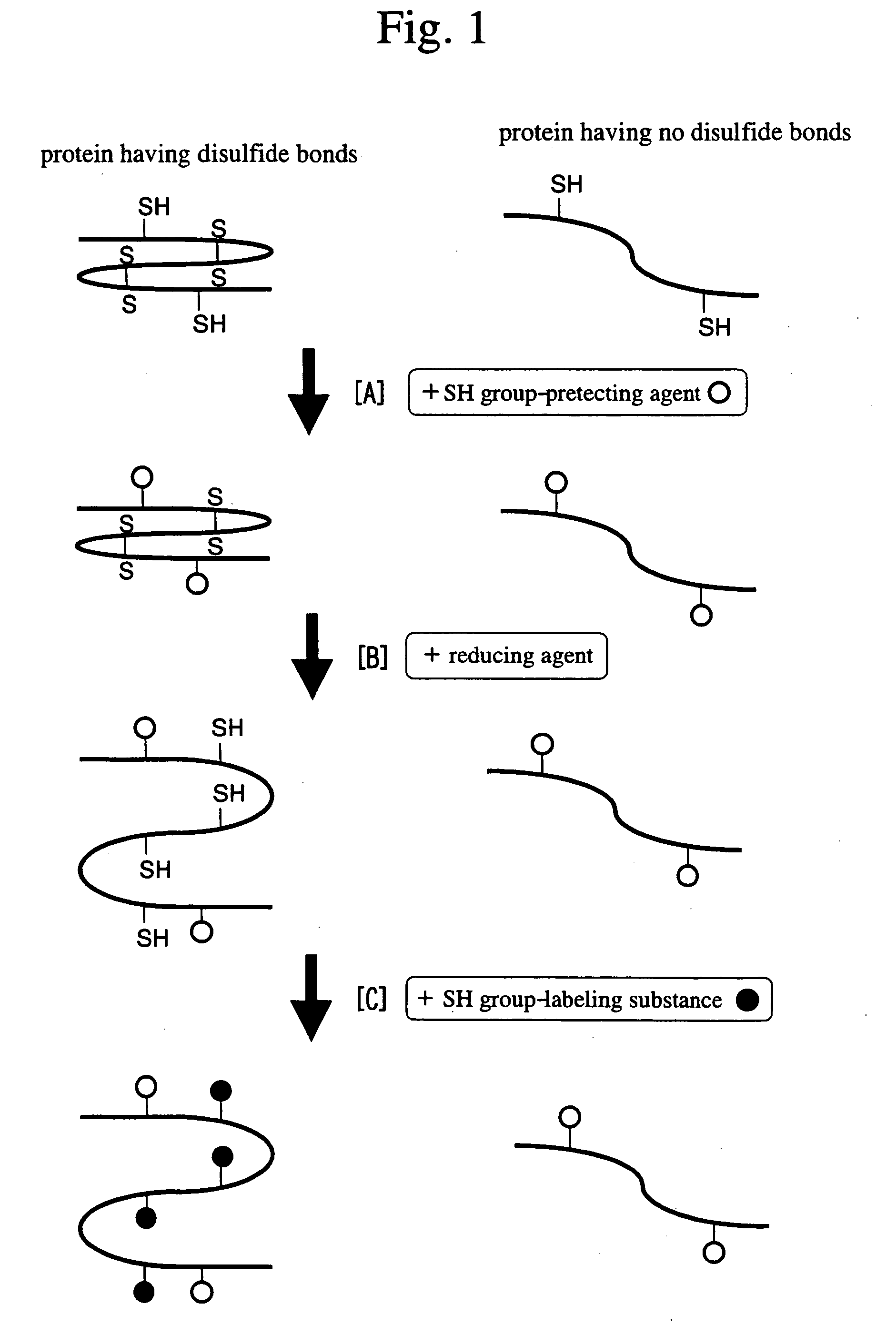
[0061] In the method of the present invention, detection of exposed SH groups using an SH group-detecting substance may be carried out by ordinary methods. When exposed SH groups are detected using such SH group-detecting substance, exposed SH groups are caused to react with an SH group-detecting substance, and then detection can be directly carried out for a sample to be tested containing a number of proteins. However, after the reaction of exposed SH groups with an SH group-detecting substance, a sample to be tested may be previously separated by a conventionally known protein separation method, and then the exposed SH groups may be detected. For example, proteins in the above sample to be tested may be separated by a method known by persons skilled in the art, such as one-dimensional electrophoresis, two-dimensional electrophoresis, high performance liquid chromatography (HPLC), column chromatography, or mass spectrometry. After separation of proteins by subjecting a sample to be tested to electrophoresis, detection may be carried out on the electrophoretic gel. After separation of proteins by subjecting sample to be tested to high performance liquid chromatography (HPLC), detection may be carried out in the eluted molecular weight fractions. In the method of the present invention, it is particularly preferable to separate proteins by two-dimensional electrophoresis and then carry out detection thereof. With the use of two-dimensional electrophoresis, there is an advantage in that not only are proteins isolated with high resolution and high accuracy, but also the following detection for one sample to be tested can be completed in a single detection.
[0082] This method particularly has the following advantages compared with conventional methods.
[0084] Separation by two-dimensional electrophoresis leads to higher analytical resolution of proteins.
[0085] No transfer to a membrane is needed and risk of losing proteins present in a very small quantity is low.

Problems solved by technology

Allergen proteins show harmful effects resulting in food allergies, house dust allergies, pollenosis, and the like, and occasionally induce fatal symptoms, for example, of anaphylaxis.
When this method is employed, allergen proteins present in a very small quantity are easily lost upon transfer to membranes, indicating that some important allergen proteins may be overlooked and remain undetected.
However, there are no known methods whereby allergen proteins can be detected with high sensitivity.

Method used


Image

  • Method of detecting allergen protein
  • Method of detecting allergen protein
  • Method of detecting allergen protein

Examples


example 1

Detection of Proteins Having Disulfide Bonds in a Rice Extract

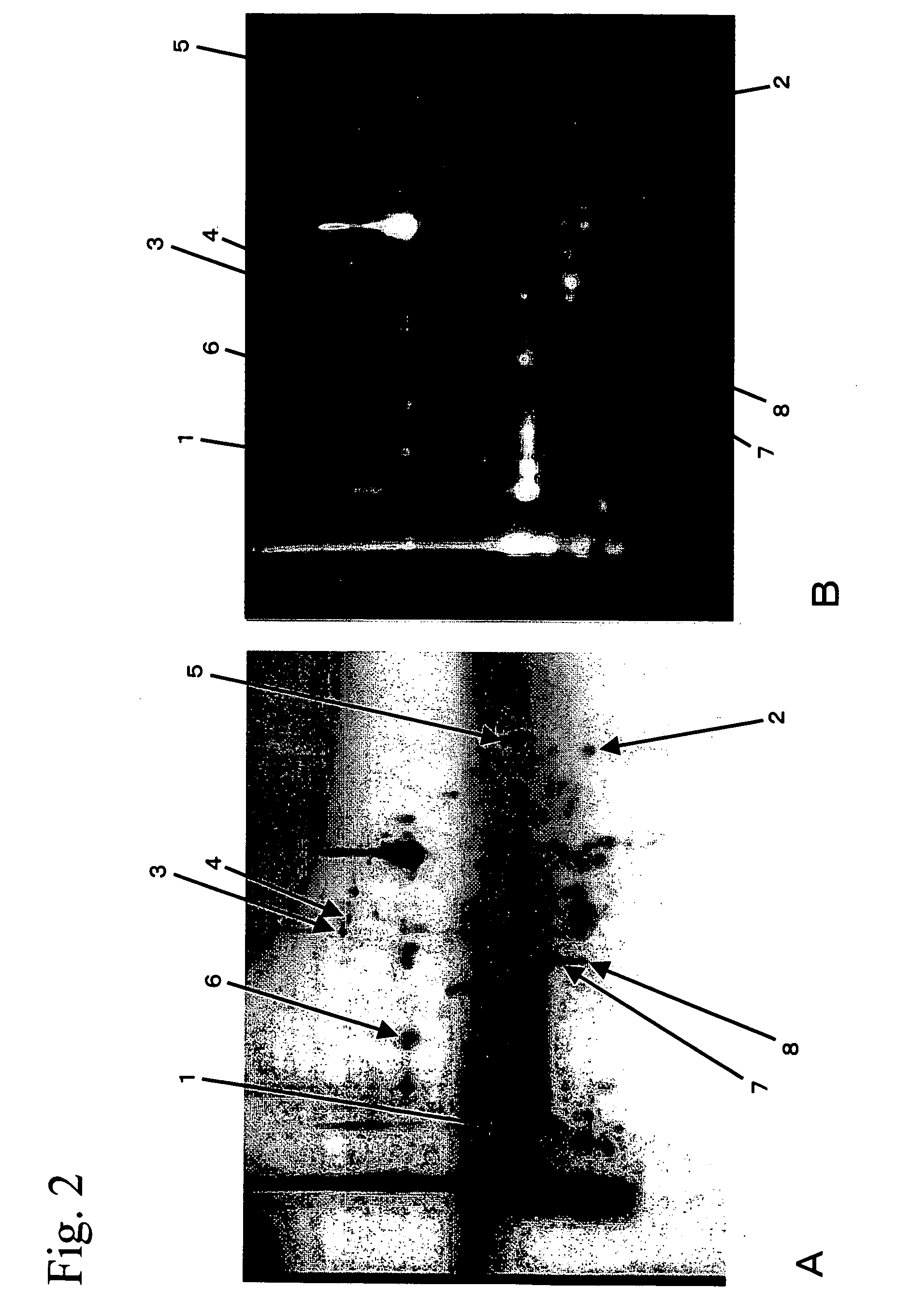
[0098] 20 g of rice seeds was ground using a mortar. The powder was put in 400 ml of 1 M sodium chloride solution and then agitated for 1 hour, so that proteins from the seeds were extracted in the buffer. After the solution was centrifuged at 14,000 g for 5 minutes to precipitate insoluble constituents, the supernatant was collected. The supernatant was desalted by dialysis and then freeze-dried. A 1 / 80 amount of the freeze-dried product (equivalent to 0.25 g of the rice seeds) was dissolved in a buffer for isoelectric focusing (8 M urea, 0.5% CHAPS, and 0.1% Bio-Lytes). lodoacetamide as an SH group-protecting agent was added (to a final concentration of 5 mM) to the solution, and then the solution was incubated at room temperature for 1 hour. Next, dithiothreitol as a reducing agent was added (to a final concentration of 5 mM) to the solution, and then the solution was incubated at room temperature for 1 hour. Furtherm...

example 2

Analysis of Allergen Proteins Contained in the Rice Extract

[0106] Spots 1 to 6 for which fluorescence signals were detected in FIG. 2B were excised from the gel and then digested within the gel using trypsin, so that the proteins in the spots were fragmented. After the fragmented proteins were subjected to HPLC for separation, the internal amino acid sequences were determined by the Edman method. The thus determined internal amino acid sequences for proteins in each spot are shown in Table 1. “CmBBr” in the internal amino acid sequences shown in Table 1 denotes cysteine residues having SH groups labeled with monobromobimane. Next, a homology search was carried out for each of the internal amino acid sequences as a query sequence using the FASTA program and the BLAST program within amino acid sequence databases (GenBank and PIR). The search results showed that the internal amino acid sequence of the protein from each spot is identical to or has high homology with a partial amino aci...

example 3

Detection of Proteins Having Disulfide Bonds in a Pollen Extract

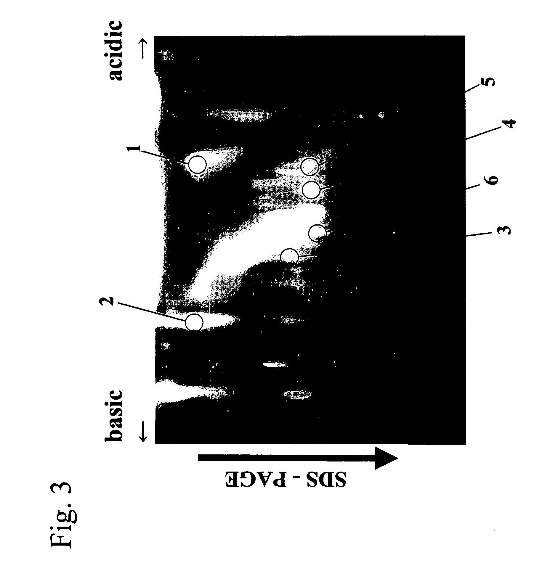
[0109] Ragweed (Ambrosia trifida) pollen was ground in a 100 mM Tris-HCl buffer (pH 8.0) containing 1 mM PASF and 1 mM EDTA using a mortar, and then the resultant was centrifuged at 14,000 g for 30 minutes. After centrifugation, the supernatant was collected, filtered through an Ultrafree-CL centrifugal filter (Millipore), and then desalted through a Microcon YM-10 centrifugal filter. The desalted residue was dissolved in a buffer for isoelectric focusing (8 M urea, 0.5% CHAPS, and 0.1% Bio-Lytes). lodoacetamide as an SH group-protecting agent was added (to a final concentration of 5 mM) to this solution, and then the solution was incubated at room temperature for 1 hour. Furthermore, dithiothreitol as a reducing agent was added (to a final concentration of 5 mM) to and mixed with the solution, and then the solution was incubated at room temperature for 1 hour. Furthermore, monobromobimane as an SH group-labeling subst...


PUM


Abstract

The present invention relates to a method for detecting a protein having a disulfide bond or an allergen protein, comprising: protecting by chemically modifying a free SH group of a protein in a sample to be tested; cleaving a disulfide bond of the free SH group-protected protein to expose SH groups; and detecting the exposed SH groups.

Description

TECHNICAL FIELD [0001] The present invention relates to a method for detecting proteins, and particularly relates to a method for detecting allergen proteins. BACKGROUND ART [0002] Allergen proteins are broadly present in our living environment. Allergen proteins show harmful effects resulting in food allergies, house dust allergies, pollenosis, and the like, and occasionally induce fatal symptoms, for example, of anaphylaxis. To elucidate the allergy onset mechanism and to develop a protective method against allergies, it is an important object to screen for allergen proteins comprehensively. [0003] As a conventional screening method for allergen proteins, a detection method based on the immunoblotting method, which involves transferring proteins to membranes such as nitrocellulose or PVDF, reacting the proteins with IgE antibodies in the blood serum of an allergy patient, and then detecting the antibodies binding to the proteins, is employed (e.g., Weiss, W., et al., Electrophores...

Claims


Application Information

Patent Timeline
IPC IPC(8): C12Q1/26G01N33/00G01N27/447G01N33/68
CPCG01N33/6815
Inventor YANO, HIROYUKIKURODA, SHIGERU
Owner INC ADMINISTRATIVE AGENCY NAT AGRI & BIO ORIENTED RES ORG
Learn More