

Tissue engineered blood vessels
a technology of tissue engineering and blood vessels, applied in the field of tissue engineering blood vessels, can solve the problems of poor mechanical and burst strength of the substitute, failure of the graft,
- Summary
- Abstract
- Description
- Claims
- Application Information
AI Technical Summary
Benefits of technology
Problems solved by technology
Method used
Image

Examples
example 1
Methods and Materials
Materials
[0166] Unless otherwise stated, all chemicals were purchased from Sigma (St. Louis, Mo.). ECL buffers and 125I-Sodium were purchased from PerkinElmer NEN (Boston, Mass.). Collagenase type II (1 mg / ml) was purchased from Boehringer Mannheim (Mannheim, Germany). Endothelial medium (EBM-2) was purchased from Cambrex Bio Science (Walkersville, Md.). PCR reagents and primers, M199 medium, fetal bovine serum (FBS) and penicillin were purchased from Life Technologies (Gaithersburg, Md.). Basic Fibroblast Growth Factor (bFGF) was a gift from Judith Abraham, (Scios Nova, Calif.). FITC-labeled monoclonal anti human CD31 antibodies were purchased from Santa Cruz (Santa Cruz, Calif.). Rabbit polyclonal anti vonWillebrand factor (vWF), anti smooth muscle actin antibodies, and FITC labeled anti-rabbit IgG antibodies were purchased from DAKO (Glostrup, Denmark). Monoclonal anti human CD31, Goat polyclonal anti-VE Cadherin (VE-Cad), Goat anti-vWF and Rabbit polyclon...
example 2
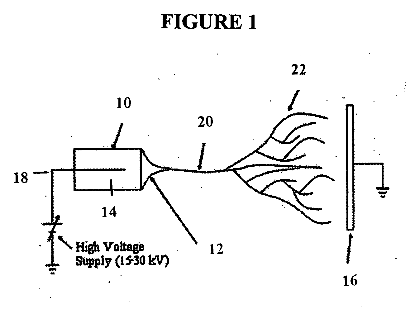
Electrospun Matrices
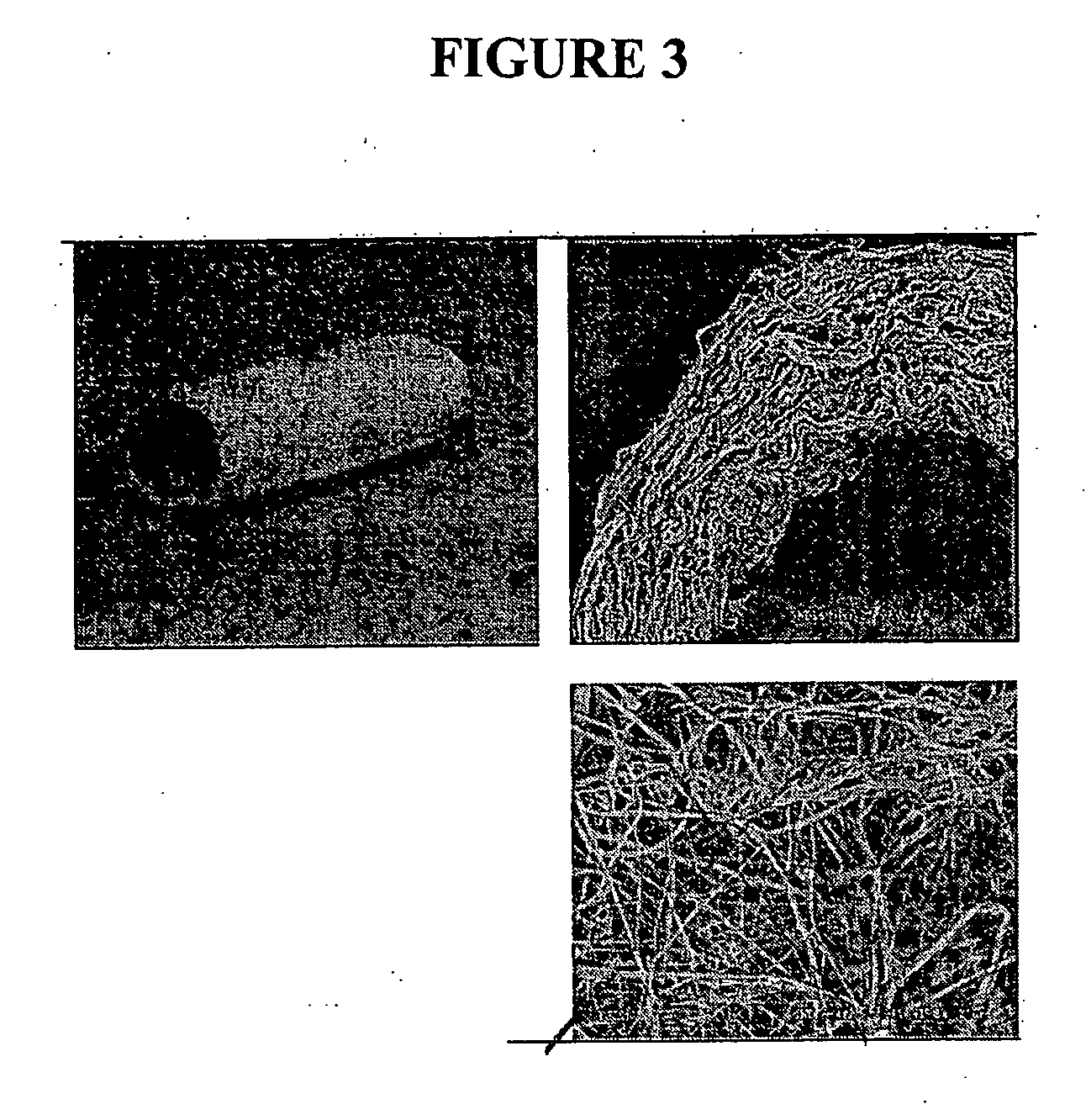
[0186] An electrospun matrix was formed using the methods outlined in Example 1. A solution of collagen type I, elastin, and PLGA, were used. The collagen type I, elastin, and PLGA were mixed at a relative concentration by weight of 45% collagen, 40% PLGA, and 15% elastin.
[0187] The resulting fibrous scaffold had a length of 12 cm with a thickness of 1 mm. A 2 cm representative sample is depicted in FIG. 3. This demonstrates the feasibility of spinning Type I Collagen and elastin into fibers from nanometer to micrometer diameter using concentrations from 3% to 8% by weight in solution. These results also show that by adding PLGA (MW 110,000) to the mixture, solutions with higher viscosity and improved spinning characteristics could attained. By increasing the solution concentration to 15%, thicker, stronger scaffolds were able to be built while maintaining the collagen and elastin components.
[0188] Collagen type I stained positively on the decellularized scaff...
example 3
Cross-Linking of Electrospun Matrices
[0195] This example demonstrates how to increase the strength and stability of the electrospun scaffold by chemical cross-linking. The scaffolds were soaked in 20% dextran solution in phosphate buffered saline prior to crosslinking to reduce hydration-induced swelling and contraction of the scaffold. The scaffolds were crosslinked by immersion in EDC / NHS in MES / EtOH solution for 2 hours at room temperature. Scanning electron micrographs of the resulting fibers showed fiber diameters of 500 nm or less and a random orientation of fibers. Atomic force microscopy of the scaffold and a confocal image of nanofibers with an adhering endothelial cell demonstrate the scaffold structure. These data show that it is possible to fabricate vascular scaffolds from biological polymers with mechanics and structure similar to decellularized scaffolds and native arteries.
PUM
 Abstract
Description
Claims
Application Information