Method and system for assessment of biomarkers by measurement of response to surgical implant

a biomarker and surgical implant technology, applied in the field of biomarker assessment by surgical implant measurement, can solve the problems of inability to accurately describe complex topology or shape in an accurate manner, limited resolution, and inability to accurately assess and quantify conventional measurements, etc., to achieve improved structure or function, improve the effect of function and high resolution

Inactive Publication Date: 2006-11-02
VIRTUALSCOPICS
View PDF11 Cites 17 Cited by
  • Summary
  • Abstract
  • Description
  • Claims
  • Application Information

AI Technical Summary

Benefits of technology

[0027] To achieve the above and other objects, the present invention is directed to a system and method for accurately and precisely identifying important structures and sub-structures, their normalities and abnormalities, and their specific topological and morphological characteristics—all of which are sensitive indicators of disease processes and related pathology. Biomarker measurements both before and after the implantation of a surgical implant are taken, so that the response of the biomarker to the surgical implant can be determined. Surgical implants include stents, meshes, and other biocompatible or resaborbable objects that are positioned within the body to effect an improved structure or function.
[0028] The preferred technique is to identify the biomarkers based on automatic techniques that employ statistical reasoning to segment the biomarker of interest from the surrounding tissues (the statistical reasoning is given in Parker et al., U.S. Pat. No. 6,169,817, whose disclosure is hereby incorporated by reference in its entirety into the present disclosure). This can be accomplished by fusion of a high resolution scan in the orthogonal, or out-of-plane direction, to create a high resolution voxel data set (Peña, J.-T., Totterman, S. M. S., Parker, K. J. “MRI Isotropic Resolution Reconstruction from Two Orthogonal Scans,”SPIE Medical Imaging, 2001). In addition to the assessment of subtle defects in structures, this high-resolution voxel data set enables more accurate measurement of structures that are thin, curved, or tortuous. More specifically, this invention improves the situation in such medical fields as oncology, neurology, and orthopedics. In the field of oncology, for example, the invention is capable of identifying tumor margins, specific sub-components such as necrotic core, viable perimeter, and development of tumor vasculature (angiogenesis), which are sensitive indicators of disease progress or response to therapy. Similarly, in the fields of neurology and orthopedics, the invention is capable of identifying characteristics of both the whole brain and prosthesis wear, respectively.

Problems solved by technology

Currently, medical imaging techniques such as MRI, CT, and ultrasound are used to assess biological structures and sub-structures and offer a limited degree of resolution.
However, the conventional measurements are not well suited to assessing and quantifying subtle changes in lesion size, and are incapable of describing complex topology or shape in an accurate manner or of addressing finer details of biological structure(s).
In consideration of current medical imaging and tracking techniques, it becomes apparent that there are many disadvantages in using such technologies.
The need for intensive and expert manual intervention is a disadvantage, since the demarcations can be tedious and prone to a high inter- and intra-observer variability.
Furthermore, the typical application of manual measurements within two-dimensional (2D) slices, or even sequential 2D slices within a 3D data set, is not optimal since tortuous structures, curved structures, and thin structures are not well characterized within a single 2D slice, leading again to operator confusion and high variability in results.
If these measurements are repeated over time on successive scans, then inaccurate trend information can unfortunately be obtained.
Yet another problem with conventional methods is that they lack sophistication and are based on “first order” measurements of diameter, length, or thickness.
These traditional measurements can be insensitive to small but important changes.
As previously mentioned, the manual and semi-manual tracings of images lead to high intra- and inter-observer variability, and also lead to uneven or “ragged” 3D structures.
Unfortunately, most CT, MRI, and ultrasound systems have poor resolution in the out-of-plane, or “z” axis.
However, this work concerns changes in intensity as contrast agents flow into and out of a tumor.

Method used


Image

  • Method and system for assessment of biomarkers by measurement of response to surgical implant
  • Method and system for assessment of biomarkers by measurement of response to surgical implant
  • Method and system for assessment of biomarkers by measurement of response to surgical implant

Examples


Embodiment Construction

[0148] A preferred embodiment of the present invention will now be set forth in detail with reference to the drawings.

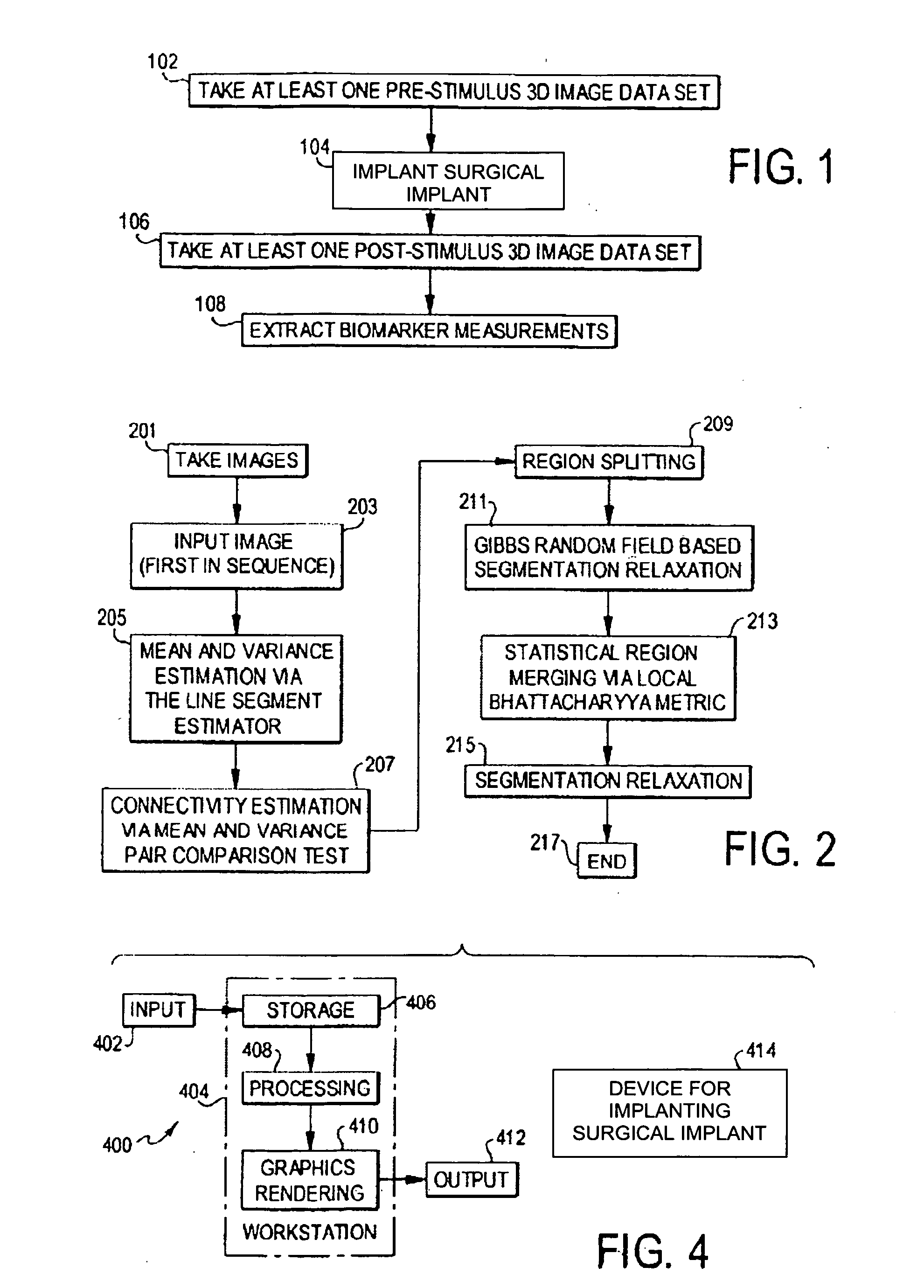
[0149] An overview of the operational steps carried out in the preferred embodiment is shown in FIG. 1. In step 102, one or more 3D image data sets are taken in a region of interest in the patient before the implantation of a surgical implant. The 3D image data sets can be taken by any suitable technique, such as MRI; if there are more than one, they are separated by time to form a time sequence of images. In step 104, a surgical implant is implanted into a portion within the region of interest constituting the biomarker. In step 106, one or more 3D image data sets are taken again, as in step 102, except after the implantation. In step 108, the biomarker measurements are extracted from the image data sets taken before and after the implantation. With those measurements, the reaction of the biomarker to the surgical implant can be determined.

[0150] The extraction of...


PUM


Abstract

In a human or animal organ or other region of interest, specific objects, such as liver metastases and brain lesions, serve as indicators, or biomarkers, of disease. In a three-dimensional image of the organ, the biomarkers are identified and quantified both before and after a surgical implant is implanted, and their reaction to the surgical implant is observed. Statistical segmentation techniques are used to identify the biomarker in a first image and to carry the identification over to the remaining images.

Description

FIELD OF THE INVENTION [0001] The present invention is directed to the assessment of certain biologically or medically significant characteristics of bodily structures, known as biomarkers, and more particularly to the assessment of biomarkers by quantitative measurement of their response to surgical implant. DESCRIPTION OF RELATED ART [0002] The measurement of internal organs and structures from computed tomography (CT), magnetic resonance imaging (MRI), ultrasound, positron emission tomography (PET), and other imaging data sets is an important objective in many fields of medicine. These imaging modalities are quantitative assessments that, when used, are typically based on manual intervention by a trained technician or radiologist. Examples illustrating current applications of medical imaging include the measurement of the hippocampus in patients with epilepsy (Ashton E. A., Parker K. J., Berg M. J., and Chen C. W. “A Novel Volumetric Feature Extraction Technique with Applications...

Claims


Application Information

Patent Timeline
Patent Type & Authority Applications(United States)
IPC IPC(8): G06F19/00G06G7/48G06K9/00G16Z99/00
CPCG06F19/3437G06T2207/30004G06T7/0012G16H50/50G16Z99/00
Inventor TAMEZ-PENA, JOSETOTTERMAN, SAARA MARJATTA SOFIAASHTON, EDWARD
Owner VIRTUALSCOPICS
Why Eureka
Try Eureka