Composition and method for direct visualization of the human appendix

a human appendix and visualization method technology, applied in the field of visualization and visualization can solve the problems of appendicitis, the appendix is particularly difficult to visualize and detect the pathology of the human appendix, and the appendix is an added diagnostic dilemma, so as to reduce the amount of undesirable patient side effects, and facilitate the release of the active enhancing agent.

Inactive Publication Date: 2006-11-09
VINCON RESEARC ENTERPRISES
View PDF4 Cites 4 Cited by
  • Summary
  • Abstract
  • Description
  • Claims
  • Application Information

AI Technical Summary

Benefits of technology

[0033] Another object of the present invention is to provide a composition that is a positive contrast agent that produces an organ-specific positive contrast effect.
[0040] The present invention fulfills the above and other objects by providing an oral crystalline composition having a combination of diatrizoate salts, simethicone and famotidine in predetermined amounts to result in rapid, direct and unambiguous visualization of the human appendix in conjunction with computerized tomography (CT scans). The composition fulfills all of the pertinent clinical criteria for an ideal practicable diagnostic pharmaceutical agent for appendicitis diagnosis as it is organ-specific, purpose-limited, safe for extensive outpatient usage and has negligible side effects. In addition, the composition is highly-effective based on a favorable bioavailability in sufficient quantity to facilitate passive diffusion and concentration within the appendix within a short enough time period for imaging and has a favorable release profile for the active enhancing agent, diatrizoate. The composition would be orally administered at a pre-determined time period prior to conducting the computerized axial tomographic examination.

Problems solved by technology

These differences make it particularly challenging to visualize and detect pathology of the appendix in humans.
A build-up of purulent collection builds up within the appendix and can perforate freely into body cavities, resulting in sepsis, which is widespread dissemination of infection throughout the body, and potential death.
Delayed or missed appendicitis is among the leading causes of medical liability, which involve principally the morbidity and mortality complications associated with perforation and sepsis, which can occur rapidly, without warning, proving potentially catastrophic to the patient.
Appendicitis also presents an added diagnostic dilemma, and can evade timely medical diagnosis.
One of the difficulties with appendicitis diagnosis is that appendicitis is not part of the gastrointestinal tract and has no known gastrointestinal function.
In diseases of the bowel, the bowel can become obstructed, leading to abdominal distention and changes in bowel function, such as changes frequency or consistency of bowel movements.
In the common practice of performing CT examinations, there also can be a significant time lag between the imaging findings and clinical manifestations of appendicitis.
Further compounding this diagnostic dilemma is the lack of a unified medical consensus regarding the optimal technique for CT scans of the appendix, including the optimal response time between performing a CT scan and obtaining reliable diagnostic results.
In actual practice, this response time can lag by as much as 4 to 6 hours or more, an unacceptable standard.
Many appendicitis patients are encountered in the outpatient setting in doctor's offices and clinics, but cannot be evaluated in the typical outpatient radiology facility because of significant time limitations and the lack of an available safe diagnostic pharmaceutical agent to facilitate rapid diagnosis.
The potential dosing possibilities of these solutions are infinite but limited by the concentration of diatrizoate salts in solution.
However, barium suspension is contraindicated in clinical evaluations for appendicitis because of the risk of barium-induced peritonitis when barium suspension spills freely into the peritoneal cavity.
Barium suspension cannot be used as a practicable alternative contrast media when dealing with bowel perforations or indirect evaluation of the appendix.
However, the appendix can form an impression or indentation on the bowel during an examination, resulting in an indirect method of visualizing the appendix, which is also known as a negative contrast effect.
However, there are no positive contrast agents available or known commercially that produce an organ-specific positive contrast effect, i.e., concentration within the appendix.
The use of negative contrast agents in the prior art has been well studied and lead to the same conclusion: there are significant inherent technical limitations in the evaluation of appendicitis.
One of the major drawbacks of negative contrast agents is that they produce a weak bowel opacification that renders the agent useless for direct visualization of the appendix.
Significant clinical side effects are also problematic and include diarrhea and patient non-compliance related to bowel distention and discomfort from excessive oral fluid intake, in addition to gaseous distention produced by the addition of mannitol, sorbitol, and sugar-based flavoring agents.
The work of Megibow has further shown that a negative contrast effect is not considered sufficient clinically to allow for a confident diagnosis of appendicitis.
Megibow has investigated the extensive use of negative contrast agents available in the prior art, also encompassing those described herein, and concluded that negative contrast agents are not reliable in the diagnosis of appendicitis.
One of the principal reasons that indirect methods fail to provide direct or organ-specific appendiceal visualization is that the primary goal of negative contrast agents is to visualize the bowel, not the appendix.
However, Neutrospect is not site or organ specific for appendicitis and can be associated with any type of infection.
There are a number of inherent technical and therapeutic limitations of using Neutrospect in the clinical setting.
First, it is of limited value clinically because it is not organ specific and has limited specificity.
A positive Neutospect scan can produce an accumulation of radioactivity, or a spot on the radiographic film, practically anywhere in the body where there is an infection, confounding the clinician and further rendering a diagnosis of appendicitis as ambiguous and not obvious.
In addition, significant safety issues preclude the safe administration of Neutrospect in human subjects for appendicitis diagnosis.
The United States Food and Drug Administration recently suspended clinical use of Neutrospect following documented incidents of fatal anaphylactic reactions leading to cardiopulmonary demise in several hospitalized patients.
However, these significant safety issues, in addition to limited specificity for appendicitis diagnosis, preclude Neutrospect as a practicable diagnostic pharmaceutical for clinical use, particularly in the outpatient setting.
No such ideal pharmaceutical agent is available commercially for routine clinical application.
Because of the possibilities for large numbers of patients undergoing diagnostic evaluations for appendicitis, particularly in the outpatient setting, a further need exists for a safe diagnostic pharmaceutical agent, in addition to being organ-specific or purpose-limited for appendicitis.

Method used


Image

  • Composition and method for direct visualization of the human appendix
  • Composition and method for direct visualization of the human appendix
  • Composition and method for direct visualization of the human appendix

Examples


example 1

Case Study

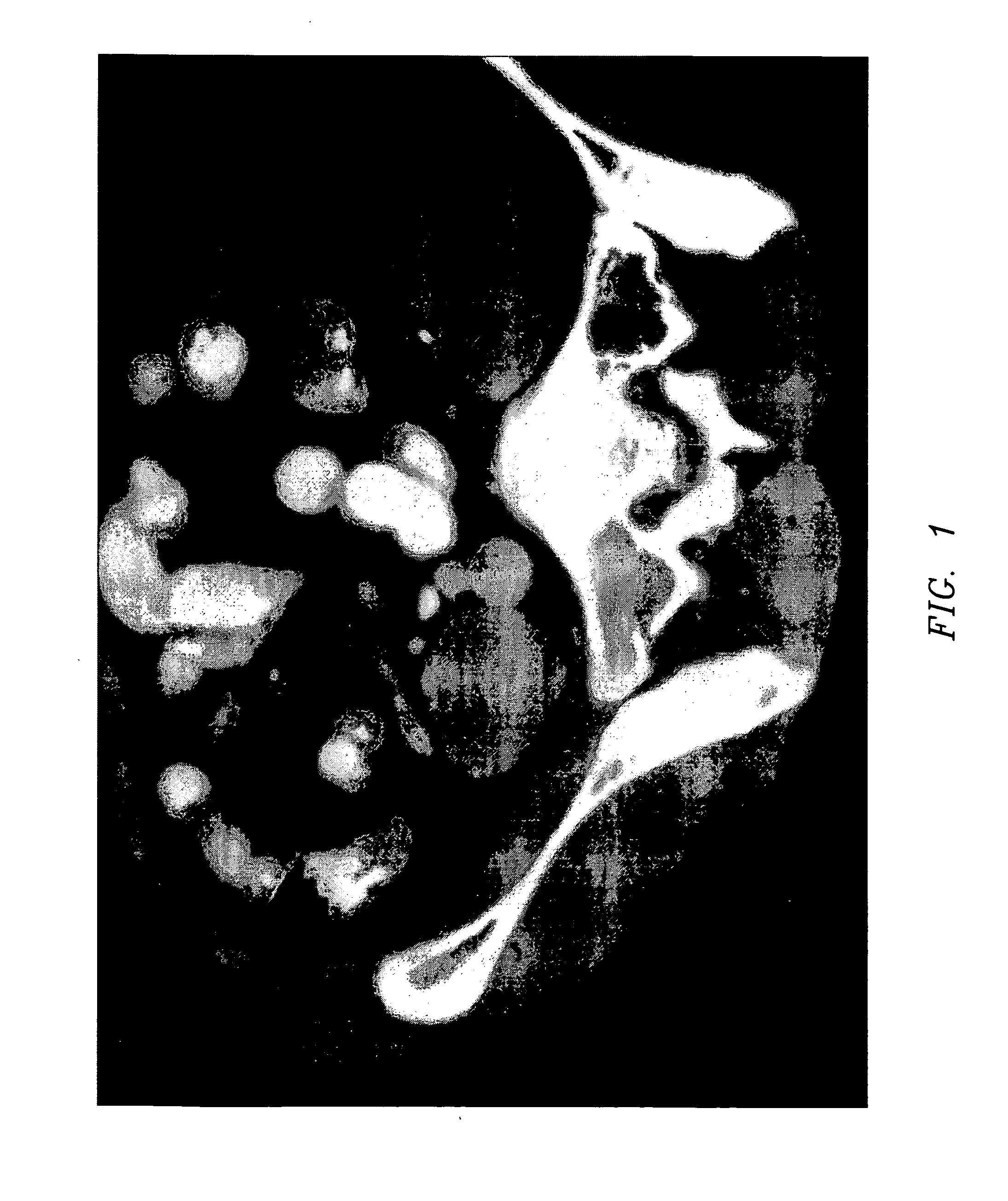
[0082] A 26-year-old woman presented with vague right lower quadrant pain and tenderness, with equivocal peritoneal signs. A prepared solution of the invention was administered as a single 224-mL oral bolus. Subsequent helical CT scans of the right lower quadrant were performed at 50-min post administration, utilizing 5 mm and 7 mm collimation, 120 kV, 250 mA, and 1.0 s scans, reconstructed in the soft tissue algorithm at a window level of +50 HU and window width of +450. A contrast opacified appendix was demonstrated at 50 minutes, designated as FIG. 1.

example 2

Case Study

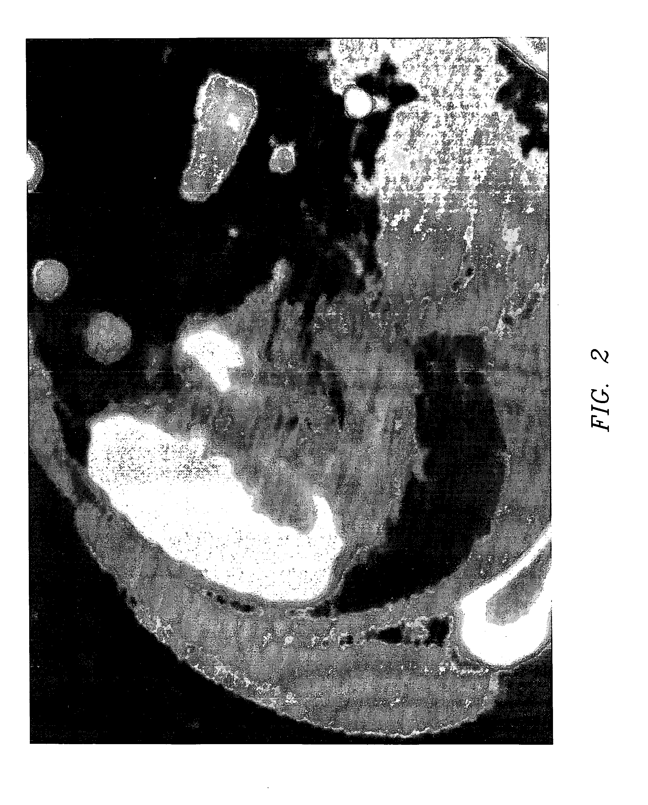
[0083] A 47-year-old man presented with focal right lower quadrant pain, peritoneal tenderness, and fever. A prepared solution of the invention was administered as a single 224-mL oral bolus. Subsequent helical CT scans of the right lower quadrant were performed at 50-min post administration, utilizing 5 mm and 7 mm collimation, 120 kV, 250 mA, and 1.0 s scans, reconstructed in the soft tissue algorithm at a window level of +50 HU and window width of +450. The appendiceal orifice was obstructed, revealing a classic ‘beak sign’ of appendicitis, with surrounding peritoneal inflammatory inflammatory changes, consistent with appendicitis, designated as FIG. 2.

example 3

Case Study

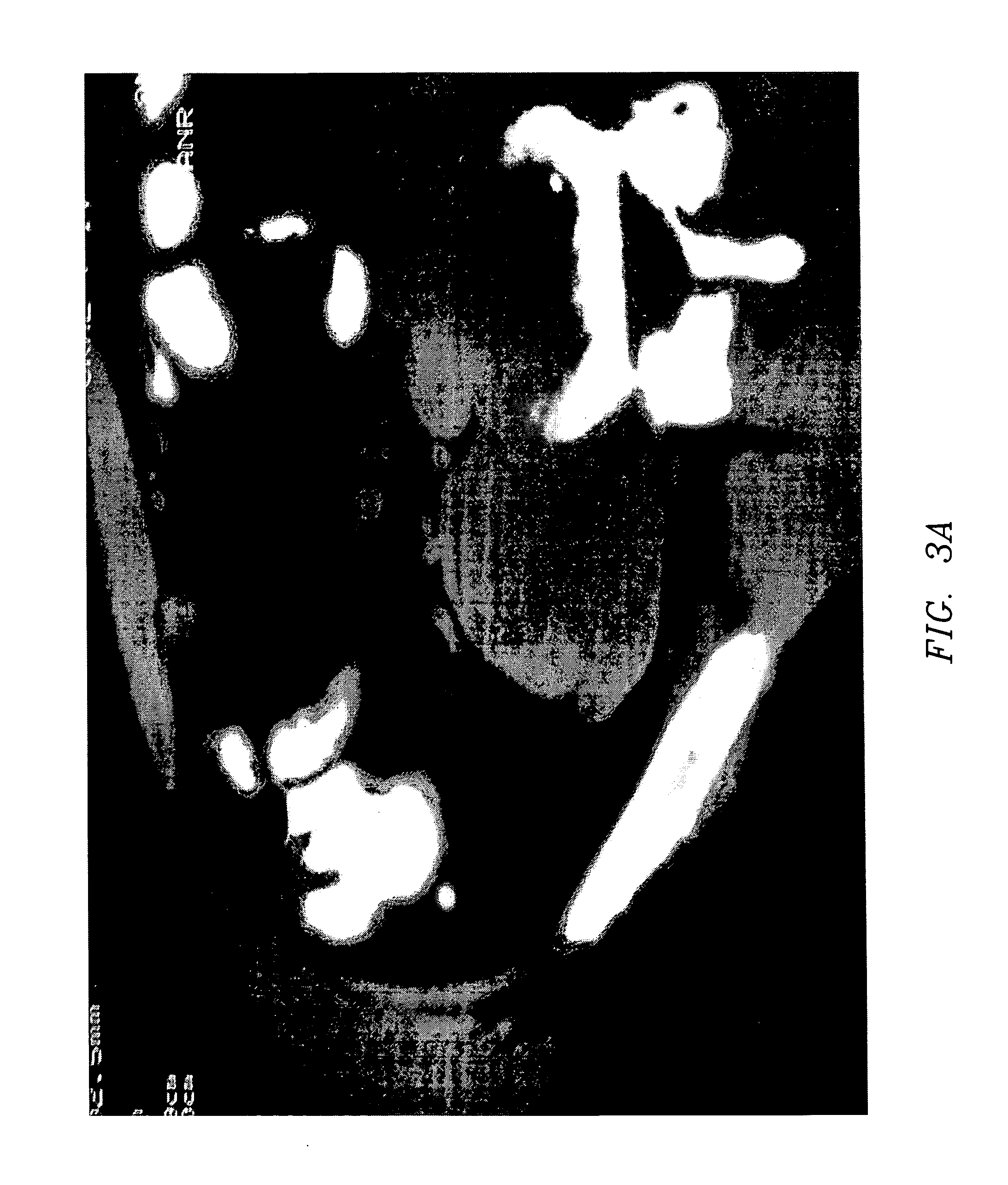
[0084] A 47-year-old man presented with intermittent right lower quadrant pain on a chronic basis over a period of greater than one month. No clinical signs of appendiceal inflammation were present, including fever, peritoneal tenderness, or leukocytosis. A prepared solution of the invention was administered as a single 224-mL oral bolus. Subsequent helical CT scans of the right lower quadrant were performed at 50-min post administration, utilizing 5 mm and 7 mm collimation, 120 kV, 250 mA, and 1.0 s scans, reconstructed in the soft tissue algorithm at a window level of +50 HU and window width of +450. The appendiceal orifice was obstructed, revealing a thickened appendix and adjacent small calculus, an appendicolith, considered diagnostic for chronic adult appendicitis, designated as FIG. 3.


PUM


Abstract

A positive contrast agent composition containing meglumine diatrizoate, sodium diatrizoate, simethicone, famotidine and aspartame in predetermined amounts that is orally administered to a patient for clinical evaluations of appendicitis wherein a positive contrast effect is achieved. Methods of use include orally administering individual doses of the composition approximately 50 minutes prior to appendix visualization using computerized axial tomography.

Description

CROSS-REFERENCE TO RELATED APPLICATION [0001] This is a continuation-in-part application of Ser. No. 11 / 104,700 filed Apr. 13, 2005, which was a continuation-in-part application of Ser. No. 10 / 354,644 filed Jan. 29, 2003, now abandoned.BACKGROUND OF THE INVENTION [0002] This invention relates to a composition and method for direct visualization of the human appendix and method, more particularly, to a composition for use in direct visualization of the human appendix and method for diagnosing appendicitis that provides direct and unambiguous visualization of the human appendix, separate and distinct from the gastrointestinal tract. [0003] The gastrointestinal tract is a long hollow tube, or viscus, extending from the mouth to the anus, and includes the stomach, small intestine, colon, and large intestine. The gastrointestinal tract has an important and necessary physiologic function in the digestion of food into biologically usable form to sustain life. [0004] On the other hand, unli...

Claims


Application Information

Patent Timeline
Patent Type & Authority Applications(United States)
IPC IPC(8): A61K51/00A61K49/04
CPCA61K49/0438A61K49/0447
Inventor GIULIANO, VINCENZOGIULIANO, CONCETTA
Owner VINCON RESEARC ENTERPRISES
Why Eureka
Try Eureka