Methods of increasing proliferation of adult mammalian cardiomyocytes through p38 map kinase inhibition

a technology of fetal cardiomyocytes and p38 map, which is applied in the direction of biocide, drug composition, peptide/protein ingredients, etc., can solve the problems of cardiac muscle loss, slow down, reduce or prevent the onset of cardiac damage, and increase the proliferation of fetal cardiomyocytes. , to achieve the effect of blocking the proliferation of fetal cardiomyocytes, slowing down the onset of cardiac damage, and slowing down the de-differen

Inactive Publication Date: 2006-12-14
CHILDRENS MEDICAL CENT CORP
View PDF8 Cites 30 Cited by
  • Summary
  • Abstract
  • Description
  • Claims
  • Application Information

AI Technical Summary

Benefits of technology

[0005] The present invention provides compositions and methods for increasing proliferation and / or de-differentiation of postmitotic mammalian cardiomyocytes. The invention can be used to slow, reduce, or prevent the onset of cardiac damage caused by, for example, myocardial ischemia, hypoxia, stroke, or myocardial infarction. In addition, the methods and compositions of the invention can used to produce de-differentiated cardiomyocytes, which can then be used in tissue grafting.
[0006] The invention is based, in part, on the discovery that postmitotic mammalian cardiomyocytes can proliferate. One mechanism of cell cycle regulation for mammalian cardiomyocytes is p38 activity; that is p38 is a key negative regulator of mammalian cardiomyocyte division. p38 activity is inversely correlated with cardiac growth during development, and its overexpression blocks proliferation of fetal cardiomyocytes in vitro. Genetic activation of p38 in vivo reduces fetal cardiomyocytes proliferation, whereas targeted disruption of p38w increases neonatal cardiomyocyte mitoses. Growth factor stimulation and p38 inhibition can induce cytokinesis in adult cardiomyocytes. Growth factors useful in conjunction with p38 inhibitors in clued FGF1, IL-1β, and NRG-1-β1 as well as factors listed in Table S-2. These results indicate that the inhibitory effects of p38 on cardiomyocyte proliferation are reversible and that postmitotic, differentiated cells are capable of proliferation.

Problems solved by technology

This is a major medical problem, as ischaemic heart disease, resulting in cardiac muscle loss, is the leading cause of morbidity and mortality among adults aged 60 and older, and the second most common cause of death in ages 15 to 59.

Method used


Image

  • Methods of increasing proliferation of adult mammalian cardiomyocytes through p38 map kinase inhibition
  • Methods of increasing proliferation of adult mammalian cardiomyocytes through p38 map kinase inhibition
  • Methods of increasing proliferation of adult mammalian cardiomyocytes through p38 map kinase inhibition

Examples


example 1

De-Differentiation and Proliferation of Adult Cardiomyocytes

Animals, Cells, and Stimulation

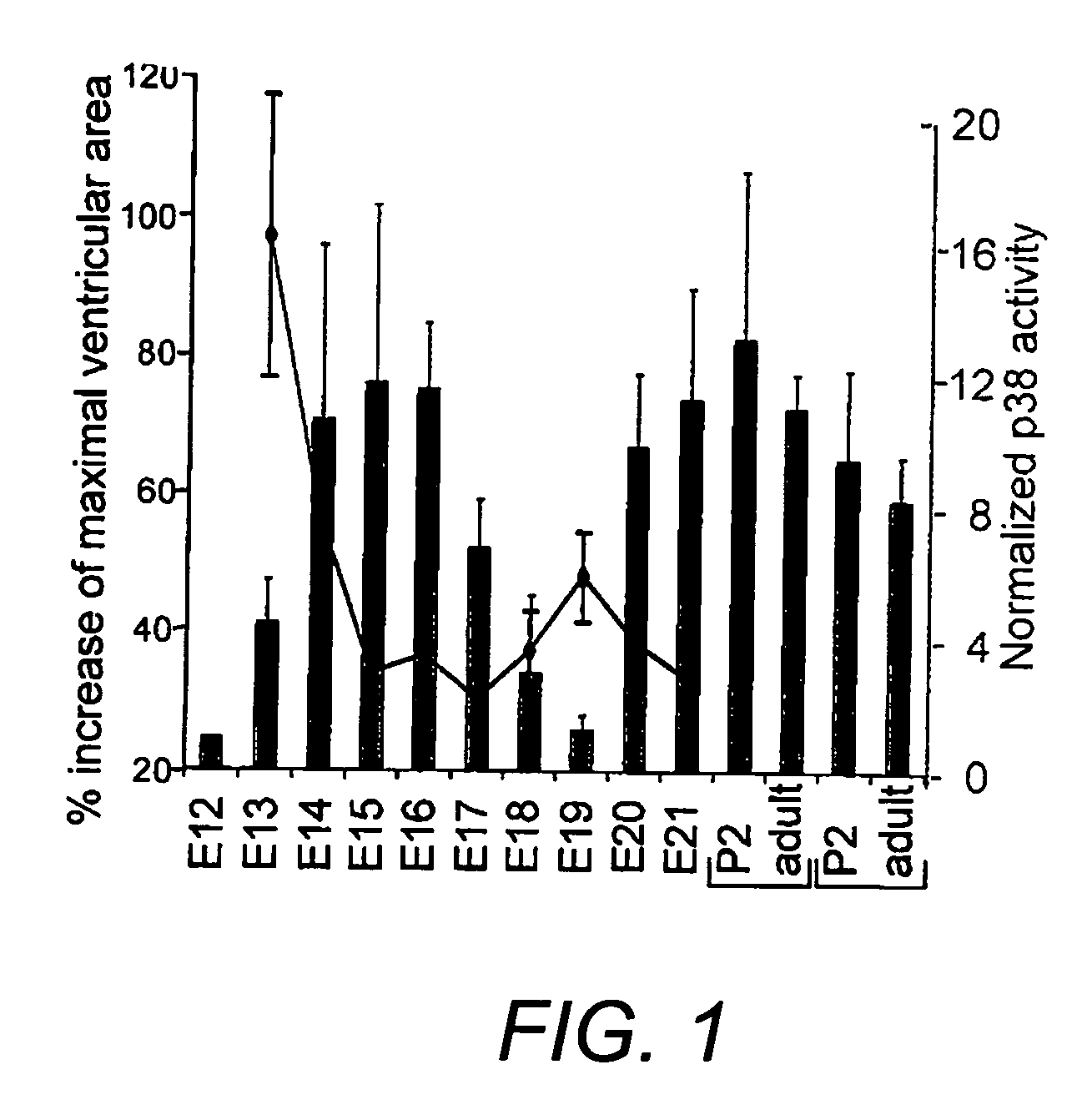
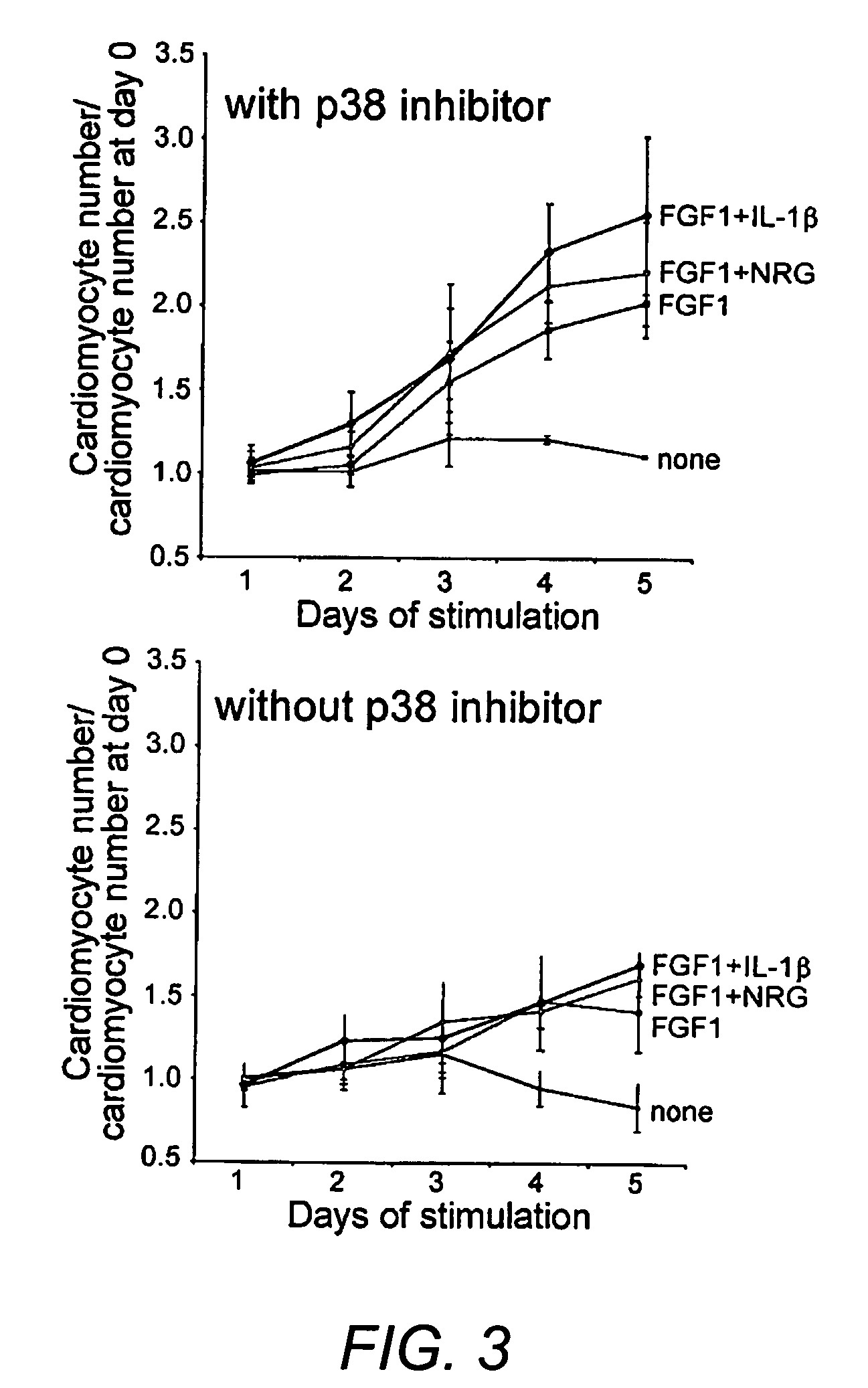
[0102] Animal experiments were performed in accordance with guidelines of Children's Hospital, Boston and UCLA. Ventricular cardiomyocytes from fetal (E19), 2-day-old (P2) and adult (250-350 g) Wistar rats (Charles River) were isolated as described with minor modifications (Engel et al. 1999; Engel et al. 2003). After digestion of fetal or neonatal hearts (0.14 mg / ml collagenase II (Invitrogen), 0.55 mg / ml pancreatin (Sigma)) cells were cultured in DMEM / F12 (GIBCO) containing 3 mM Na-pyruvate, 0.2% BSA, 0.1 mM ascorbic acid (Sigma), 0.5% Insulin-Transferrin-Selenium (100×), penicillin (100 U / ml), streptomycin (100 μg / ml), and 2 mM L-glutamine (GIBCO). Adult cardiomyocytes were cultured for 1 day in standard medium (DMEM, 25 mM Hepes, 5 mM taurine, 5 mM creatine, 2 mM L-carnitine (Sigma), 20 U / ml insulin (GIBCO), 0.2% BSA, penicillin (100 U / ml), and streptomycin (100 μg / ml)). Cells were stim...

example 2

In Vivo Effects of p38 Inhibitors Following Myocardial Infarct

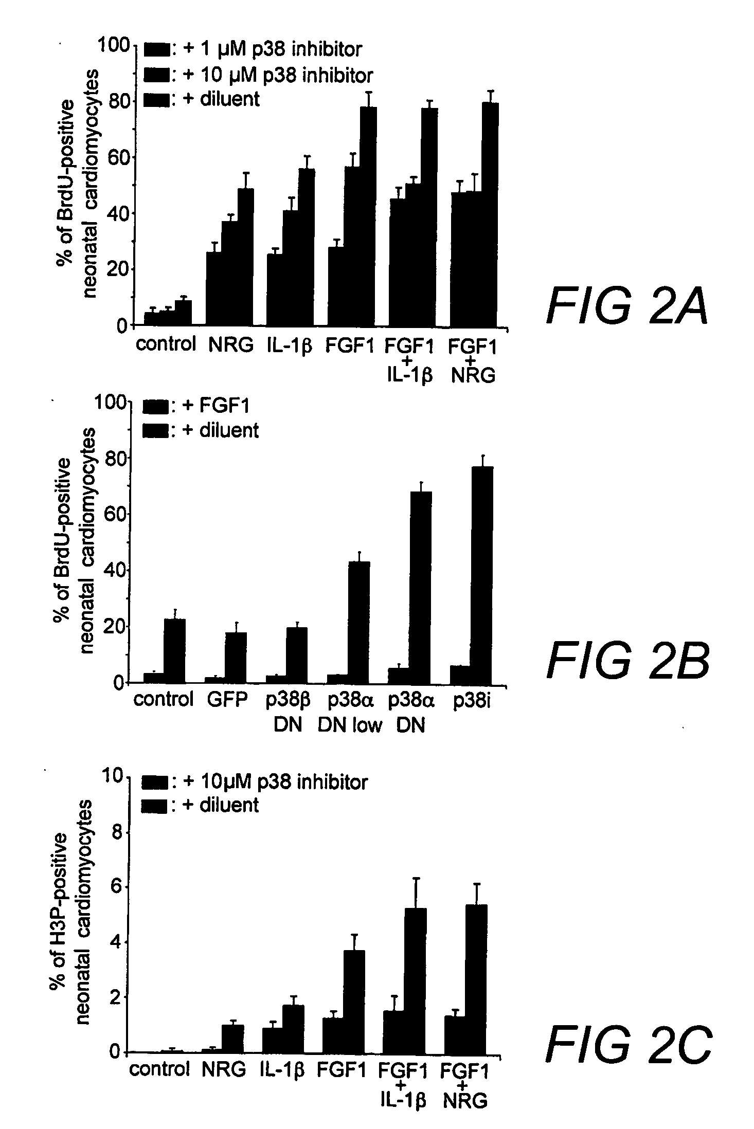
[0137] The effects of a variety of p38 inhibitors on adult rat cardiomyocytes were compared using Ki67, BrdU, and H3P (FIG. 5A shows the percentage of Ki67-positive neonatal cardiomyocytes. FIG. 5B shows the percentage of BrdU-positive neonatal cardiomyocytes and (FIG. 5C shows the percentage of H3P-positive neonatal cardiomyocytes). The compounds tested in FIGS. 5A-5C include SB203580, which has 100- to 500-fold selectivity over GSK3β and PKBα, SB203580 HCL (water insoluble), SB202474, a negative control commonly use for MAP kinase inhibition studies, and SB239063 which has >200-fold selectivity over ERK and JNK.

[0138] The p38 inhibitors were tested for in vivo effect following myocardial infarct. For the evaluation of left ventricular function, transthoracic echocardiogram can be performed on the rats after myocardial infarction 1 day or 14 days right. Rats can be anesthetized with 4-5% isoflurane in an induction cham...

example 3

Further In Vivo Effects of p38 Inhibitors Following Myocardial Infarct

[0141] To determine whether p38 inhibition / FGF1 stimulation can induce cardiomyocyte proliferation in vivo and whether it has a positive effect on cardiac function after cardiac injury we created myocardial infarctions (MI) in adult rats (250 g) by coronary artery ligation. The p38 inhibitor SB203580 HCl or its vehicle, saline, were injected intraperitoneal every three days for the first month of the study. FGF1 or its carrier BSA was injected mixed with self-assembling peptides once into the infarct border zone immediately after coronary artery ligation. We injecting a total of 80 μl of 400 ng / ml FGF1, given at 3 different injection sites, into 400 mg of infarcted myocardium estimated to deliver a FGF 1 concentration to the cardiomyocytes of approximately 50 to 100 ng / ml. Animals were analyzed 24 hours, 2 weeks, and 3 month after surgery. We performed two blinded and randomized studies using 62 rats for the 2 we...


PUM

PropertyMeasurementUnit
concentrationaaaaaaaaaa
end-diastolic diameteraaaaaaaaaa

Abstract

Compositions and methods for increasing proliferation and / or de-differentiation of postmitotic mammalian cardiomyocytes are disclosed to slow, reduce, or prevent the onset of cardiac damage. In addition, the methods and compositions of the invention can used to produce de-differentiated cardiomyocytes, which can then be used in tissue grafting, implantation or transplantation. The invention is based, in part, on the discovery that postmitotic mammalian cardiomyocytes can proliferate as a result of targeted disruption of p38 MAP kinase. p38 inhibition with optional growth factor stimulation can induce cytokinesis in adult cardiomyocytes.

Description

REFERENCE TO RELATED APPLICATION [0001] The present application claims priority to U.S. Provisional Patent Application Ser. No. 60 / 676,117 entitled “Methods Of Increasing Proliferation Of Adult Mammalian Cardiomyocytes Through P38 Map Kinase Inhibition,” filed on Apr. 29, 2005, which is herein incorporated by reference.BACKGROUND OF THE INVENTION [0002] Highly differentiated mammalian cells are thought to be incapable of proliferation. These cells have exited the cell cycle. Proteins critical for cellular specialisation have accumulated and driven these cells to their final form and function (Studzinski and Harrison 1999 Int Rev Cytol 189: 1-58). In contrast with mammals, differentiated cells in teleost fish (Poss et al. 2003 Dev Dyn 226: 202-10) and urodele amphibians (Brockes and Kumar 2002 Nat Rev Mol Cell Biol 3: 566-74) can dedifferentiate and / or proliferate, enabling regeneration. For example zebrafish hearts regenerate through cardiomyocyte proliferation (Poss et al. 2002 Sci...

Claims


Application Information

Patent Timeline
Patent Type & Authority Applications(United States)
IPC IPC(8): A61K31/4412
CPCA61K31/17A61K31/4412A61K31/4418A61K38/1825A61K2300/00A61P9/10
Inventor KEATING, MARK T.ENGEL, FELIX B.
Owner CHILDRENS MEDICAL CENT CORP
Why Eureka
Try Eureka