

Medical devices combined with diblock copolymer compositions
a technology of copolymer composition and medical devices, applied in the field of coating medical devices, can solve the problems of stenosis (or narrowing), damage to the epithelial lining of the tube and the smooth muscle cells (smcs) that make up the wall, and many devices implanted in the body are subject to a “foreign body” respons
- Summary
- Abstract
- Description
- Claims
- Application Information
AI Technical Summary
Benefits of technology
Problems solved by technology
Method used
Image

Examples
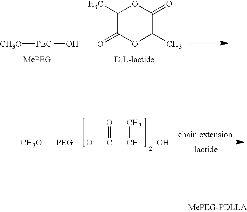
example 1
Polymerizing of MePEG-PDLLA-6535
[0320] 65 g of methoxy polyethylene glycol (MePEG) with a molecular weight of 5,000 Dalton (Polysciences, Cat # 05986) were weighed in a 250 ml flat bottom (FB) flask. 35 g of D, L-lactide (Purasorb) were weighed separately in a weighing boat. Both MePEG5000 and D, L-lactide were dried under vacuum overnight at room temperature before use.
[0321] An oil-bath with light or heavy mineral oil (Aldrich, CAT# 33076-0) was heated to and maintained at 135° C. by using a thermo-controller (VWR, Model, LN: 002392, PN: 400188-REV A).
[0322] 0.3-0.5 ml (Appr. 300-500 mg) of stannous 2-ethyl-hexanoate catalyst (Sigma, >95%, CAT# 33076-0) was added into the FB flask and then the flask was purged slowly with N2 (oxygen free, Praxair, Grade 4.8) for 5 minutes.
[0323] The flask was stoppered with a glass stopper and placed into the oil-bath, and the magnetic stirrer was gradually turned on to a setting at 6 (Corning Thermo Stirrer / Hot Plate, Model PC-620). After 30 ...
example 2
Purification of MePEG-PDLLA-6535
[0327] To about 75 g of MePEG-PDLLA (65:35) in a 1000 ml flat-bottom titration and culture flask, isopropanol (HPLC grade) was added until it reached the 1000 ml mark.
[0328] The flask was placed in a 60° C. water bath (a 2000 ml jacket beaker connected with a VWR Isotemp Circulator, Model 1130-1) and the mixture was stirred till the MePEG-PDLLA (65:35) dissolved.
[0329] The solution was cooled down to room temperature (20-22° C.) to precipitate the diblock polymer, which was isolated by filtration. The precipitant was washed three times with 200-250 ml isopropanol each.
[0330] The polymer was first dried in the open air overnight for approximately 18 hours to remove most of the solvents. The pre-dried polymer was then transferred to a vacuum oven. The polymer was dried until the residual solvent was below the acceptable level (about 24 hours).
[0331] The dried polymer was stored in a refrigerator at 2-8° C. for use.
example 3
Coating of MePEG-PDLLA (20:80), 5% Paclitaxel on a Perivascular Wrap
[0332] The VICRYL or PLGA meshes (PolyMed) were cut into the size of 2×5 cm2. The meshes were washed using HPLC grade isopropanol and completely dried in the forced-air oven at 50° C. The weight of each bare mesh was recorded.
[0333] About 1.9 g of MePEG-PDLLA-2080 was dissolved in 10 mL of acetone or dichloromethane (Calcdon, HPLC grade) to form a target of 20% solution.
[0334] About 100 mg of paclitaxel were added into each polymer solution. Paclitaxel was dissolved completely by placing the vials on Nutator Rotor (Model 421105, SN: 1100-15989).
[0335] The mesh was coated by dipped into the polymer / paclitaxel solution. The mesh was then removed from the vial while removing any excessive amount of solution on the mesh).
[0336] The coated mesh was dried 3-5 minutes in the air. The coated mesh was thereafter placed in a PTFE petri-disk, transferred into a vacuum oven, and continued drying under vacuum overnight at r...
PUM
| Property | Measurement | Unit |
|---|---|---|
| Tg | aaaaa | aaaaa |
| Tg | aaaaa | aaaaa |
| thickness | aaaaa | aaaaa |
Abstract
Description
Claims
Application Information