Ultrasound-mediated high-speed biological reaction and tissue processing

a biological reaction and ultrasonic technology, applied in the field of ultrasonic high-speed biological reaction and tissue processing, can solve the problems of insufficient time to allow even penetration and complete reaction with the tissues, the heat as a form of tissue fixation has not been exploited in the diagnostic laboratory, and the time taken for 10 seconds is not enough

Inactive Publication Date: 2007-03-29
AMERICAN REGISTRY OF PATHOLOGY
View PDF21 Cites 20 Cited by
  • Summary
  • Abstract
  • Description
  • Claims
  • Application Information

AI Technical Summary

Benefits of technology

[0047] It is important to understand that in the practice of the current invention more power equates to a shorter time from beginning to end for each reaction step and additionally less destruction of tissue matter. The present invention is distinguished over the prior art in that the ultrasonic transducer is placed relatively close to the tissue sample being processed. The prior art uses a system in which the multipiece tissue sample is immersed in a liquid bath wherein the liquid bath is subjected to ultrasonic energy. The prior art method (low frequency ultrasound) results in much more tissue damage. The present invention can be used in a variety of histological, pathological, immunological and molecular techniques.
[0050] The invention is directed to using ultrasound in conjunction with well known techniques to decrease the time required to perform the techniques, including immunological reactions, hybridizations, tissue fixation and processing, reactions on membranes as well as in tissues. The intensity of ultrasound used depends on the particular procedure and will be in the range of about 0.001-20 W / cm2
[0051] Another aspect of the invention is that the complete process can be controlled and monitored by a feedback sensor-central processor unit (CPU)-ultrasound generation system. All of the steps of fixation and processing can be controlled by the CPU-ultrasound generation system. Use of a feedback sensor allows for optimization of the processes.

Problems solved by technology

(Drury and Wallington, 1980), heat as a form of tissue fixation has not been exploited in the diagnostic laboratory.
However, most chemical fixatives produce denaturation or masking of many antigens and degradation of RNA and DNA.
However, while the optimal time for fixation varies with the chemical agent employed, this generally takes hours, approximately a day, to accomplish.
The 10 seconds was not enough time to allow even penetration and complete reaction with the tissues.
Also, 70° C. is not hot enough to inhibit all enzymes such as RNases (Sambrook et al., 1989).
The exact amount of MW energy received by tissue was very difficult to control (Login and Dvorak, 1985; Azumi et al., 1990).
Therapeutic ultrasound (800-880 kHz frequency and 1.4-2 W / cm2 intensity) did not significantly improve the quality and time of fixation (Drakhli, 1967; Botsman and Bobrova, 1968; Obertyshev, 1987; Rozenberg, 1991) even when combined with MW energy (Shmurun, 1992).
However, the tissue blocks or sections used for analysis of molecular information of LCM and tissue microarray techniques have not fitted well with the classic method of tissue fixation—formalin fixed paraffin embedded (FFPE) tissue, which has provided the best morphology for pathologists throughout this century (Fend et al., 1999; Goldsworthy et al., 1999).
However, there are many drawbacks in using FFPE tissue for molecular pathology, such as inconsistency in fixation condition, antigen masking, and RNA / DNA degradation.
All these drawbacks limit the use of LCM and tissue microarray techniques with FFPE tissues (Fend et al., 1999a; Goldsworthy et al., 1999).
However, these technologies have not been successfully adopted in clinical diagnostic laboratories and controversial observations of these techniques have been reported (Azumi et al., 1990; Login et al., 1991; Azumi et al., 1991).
Clearly there are many individual steps involved and each step takes time.
The procedure possesses certain disadvantages in that the prepared slide does not possess the uniformity of quality of morphology prepared by other methods.
Moreover, it is technically more difficult for serial sections of the same specimen to be examined by this procedure, and extreme caution must be exercised in cutting the specimen in order to ensure a sufficiently thin section and to avoid the possibility of damaging details of the specimen.
The most serious objection to using the frozen section procedure is the necessity of preparing all the slides required for special stains and / or consultation and teaching purposes while the tissue is in the initial frozen state.
If the tissue is thawed and refrozen for sectioning, it is severely damaged.
Although the slides produced according to this procedure are of higher quality of morphology than those produced by the frozen-section technique, the length of time required to process specimens is too great to enable this procedure to be used in situations where time is of the essence.

Method used


Image

  • Ultrasound-mediated high-speed biological reaction and tissue processing
  • Ultrasound-mediated high-speed biological reaction and tissue processing
  • Ultrasound-mediated high-speed biological reaction and tissue processing

Examples


example 1

Tissue Fixation and Processing

[0083] A general procedure for preparing tissue fixed by NBF, processed and imbedded in paraffin using ultrasound as part of the process to decrease the required time to prepare the sample for use follows. Specifics are set out in the Examples which follow the general procedure. Those of skill in the art recognize that many variations can be made in the following procedures and the values set forth in the following procedures and Examples are not meant to be limiting.

A) Fixation

[0084] Step 1: A fresh tissue sample is cut in a size range from 3 to 5 mm thick, preferably less than 5 mm thick.

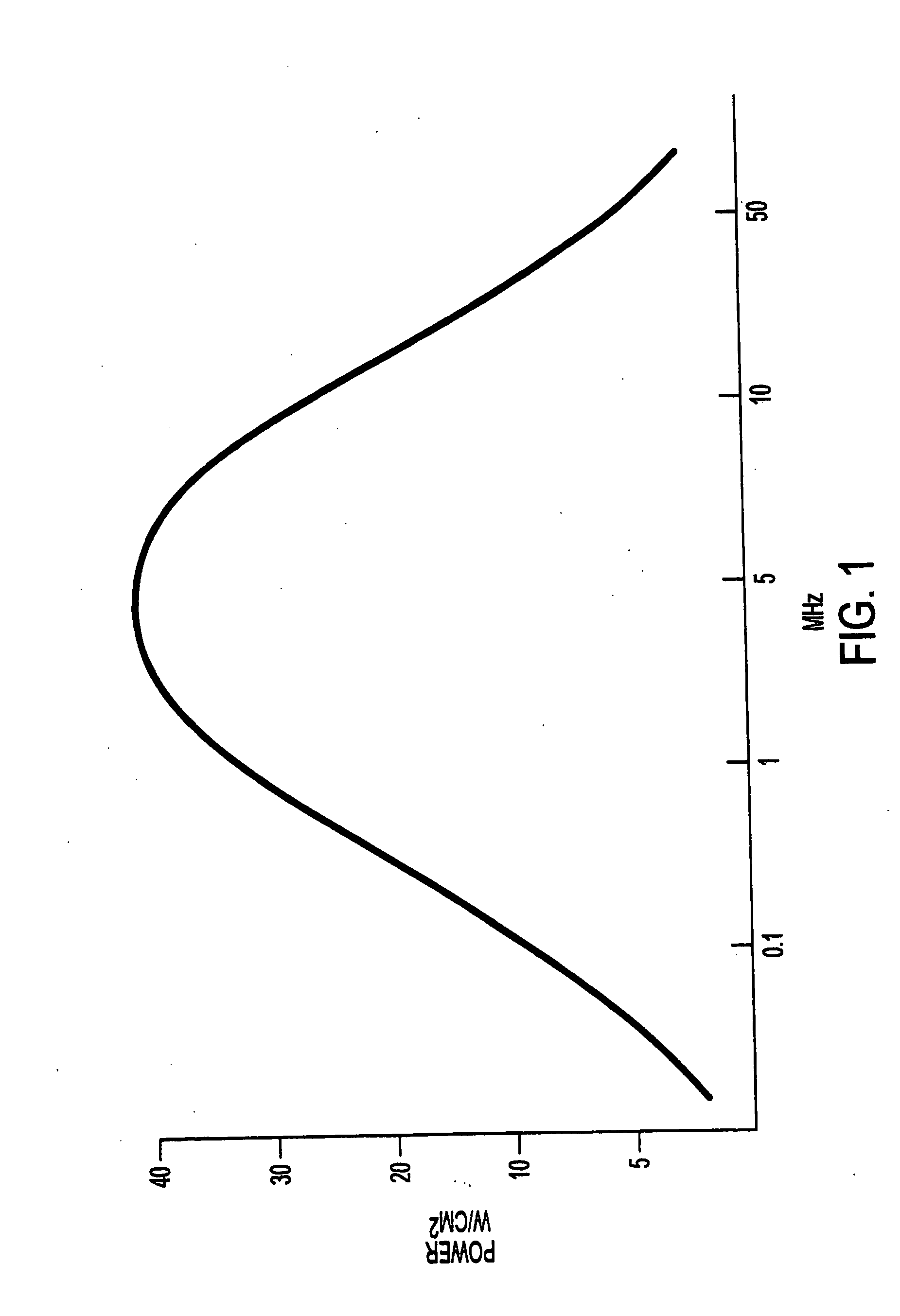
[0085] Step 2: The tissue sample (a single piece) is immersed in a 10% formalin solution or other acceptable fixative. Depending on the type of tissue, the sample is immersed from 5 to 30 minutes at 37° C., preferably for 15 minutes. During the immersion the sample is subjected to ultrasonic energy. When using a fixed power setting, the frequency of the ultrasoni...

example 2

Ultrasonic Apparatus and Application

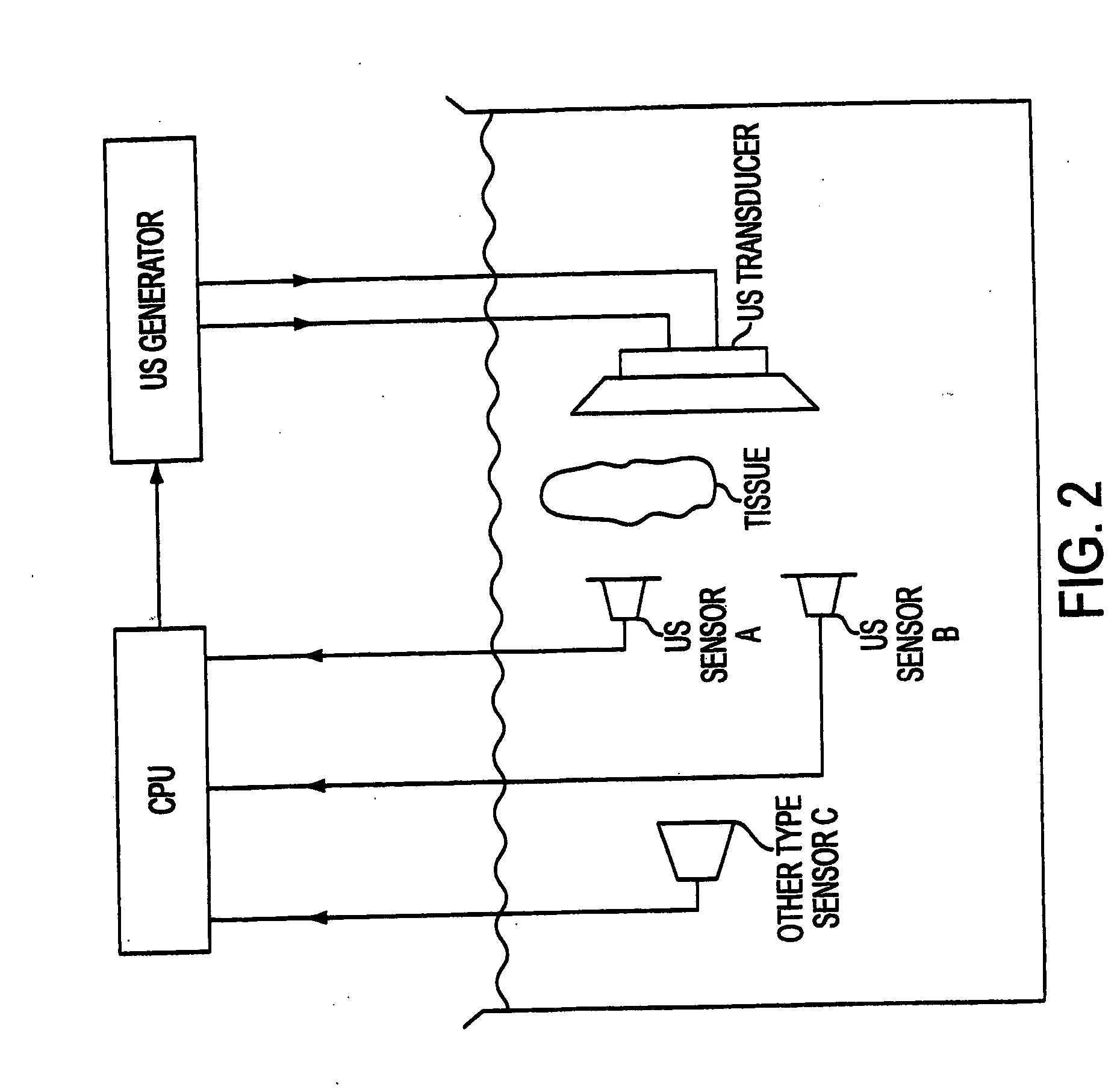
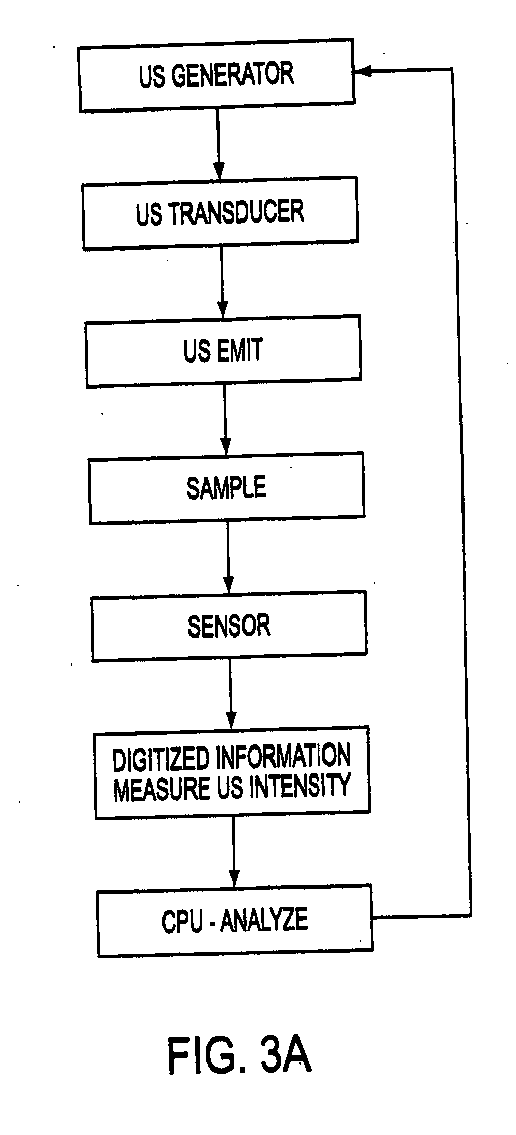
[0097] The ultrasonic apparatus used in the present study was specifically designed by the inventor and built by Bio-Quick, Silver Spring, MD. It consisted of an ultrasonic generator and a 1.6-1.7 MHz ceramic transducers (1.5 cm diameter) with adjustable output intensity range of 1 to 22 W / cm2. The tissue (only one sliced tissue at time), which was irradiated by ultrasound in 200 mL of NBF, grades of alcohol, xylene and 60° C. paraffin, was directly faced and aimed at the transducers. The distance between the transducers and irradiated tissue was within 3 cm to insure that the tissue received even and accurate ultrasound energy. The application of ultrasound was always continuous rather than pulsed. The tissue receiving ultrasound energy was monitored by means of a UW-3 ultrasound watt meter (Bio-Tek Instruments Inc., Winooski, Vt.). The temperature of the fixative inside the container was limited to 37° C. during the exposure to ultrasound.

example 3

Immunohistochemistry with Ultrasound

[0098] Tissue sections from each experiment were stained with a panel of seven primary antibodies (CD20, CD45, CD3, CD5, Bcl-2, kappa and lambda; Table 1). The primary antibodies were demonstrated using the ABC method (Hsu et al., 1981; Chu et al., 1992) as follows: sections were deparaffinized and taken through to 70% alcohol before being placed into water. Sections were then pretreated with / without antigen retrieval by MW (Shi et al., 1991) or pepsin (Chu et al., 1999) as required for each antibody (Table 1). Sections were rinsed in phosphate buffered saline (PBS) and incubated in primary antibody for 5-10 minutes at room temperature with a higher single frequency (1.6-1.7 MHz) setting and lower power (0.01-5 W / cm2) setting of ultrasound. After three rinses in PBS for 5 seconds with ultrasound, sections were incubated in biotinylated second antibody for 2.5 or 5 minutes at room temperature with ultrasound. Sections were rinsed in PBS with ultra...


PUM

PropertyMeasurementUnit
temperatureaaaaaaaaaa
frequencyaaaaaaaaaa
frequencyaaaaaaaaaa

Abstract

Methods of fixing and processing tissue and samples on a membrane by using ultrasound radiation as a part of the method are presented. Ultrasound of a frequency in the range of 0.1-50 MHz is used and the sample or tissue receives 0.1-200 W / cm2 of ultrasound intensity. The use of ultrasound allows much shorter times in the methods. Also presented are apparati comprising transducers of one or of multiple heads for producing the ultrasound radiation and further comprising a central processing unit and optionally comprising one or more sensors. Sensors can include those to measure and monitor ultrasound and temperature. This monitoring system allows one to achieve accurate and optimum tissue fixation and processing without overfixation and tissue damage. The system also allows the performance of antigen-antibody reactions or nucleic acid hybridizations to be completed in a very short time while being highly specific and with a very low or no background.

Description

CROSS REFERENCE TO RELATED APPLICATIONS [0001] This application is a continuation of application Ser. No. 09 / 901,120, filed Jul. 10, 2001, which is a divisional of application Ser. No. 09 / 407,964, filed 29 Sep. 1999 (U.S. Pat. No. 6,291,180) to which priority is claimed and which are incorporated herein by reference.BACKGROUND OF THE INVENTION [0002] The process of fixation forms the foundation for the preparation of tissue sections. It prevents or arrests autolysis and putrefaction, coagulates and stabilizes soluble and structural proteins, fortifies the tissues against the deleterious effects of subsequent processing and facilitates staining. Current methods of fixation rely on chemical agents, the most widely used being formaldehyde. Although autolysis is known to be retarded by cold and almost inhibited by heating to 60° C. (Drury and Wallington, 1980), heat as a form of tissue fixation has not been exploited in the diagnostic laboratory. [0003] The use of routinely fixed, paraf...

Claims


Application Information

Patent Timeline
Patent Type & Authority Applications(United States)
IPC IPC(8): G01N1/30G01N1/44
CPCG01N1/30G01N1/38G01N1/44Y10T436/11G01N2291/02475G01N2291/02881Y10T436/113332G01N29/223
Inventor CHU, WEI-SING
Owner AMERICAN REGISTRY OF PATHOLOGY
Why Eureka
Try Eureka