Living Body Observing Apparatus, Intraoral Imaging Apparatus and Medical Treatment Appliance

a technology of intraoral imaging and living body, applied in the direction of radiation diagnostic clinical applications, optical radiation measurement, lighting support devices, etc., can solve the problems of insufficient recognition of the condition inside the surface layer, inability to further adequately recognize the condition, tooth caries, etc., to improve the accuracy of subsequent treatment, improve the diagnostic and clinical value, and natural color tone

Inactive Publication Date: 2008-01-24
MORITA MFG CO LTD
View PDF4 Cites 50 Cited by
  • Summary
  • Abstract
  • Description
  • Claims
  • Application Information

AI Technical Summary

Benefits of technology

[0024] According to the present invention of claim 1, an illumination light and an exciting light can be irradiated simultaneously, thus permitting simultaneously to visually recognize and observe a visible light image of the periphery of the lesion obtained through the irradiation of the illumination light and a fluorescence image of the lesion obtained through the irradiation of the exciting light. Therefore, the position of the lesion relative to healthy normal living tissue at the periphery of the lesion and the degree of the lesion can be adequately recognized, thus providing image information of high diagnostic and clinical values. Especially in application to dental caries diagnosis, the irradiation of the illumination light permits the gum and the outline of a tooth around the caries to be clearly visually recognized, and also the irradiation of the exciting light permits a fluorescence image specific to the caries to be clearly recognized, thus largely contributing to an improvement in the accuracy in subsequent treatment.
[0025] In addition to the above, as with the present invention of claim 2, if the irradiation means includes light amount adjustment means for adjusting an irradiation amount of at least one of the illumination light and the exciting light, for example, the irradiation amount of the exciting light can be so adjusted as to be relatively larger than that of the illumination light in order to stress the fluorescence image of the lesion, while the irradiation amount of the illumination light can be so adjusted as to be relatively larger than that of the exciting light in order to stress the visible light image of healthy normal tissue at the periphery of the lesion and observe the image in the state close to an image with a natural color tone. This permits easily obtaining a useful image in accordance with application and diagnostic purposes. Then, as with the present invention of claim 3, if selection can be made from among three types of modes, flexible measures can be taken smoothly in accordance with application and diagnostic purposes, such as simultaneously obtaining an image of healthy normal tissue at the periphery of the lesion and an image of the lesion, obtaining only the image of the lesion, obtaining only the image of the healthy normal tissue at the periphery of the lesion, and the like.
[0026] As with the present invention of claim 4, if an illumination light emission section emits one or more of an white light, a monochromatic light, an infrared light, and an ultraviolet light, the image of healthy normal tissue at the periphery of the lesion results in an image having a natural color tone with favorable texture especially when the white light is emitted, whereas, when the visible monochromatic light in r...

Problems solved by technology

However, the capability of this intraoral imaging apparatus is limited to imaging a tooth or the surface condition in the oral cavity under the irradiation of a visible light (o...

Method used


Image

  • Living Body Observing Apparatus, Intraoral Imaging Apparatus and Medical Treatment Appliance
  • Living Body Observing Apparatus, Intraoral Imaging Apparatus and Medical Treatment Appliance
  • Living Body Observing Apparatus, Intraoral Imaging Apparatus and Medical Treatment Appliance

Examples


fourth embodiment

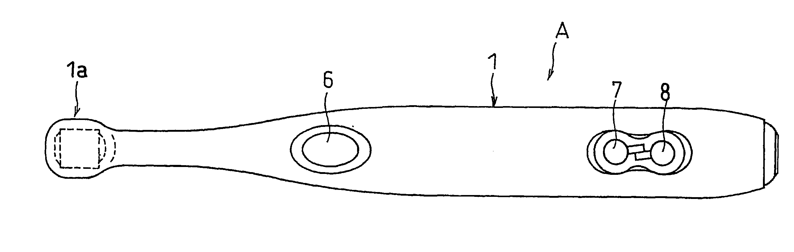
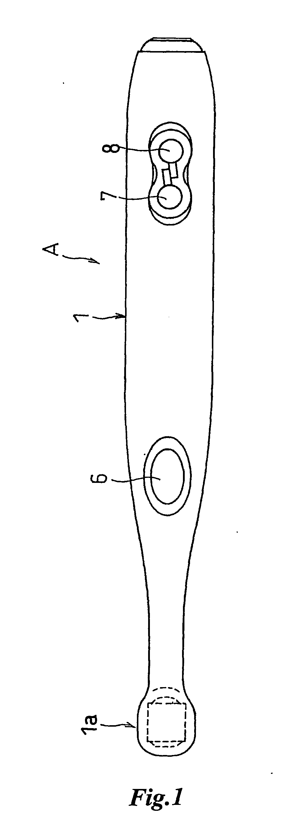
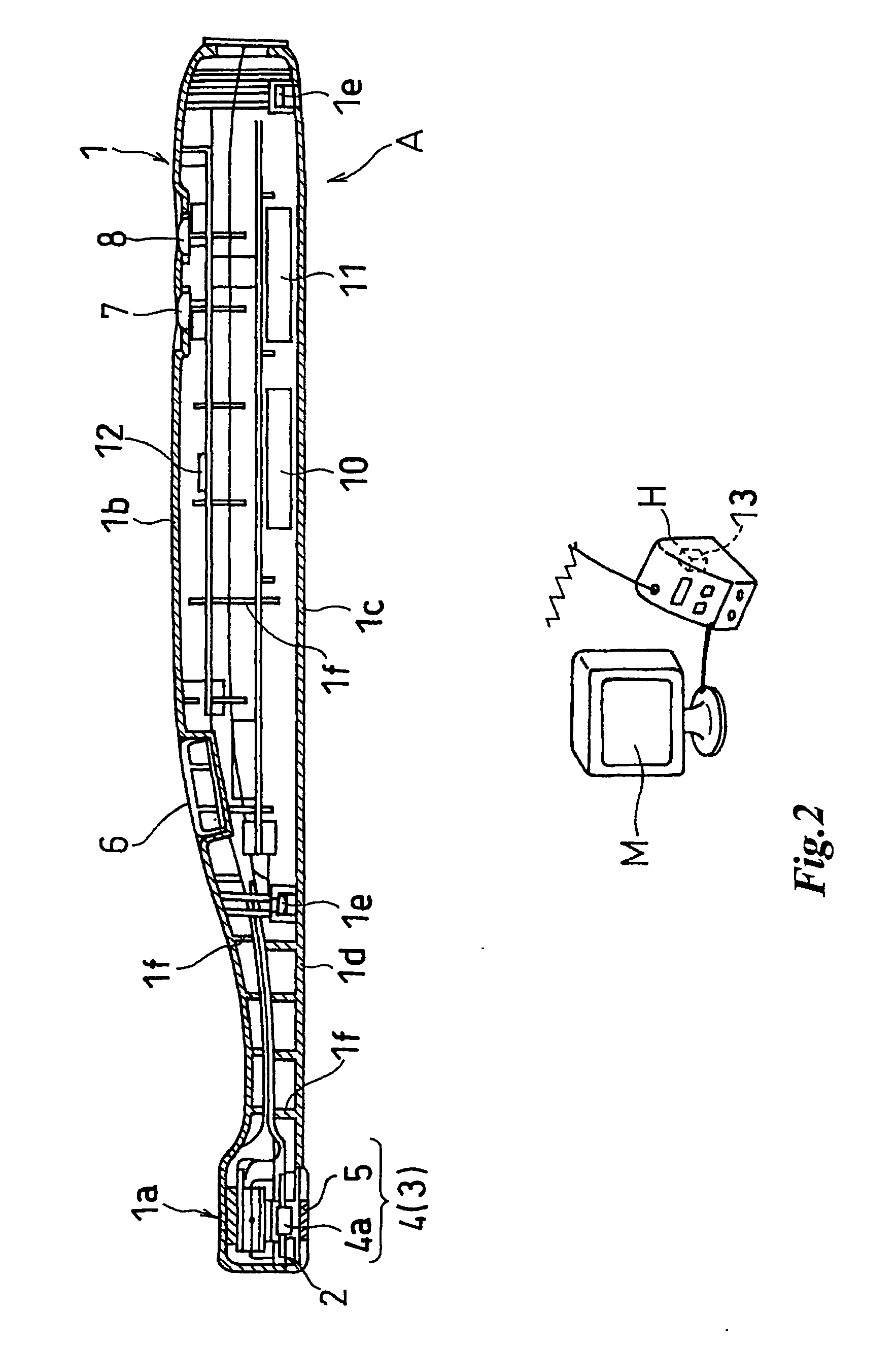
[0124]FIG. 12 shows an example of an intraoral imaging apparatus A3 having, as with the third embodiment, imaging means provided with optical path changing means and having the head part 1a separable into a head part 1a1 and a base part 1a2, with portions in common with the third embodiment provided with the same numerals. Specifically, on the inner surface of the cylindrical head part 1a1, the mirror (or prism) 18 as the optical path changing means is so mounted as to form an angle of substantially 45 degrees with respect to the optical axis described above, while, in the cylindrical base part 1a2 of the head part 1a, a solid state imaging device 4a constituting the imaging means 4 as the observation section 3 is installed. When the head part 1a1 and the base part 1a2 are linked together via link means 26 to be described later, an inner cylindrical part between this mirror 18 and the solid state imaging device 4a is provided as the light guide path 19 for the imaged light.

[0125] A...

fifth embodiment

[0129]FIGS. 13A to 13C are diagram showing examples of a living body observing apparatus of the present invention embodied in a medical irradiation head device, with 13A as a conceptual diagram with the device in use, 13B as an enlarged sectional view of irradiation means, and 13C as the same diagram of a modified example. The medical irradiation head device B in the figure is composed of head fitting means 29 fitted on the head of an operator D, and irradiation means 2 mounted on this head fitting means 29. The head fitting means 29 in the illustrated example is shown as a fastening belt or an elastic belt, although not limited thereto, and thus head fitting means of this type heretofore known in the medical field may be applicable.

[0130] The irradiation means 2 is oscillatablly fitted to a mounting base 34 provided at a position in the head fitting means 29 corresponding to the forehead portion of the operator D, and its irradiation angle with respect to an irradiation field is a...

sixth embodiment

[0137]FIGS. 14A and 14B show examples of a medical irradiation head device B1 having packaged irradiation means 2 composed of a plurality of types (three types in the illustrated example) of LEDs (or bare chips) 35c, 35d, and 35e as emission sections 35. FIG. 14A is a conceptual diagram showing the device in use, and FIG. 14B is a partially cut-away enlarged perspective view showing the irradiation means 2. Specifically, the irradiation means 2 is, as described above, composed of the exciting light emitting LED (exciting light emission section) 35c, and the white light LED 35d and the red light LED 35e as illumination light emission sections, which are disposed at the bottom of a cup-like casing 30a, and is oscillatably mounted on the mounting base 34. A light switch 34b is provided with three switch sections corresponding to these emission sections 35, and the operator D can selectively operate these switch sections to irradiate a desired light in accordance with a diagnostic purpo...


PUM


Abstract

A medical living body observation apparatus, an intraoral photography system and a medical treatment appliance which can be used conveniently by a general user at home as well as a technician, e.g. a dentist or a physician. There is provided an irradiation means (2) comprising an exciting light emission section (2a) emitting an exciting light for causing a lesion to radiate fluorescence, and an illumination light emission section (2b) emitting a light for illuminating the periphery of the lesion. The irradiating means (2) can simultaneously irradiate the illumination light and the exciting light.

Description

TECHNICAL FIELD [0001] The present invention relates to a living body observing apparatus including a medical observing apparatus for an operator to use in diagnosis of caries condition of a tooth, a missing part, or lesion, deposition condition of tartar or plaque, or lesion of root canal, gum, cheek, or tongue in the oral cavity; or in diagnosis of otological regions; treatment and diagnosis of rectal abscess and the like; or dermatological diagnosis. The present invention further relates to an intraoral imaging apparatus capable of sharply visually recognizing and further imaging the condition of the lesion. Moreover, the present invention relates to a living body observing apparatus, an intraoral imaging apparatus, and a medical treatment appliance which permit a general user to, for example, observe his (her) or his (her) family member's tartar deposition condition, progress of caries of a tooth in the oral cavity, and further skin condition. BACKGROUND ARTS [0002] For example,...

Claims


Application Information

Patent Timeline
IPC IPC(8): A61B6/14A61B1/00A61B1/24A61B5/00
CPCA61B1/0615A61B1/0623A61B1/0676A61B1/0692A61B1/24A61B5/0088A61B1/043A61B1/0684A61B1/0605A61B1/00
Inventor OKAWA, SHINICHIKINO, KENJIMATOBA, KAZUNARI
Owner MORITA MFG CO LTD
Why Eureka
Try Eureka