
In Vitro Method for Isolating, Proliferating and Differentiating Germ-Line Stem Cells

a germ-line stem cell and in vitro culture technology, which is applied in the field of isolating, proliferating and differentiating mammalian male germ-line stem cells, can solve the problems of insufficient completion of the technical system allowing the mass proliferation of germ-line stem cells by in vitro culture, potential risks in the clinical use of round spermatids produced from immortalized cells, and insufficient isolation of germ-line stem cells. to achieve the effect of isolation and proliferation of germ-line stem

Inactive Publication Date: 2008-02-21
COLLEGE OF MEDICINE POCHON CHA UNIV IND
View PDF4 Cites 19 Cited by
  • Summary
  • Abstract
  • Description
  • Claims
  • Application Information

AI Technical Summary

Benefits of technology

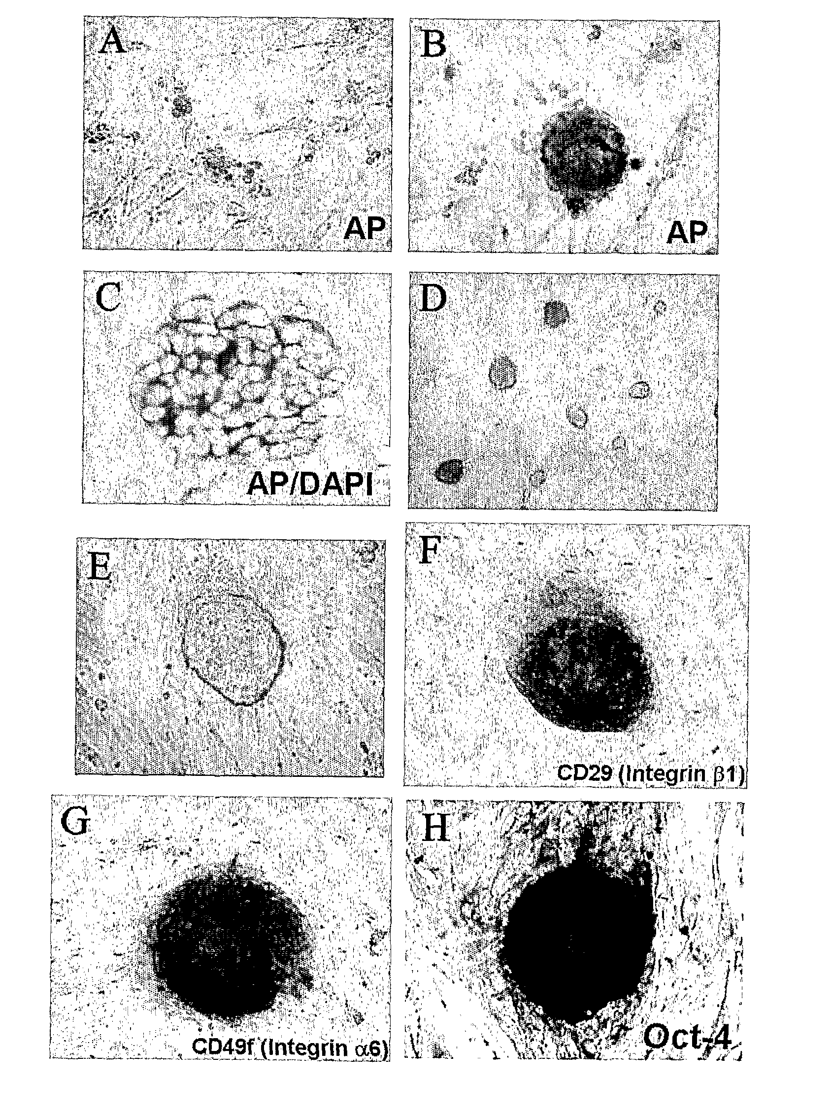
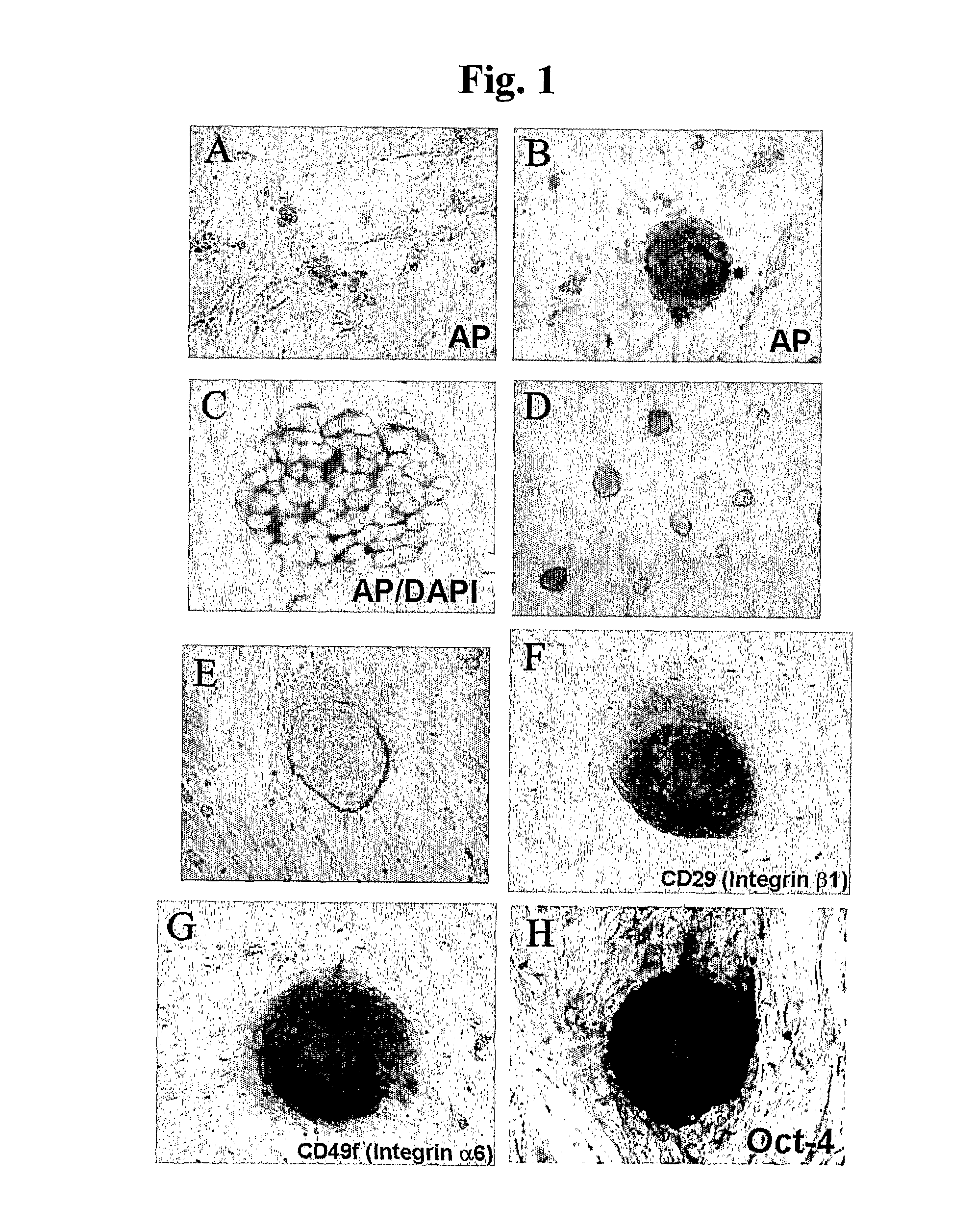
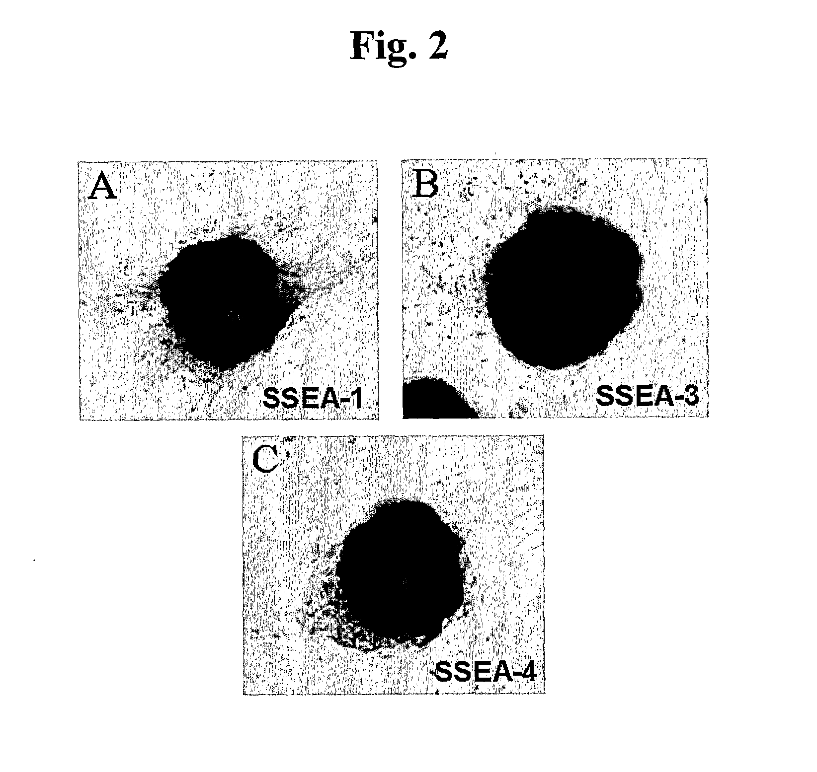
[0009] During extensive studies on methods for isolating, proliferating and differentiating germ-line stem cells from mammalian testis, the present inventors have found that when cells isolated from mammalian testis are cultured in an embryonic stem cell culture medium, the germ-line stem cells will form a mass of cell colonies so as to effectively achieve the isolation and proliferation of the germ-line stem cells at the same time. The present inventors have also found that when the isolated germ-line stem cells are encapsulated with calcium alginate and co-cultured with peritubular cells, the differentiation of the germ-line stem cells is effectively achieved. On the basis of these findings, the present invention was completed.
[0011] Another object of the present invention is to provide a method for effectively differentiating germ-line stem cells in vitro.

Problems solved by technology

However, the clinical use of the round spermatids produced from the immortalized cells has a potential risk.
However, despite of the above-described efforts, there is a difficulty in the isolation of germ-line stem cells since the amount of germ-line stem cells present in vivo is very small and the development of a specific marker for the isolation of germ-line stem cells is not yet made.
Also, because of the difficulty in isolation, a technical system allowing the mass proliferation of germ-line stem cells by in vitro culture is not yet completed.

Method used


Image

  • In Vitro Method for Isolating, Proliferating and Differentiating Germ-Line Stem Cells
  • In Vitro Method for Isolating, Proliferating and Differentiating Germ-Line Stem Cells
  • In Vitro Method for Isolating, Proliferating and Differentiating Germ-Line Stem Cells

Examples


example 1

Isolation and Proliferation of Germ-Line Stem Cells from Male Mice

[0080] Germ-line stem cells were isolated from the testis of thirty 3˜5-day-old male C57BL / 6 or ICR mice (Korea Biolink Co.) and proliferated.

[0081] First, in order to isolate seminiferous tubules from male mice, the following two-step enzymatic digestion process (Ogawa et al., Int. J. Dev. Biol., 41:111-122, 1997) was performed.

[0082] Namely, testis were extracted from the mice and washed with PBS, and the tunica albuginea was removed from the testis. Seminiferous tubules, which are exposed in the testis, were collected and incubated in 10 ml of a first enzyme solution containing 0.5 mg / ml of collagenase (Type I, Sigma), 10 μg / ml of DNase I, 1 μg / ml of soybean trypsin inhibitor (Gibco, Grand Island, N.Y.), 1500 U / ml of leukemia inhibitory factor (ESGRO) and 1 mg / ml of hyaluronidase (Sigma) in Ca2+ and Mg2+-free PBS at room temperature for 20 minutes. Then, PBS was added to the first enzyme solution and it was cent...

example 2

Isolation and Proliferation of Germ-Line Stem Cells from Human Azoospermia Patients

[0097] Germ-line stem cells were isolated from the testis of 13 human non-obstructive azoospermia patients and proliferated.

[0098] First, in order to isolate seminiferous tubules from the 13 patients, a portion of the testicular tissue was extracted by biopsy and subjected to the two-step enzymatic digestion process in the same manner as in Example 1.

[0099] The pellets produced by above method were attached and cultured on a culture dish coated with 0.2% gelatin. A culture medium was DMEM (Dulbecco's modified Eagle's medium; GIBCO) supplemented with 15% fetal bovine serum (HyClone), 1% nonessential amino acid (GIBCO), 10 μM 2-mercaptoethanol, 1500 U / ml of human leukemia inhibitory factor (ESGRO), 4 ng / ml of bFGF(R&D) and 10 μM forskolin (Sigma). Also, the culture was performed in an incubator maintained at 5% CO2 and 95% humidity. After 2-4 week culturing, a number of multicellular colonies and Ser...

example 3

In Vitro Differentiation from Germ-Line Stem Cells into Haploid Germ Cells

[0113] The isolated and proliferated germ-line stem cells were differentiated into germ cells by in vitro culture.

[0114] Namely, the germ-line stem cells and Sertoli cells of mice and human non-obstructive azoospermia patients, which have been isolated and proliferated in Examples 1 and 2, respectively, were separated into single cells by treatment with trypsin, and resuspended, followed by encapsulation with sodium alginate (Lee et al., Biol. Reprod. 65: 873-878, 2001).

[0115] Then, the encapsulated cells were transferred to 1.0 ml of a culture medium without peritubular cell monolayers as feeder cells in 24-well dish, and cultured in an incubator (maintained at 5% CO2 and 95% humidity) at 32° C. for 7 weeks. During culture, the culture medium was replaced every two day. The culture medium was a HEPES-buffered DMEM / F12 medium supplemented with 10 μg / ml of insulin-transferrin-selenium solution (Gibco), 10−4 ...


PUM

PropertyMeasurementUnit
diameteraaaaaaaaaa
humidityaaaaaaaaaa
humidityaaaaaaaaaa

Abstract

The present invention discloses methods for isolating, proliferating and differentiating germ-line stem cell in vitro. More specifically, the present invention discloses a method for the in vitro isolation and proliferation of mammalian germ-line stem cells, characterized in culturing cells isolated from mammalian testis in an embryonic stem cell culture medium, and for the in vitro differentiation, characterized in encapsulating mammalian germ-line stem cells and Sertoli cells with calcium alginate and co-culturing with peritubular cells. Also, the present invention discloses germ-line stem cells obtained by above methods, a composition for the treatment of male infertility, comprising the germ-line stem cells, and a method for the treatment of male infertility by the use of the germ-line stem cells.

Description

TECHNICAL FIELD [0001] The present invention relates to methods for isolating, proliferating and differentiating mammalian male germ-line stem cells (GSCS) in vitro. BACKGROUND ART [0002] Male germ-line stem cells are a kind of adult stem cells and known to have unipotency devoted solely to the generation of sperm by self-renewal and differentiation. The germ-line stem cells are rare relatively quiescent population that lies in a protected region in the testis among supporting cells, and their proliferation and differentiation are regulated by communications with external signals. [0003] The processes of development and differentiation of the germ-line stem cells are well known. For example, the primordial germ cells of mice appear in the epiblast at 7.5 dpc (days post coitum), proliferate and migrate to the genital ridge. Then, these cells form gonads at 12.5 dpc and differentiate into gonocytes. The gonocytes continue to proliferate until 16 dpc, and then stop to proliferate. Afte...

Claims


Application Information

Patent Timeline
Patent Type & Authority Applications(United States)
IPC IPC(8): A61K48/00A61P15/08C12N5/00C12N5/08C12N5/02C12N5/074
CPCC12N5/0611C12N2500/33C12N2502/02C12N2501/115C12N2501/235C12N2500/44A61P15/08
Inventor KIM, KYE-SEONGLEE, DONG-RYUL
Owner COLLEGE OF MEDICINE POCHON CHA UNIV IND
Learn More