Method of measuring propulsion in lymphatic structures

a lymphatic structure and propulsion technology, applied in the field of biomedical imaging, can solve the problems of inability to predict or manage lymphedema, inability to accurately measure lymphedema,

Inactive Publication Date: 2008-03-13
BAYLOR COLLEGE OF MEDICINE
View PDF16 Cites 78 Cited by
  • Summary
  • Abstract
  • Description
  • Claims
  • Application Information

AI Technical Summary

Problems solved by technology

The impairment of lymphatic transport capacity occurs due to either 1) lymph vessel damage and subsequent insufficient repair processes, or 2) congenital defects leading to abnormal lymph vessel development.
Regardless of the cause, the impairment causes fluid and protein accumulation, which in turn leads to lymphedema.
There is a paucity of strategies for predicting or managing lymphedema due in part to the lack of diagnostic imaging approaches to noninvasively and routinely measure lymphatic function.
However, there are presently very few technologies with the ability to non-invasively image the lymphatic system in vivo and in real time.

Method used


Image

  • Method of measuring propulsion in lymphatic structures
  • Method of measuring propulsion in lymphatic structures
  • Method of measuring propulsion in lymphatic structures

Examples


example 1

Material and Methods

Animal Models

[0067] Four, two-month old, 60 lb white Yorkshire swine (K Bar Livestock, HC 69 Box 270 Sabinal, Tex.) were imaged using protocols which were approved by the Baylor College of Medicine Institutional Animal Care and Use Committee. The animals were anesthetized, intubated, and maintained with isoflurane. Animal body temperature was maintained at 100° F. using a warming blanket. At the end of the procedure, the animals were euthanized and lymph nodes resected with fluorescence guidance. Swine were chosen for the lymph mapping study because swine dermis and lymphatic plexus of swine is considered most comparable to humans.

NIR Fluorescence Enhanced Imaging

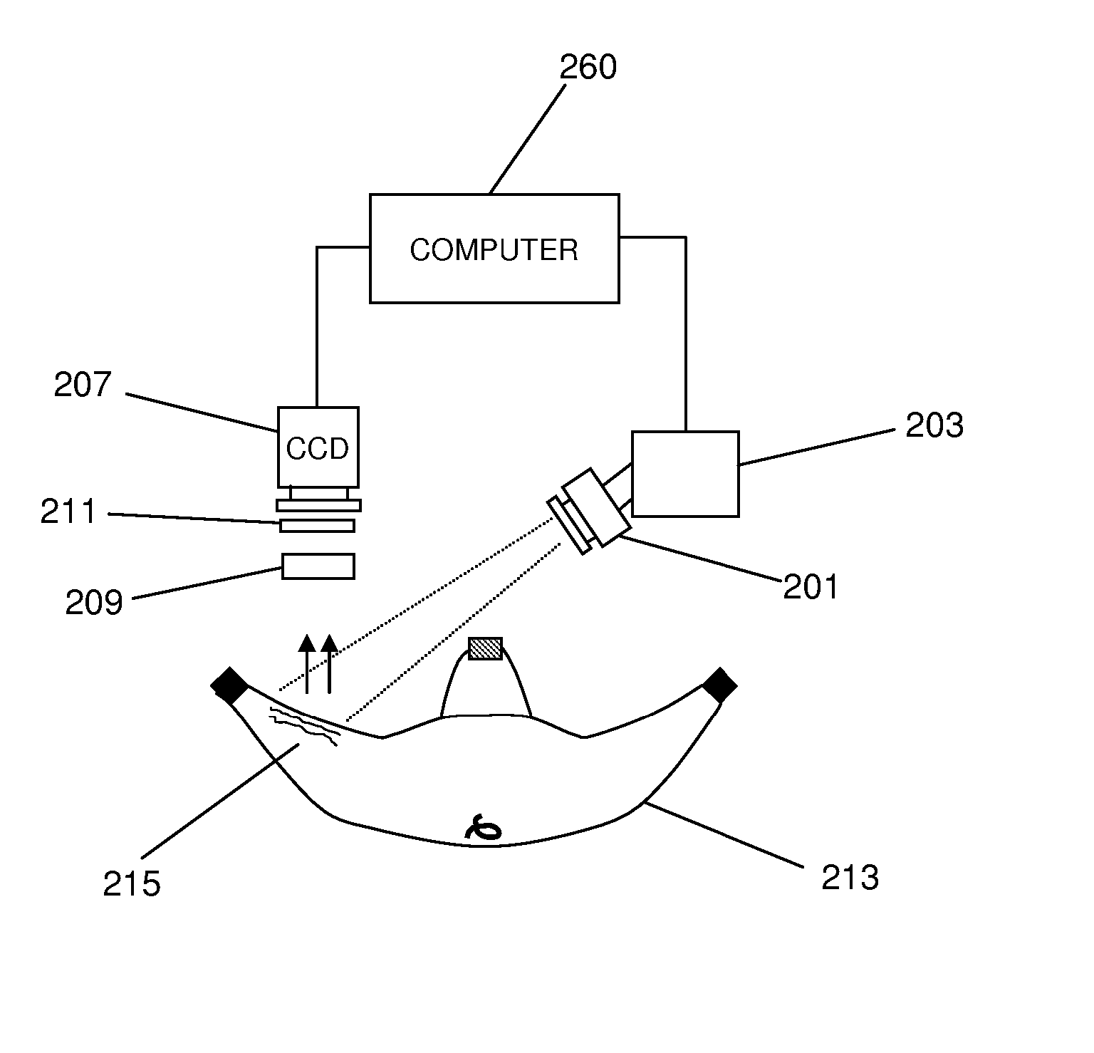
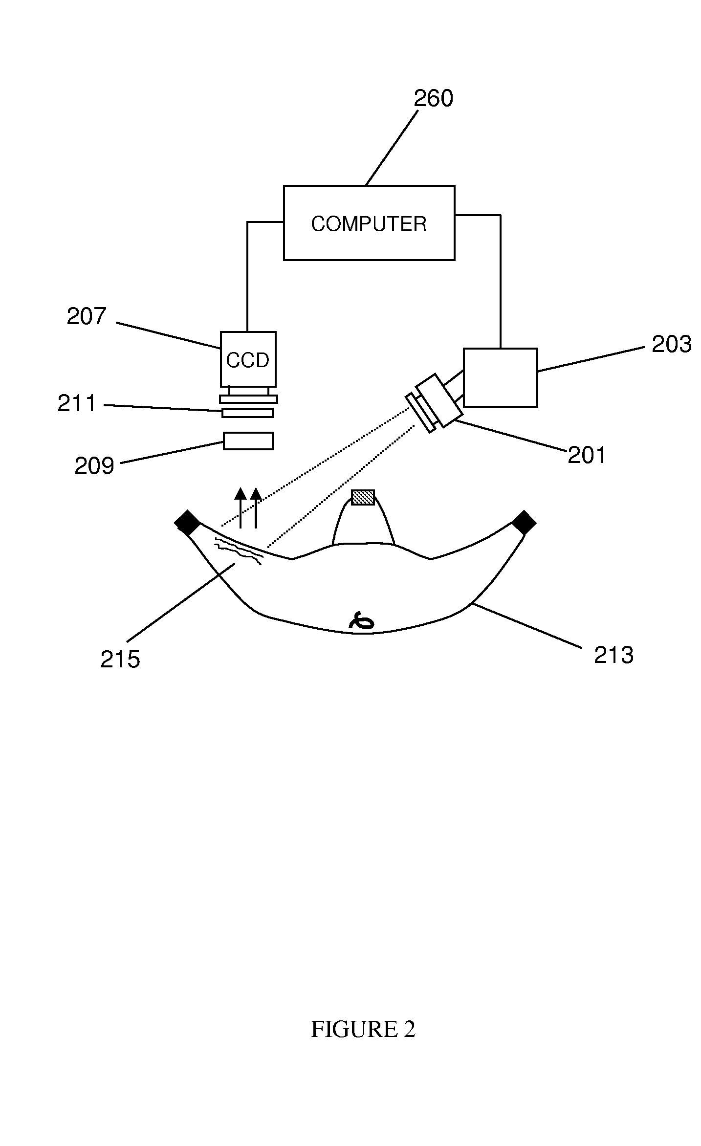
[0068] Continuous-wave optical imaging of the fluorescent NIR dyes was performed with a custom built intensified charged-coupled device (CCD). Reynolds J S, Troy T L, and Sevick-Muraca E M. Multipixel techniques for frequency-domain photon migration imaging. Biotechnol Prog 13: 669-680, 1997., inco...

example 2

Modification of IR-783 Dye

[0077] IR-783 was combined with 4 mercaptobenzoic acid in presence of ethyldiisopropylamine (DIPEA). The mixture was stirred for eight hours under nitrogen atmosphere and dimethyl formic acid solvent was removed. The compound 1 (IR-783-S-Ph-COOH) was purified by flash chromatography.

[0078] Compound 1 was treated with hydroxysuccinamide (HOSu) in dimethylformamide (DMF) and purified to obtain compound 2 (IR-783-S-Ph-COOSu). Compound 2 was combined with mono-Trityl 1,6-diaminohexan acetic acid salt in DMF and DIPEA. The mixture was stirred overnight to remove solvent and the residue (compound 3) was dissolved in methanol and purified by flash chromatography. Finally, compound 3 was mixed with 40% TFA (trifluoro acetic acid) in DCM (dichloromethane). The mixture was stirred for 30 minutes to remove the solvent. The residue was washed with ether several times and purified by HPLC (high pressure liquid chromatography.

Conjugation of Modified IR-783 to HA

[007...


PUM

PropertyMeasurementUnit
excitation wavelengthaaaaaaaaaa
wavelengthaaaaaaaaaa
integration timeaaaaaaaaaa

Abstract

Novel methods and imaging agents for functional imaging of lymph structures are disclosed herein. Embodiments of the methods utilize highly sensitive optical imaging and fluorescent spectroscopy techniques to track or monitor packets of organic dye flowing in one or more lymphatic structures. The packets of organic dye may be tracked to provide quantitative information regarding lymph propulsion and function. In particular, lymph flow velocity and pulse frequency may be determined using the disclosed methods.

Description

CROSS-REFERENCE TO RELATED APPLICATIONS [0001] The present application claims the benefit of U.S. Provisional Application Ser. No. 60 / 840,256, filed Aug. 25, 2006, and U.S. Provisional Application Ser. No. 60 / 823,481, filed Aug. 24, 2006, each of which is herein incorporated by reference in their entireties for all purposes. U.S. application Ser. No. 11 / 844,807 entitled Imaging Agents for Functional Imaging of Lymphatic Structures, filed Aug. 24, 2007, Attorney Docket No. 1373-03901, is herein incorporated by reference in its entirety for all purposes.STATEMENT REGARDING FEDERALLY SPONSORED RESEARCH OR DEVELOPMENT [0002] This work was supported by the National Institutes of Health (R01 CA112679). BACKGROUND [0003] 1. Field of the Invention [0004] This invention relates generally to the field of biomedical imaging. More specifically, the invention relates to a method of functionally imaging and measuring lymphatic function by assessing lymph propulsion in lymphatic structures. [0005]...

Claims


Application Information

Patent Timeline
Patent Type & Authority Applications(United States)
IPC IPC(8): A61B6/00
CPCA61B5/0059A61B5/0064A61B5/415A61B5/418A61K47/4823A61M5/007A61K49/0034A61K49/0054A61B5/0071A61B5/41A61K49/0041A61K49/0032A61K47/61
Inventor ADAMS, KRISTENSHARMA, RUCHIRASMUSSEN, JOHNSEVICK-MURACA, EVA
Owner BAYLOR COLLEGE OF MEDICINE
Why Eureka
Try Eureka