

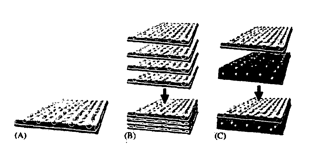
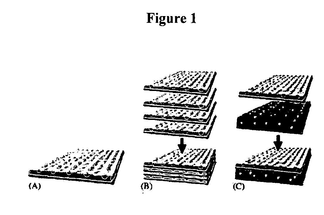
Innovative bottom-up cell assembly approach to three-dimensional tissue formation using nano-or micro-fibers
a three-dimensional tissue and bottom-up cell technology, applied in the field of bioengineered three-dimensional tissue formation, can solve the problems of limiting the application of prosthetic devices, unable to prevent the continued progression of disease, and not permanent replacement of the full and proper function of damaged tissue or organs
- Summary
- Abstract
- Description
- Claims
- Application Information
AI Technical Summary
Benefits of technology
Problems solved by technology
Method used
Image

Examples
example 1
Biomimetic Fibers for Layer-by-Layer Cell Building
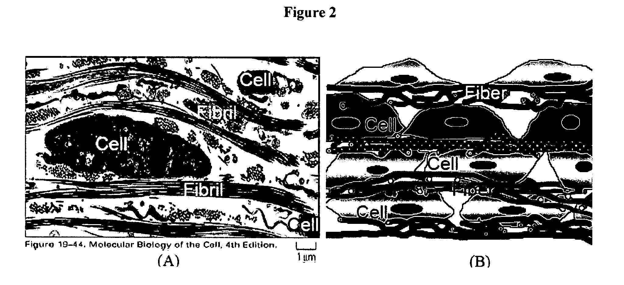
[0062] For preparation of suitable fibrous scaffolds supporting the growth and differentiation of bone cells and endothelial cells, we used an electrospinning technique, which produces fibers with similar dimensions as matrix fibrils and variable chemical compositions similar to those found in the ECM. In our preliminary study, collagen type I from calf skin was first electrospun into fibers with high mechanical properties by blending with polycaprolactone (PCL). The diameter of obtained fiber ranged from 50 nanometers to several micrometers, depending on polymer concentration and spinning conditions. Improved cell adhesion and proliferation of fibroblasts by collagen was observed, consistent with other studies (Liu, G., et al. Chin J Traumatol, 2004. 7(6): p. 358-62; Xiao, Y., et al. Tissue Eng, 2003. 9(6): p. 1167-77; Petrovic, L., et al. Int J Oral Maxillofac Implants, 2006. 21(2): p. 225-31). Fibroblasts cultured on collagen-bas...
example 2
Layer-by-Layer Alternating Assembly of Multilayer-Cell Structure with Electrospun Fibers Sandwiched in Between
[0063] To test the feasibility of forming a multilayered structure, a study was done using human dermal fibroblast. This multilayer cell sheet with alternating layers of human fibroblasts (8 layers) and layers of collagen / PCL nanofibers (9 layers) was layer-by-layer fabricated as shown in FIG. 3. The space between cell layers was adjustable and defined by the thickness of nanofibrous layer. During the alternating cell layering, fibroblast culture medium (Dulbecco's modified minimum essential medium (DMEM) (Invitrogen) supplemented with 10% foetal bovine serum (FBS), 50 U / mL penicillin, 50 μg / mL streptomycin) is used. In the prepared cell sheet, the space between cell layers was around 5-25 μm. Closer examination of the cross section of cell sheet cultured for 2 days at 37° C. in CO2 incubator stained with methylene blue under an optical microscope clearly showed the presenc...
PUM
 Abstract
Description
Claims
Application Information