High quality, continuous throughput, tissue processing

a tissue and high-quality technology, applied in the field of tissue processing for microscopic examination, can solve the problems of reducing reducing the size of the laboratory facility, and the risk of possible instrument failure and the need to monitor the instruments, so as to reduce the number of personnel required, reduce the volume of reagents used, and reduce the time required

Inactive Publication Date: 2008-06-26
UNIV OF MIAMI
View PDF99 Cites 10 Cited by
  • Summary
  • Abstract
  • Description
  • Claims
  • Application Information

AI Technical Summary

Benefits of technology

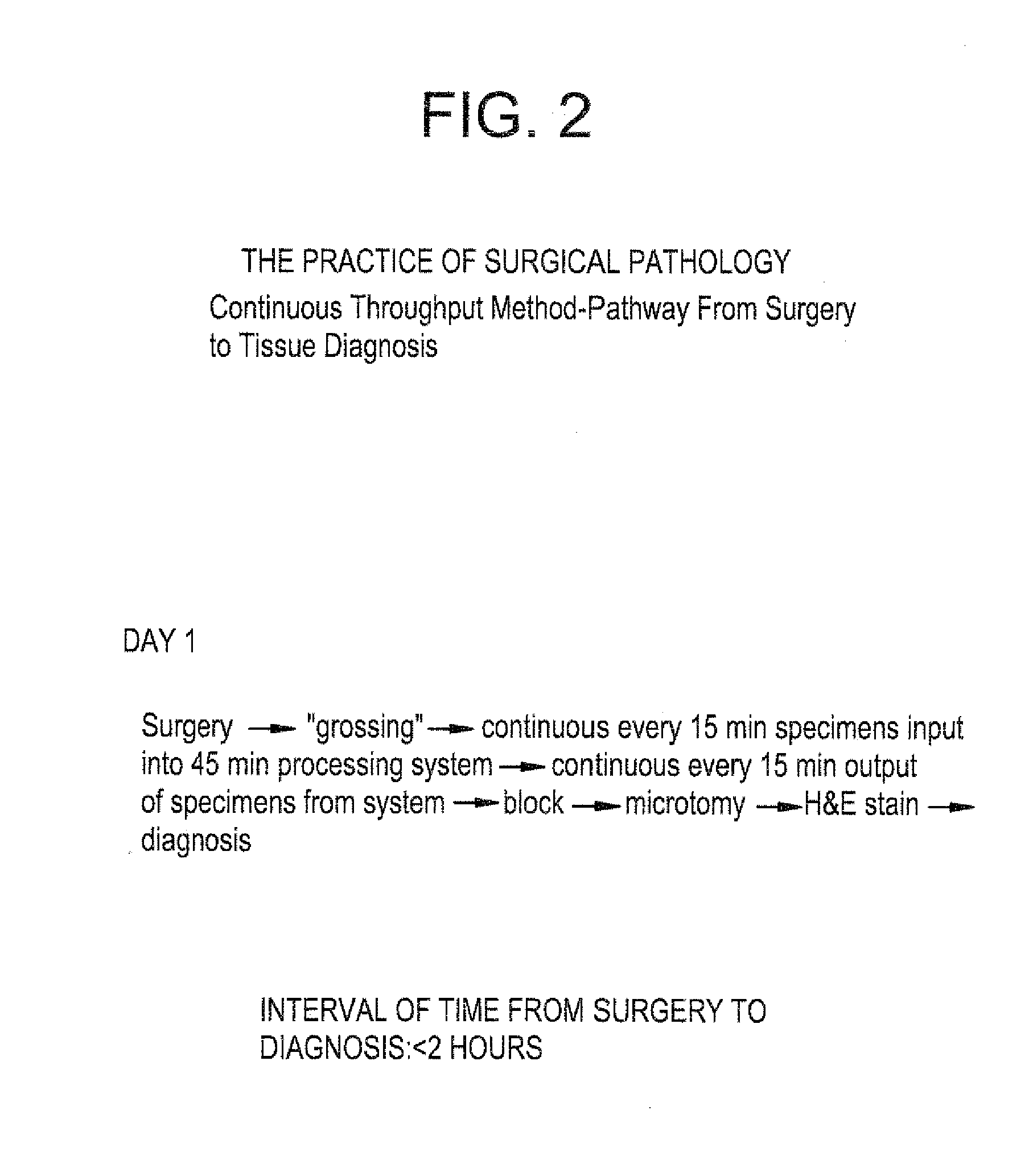
[0011]It is an object of the invention to provide compositions for tissue processing and an apparatus and system for utilizing the same that reduces the time required for tissue processing and analysis, and reduces the cost thereof by reducing time, the size of the laboratory facility, the volumes of reagents used, and the number of personnel required. This allows conversion of existing practice to rapid response surgical pathology for the patient undergoing an operation, and may even allow point-of-care diagnosis by the pathologist in the vicinity of the operating room.
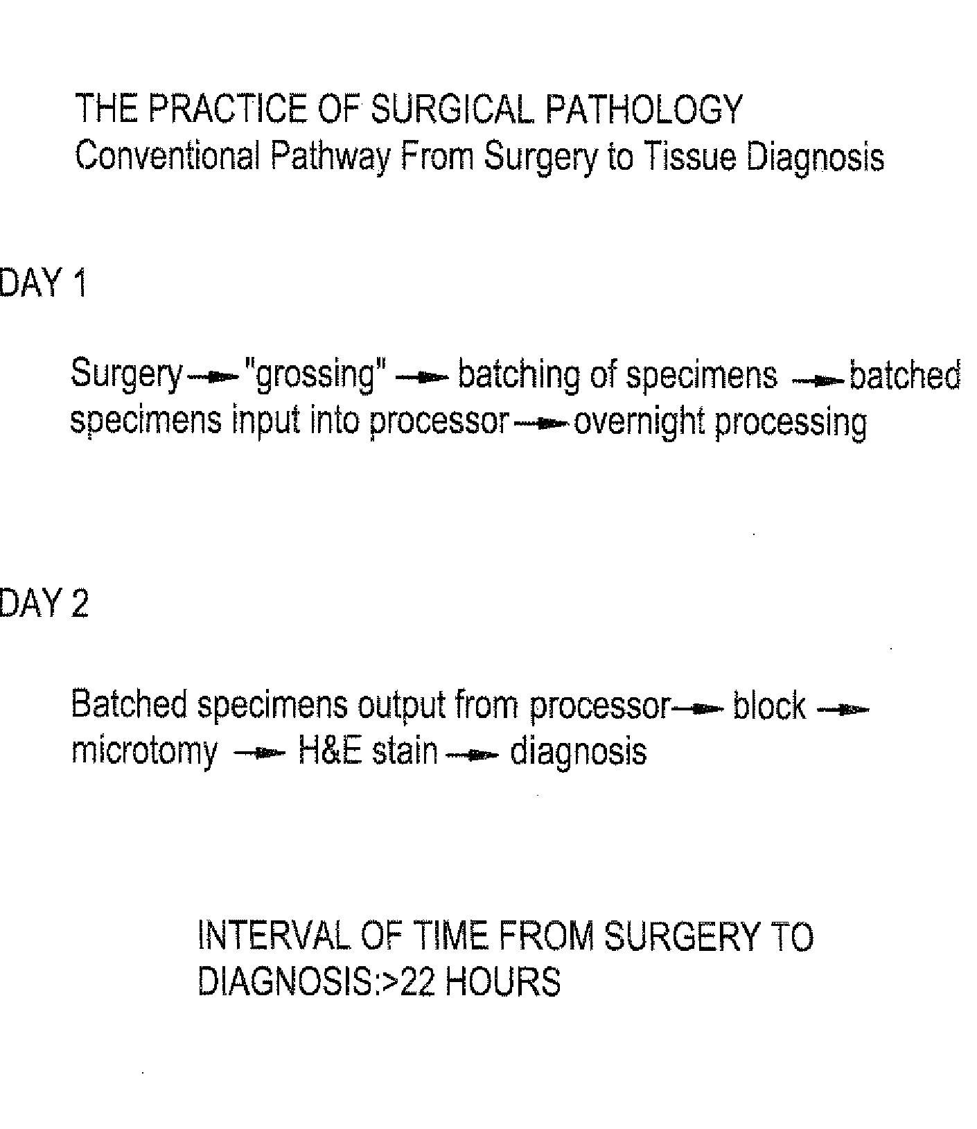
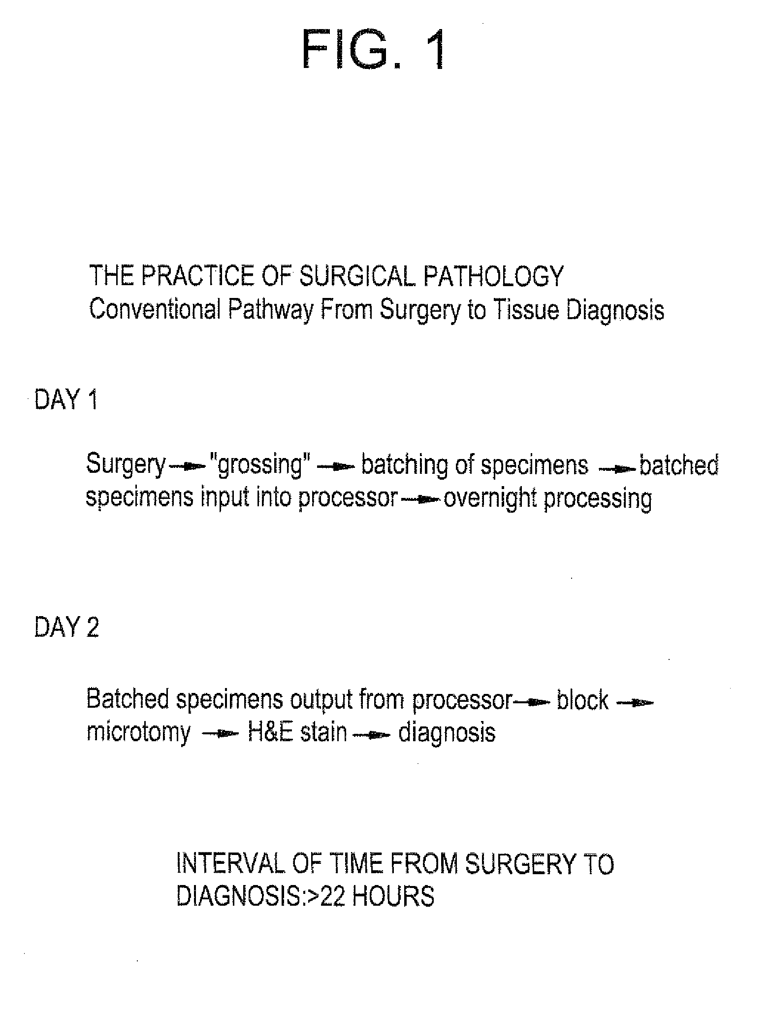
[0012]With regard to the processing and analysis of solid tissue, a tissue slice must be on the order of 4 to 6 microns to be examined under a microscope, whereas the thinnest slice of fresh tissue that can be obtained by cutting is about 1 mm with the typical slice being on the order of 3 mm. In order to produce a sufficiently thin slice from microscopic examination, it is necessary to harden the tissue so that a finer slice can be obtained, e.g., by sectioning with a microtome. The present invention greatly accelerates the tissue hardening process and thus turns the conventional overnight processing into a process which totals on the order of 40 minutes. Thus, we have developed a simple, safe, low cost, expeditious, and reliable method that permits preparation of impregnated tissue blocks suitable for microtome sectioning in less than two hours from the moment tissue is received in the pathology laboratory. This method allows continuous flow of specimens, is adaptable to automation, precludes the need for formalin and xylene with their noxious fumes, allows standardization of tissue processing, and requires considerably smaller volumes of reagents than conventional methods. Either fresh or previously fixed tissues can be processed by the present invention.
[0013]In addition to the reduction in time required for tissue processing, the rapid preparation of tissue by the present invention is capable of preserving tissue structures and morphology that were lost with conventional methods.

Problems solved by technology

In addition to the minimum one-day delay in giving a surgeon the benefit of a report from the pathologist, there are also problems associated with impeded work flow in the pathology laboratory necessitated by the requisite batch processing of specimens, the safety concerns that attend having instruments operating overnight, the risk of possible instrument failures and the need to monitor the instruments, and the waste of using large volumes of reagents for such processing when automated.
Moreover, expensive measures are required to prevent exposure of laboratory personnel to fumes and toxic substances associated with the reagents used in this process.
Also, the large volumes of solvent waste and paraffin debris produced by conventional methodology pollute the environment.
Conventional fixation and processing cause irreversible damage to the structure of DNA and particularly RNA that limits the application of genetic techniques for diagnosis and research.
Consequently, most DNA and certainly RNA analysis require special precautions with handling of material, such as immediate freezing of fresh tissues, because retrospective genetic analysis is impaired by conventional tissue processing techniques.
Histological diagnosis of a frozen section suffers from multiple disadvantages in comparison to sections prepared from paraffin blocks: the slide prepared from a frozen section “does not possess .
The costs of tissue processing are related to time, the space required for preparation and analysis, reagents (both the amount required for processing and handling discard), and the number of personnel required.
Others have recognized the need to shorten the time required for tissue processing, but they have made only modest improvements in the conventional methods.
However, the aforementioned patents do not teach or suggest that the entire process of preparing diagnostic tissue slides could be accomplished in less than two hours, starting from fixation and ending with impregnation, with continuous throughput of samples.

Method used


Image

  • High quality, continuous throughput, tissue processing
  • High quality, continuous throughput, tissue processing
  • High quality, continuous throughput, tissue processing

Examples


example 1

[0099]Two mm thick or thinner slices of fresh or previously fixed tissue were held in tissue cassettes and placed in a non-aqueous first solution of:

[0100]40% isopropyl alcohol,

[0101]40% acetone,

[0102]20% polyethylene glycol (average molecular weight 300), and

[0103]1% dimethyl sulfoxide (DMSO) (i.e., 10 ml per liter of the above mixture).

[0104]Tissues samples were incubated for 15 min at a glycerin bath temperature between 45° C. and 50° C. The 400 ml solution for fixation was placed in a 500 ml beaker in a water bath shaker (linear displacement of 5 cm / sec). Additional agitation of the fixation solution was provided by bubbling with an air pump.

[0105]Fixation, dehydration, fat removal, clearing, and impregnation are accomplished by sequential exposure of the tissue specimen to three different solutions (the second, third and fourth solutions described above), one in each of three microwave ovens from Energy Beam Sciences. A one liter solution of 70% isopropyl alcohol and 30% polyet...

example 2

[0107]Fixation, dehydration, fat removal, and paraffin impregnation of fresh or fixed tissue sections, approximately 1 mm thick, was accomplished in 40 minutes by exposing these tissue sections to four successive steps as follows.

Step 1.

[0108]In this example, the first solution consisted of:

[0109]60% isopropyl alcohol,

[0110]10% acetone,

[0111]30% polyethylene glycol (average molecular weight 300), and

[0112]dimethyl sulfoxide (DMSO) added at an approximate concentration of 1% of the total volume. One liter of this solution suffices to fix 60 samples of tissue held in tissue cassettes. The samples were incubated at 55° C. in a commercial tissue microwave processor (H12500 or H2800, Energy Beam Sciences) for 5 min each in a series of three baths containing the first solution (15 min total incubation); agitation of the solution was obtained by bubbling to accelerate solution exchange.

Step 2.

[0113]The samples were incubated in a solution of 70% isopropyl alcohol, 30% acetone, and DMSO add...

example 3

[0117]Fixation, dehydration, fat removal, and paraffin impregnation of fresh or fixed tissue sections, up to about 1 to 2 mm thick, may be accomplished in 65 minutes as follows.

Step 1.

[0118]In this example, the first solution consists of:

[0119]40% isopropyl alcohol,

[0120]40% acetone,

[0121]20% polyethylene glycol (average molecular weight 300),

[0122]glacial acetic acid added at an approximate concentration of 0.5% of the total volume, and

[0123]dimethyl sulfoxide (DMSO) added at an approximate concentration of 1% of the total volume, One liter of this solution suffices to fix 60 samples of tissue held in tissue cassettes. The samples are incubated at 65° C. in a commercial tissue microwave processor (H2500 or H2800, Energy Beam Sciences) for 15 min in a 1500 ml beaker containing the first solution: agitation of the solution is obtained by bubbling to accelerate solution exchange.

Step 2,

[0124]The samples are incubated in a solution of 55% isopropyl alcohol, 25% acetone, 10% polyethylen...


PUM

PropertyMeasurementUnit
timeaaaaaaaaaa
timeaaaaaaaaaa
thicknessaaaaaaaaaa

Abstract

A process and apparatus for rapid, continuous flow histological processing of tissues is disclosed. The steps of fixation, dehydration, clearing and impregnation are performed in less than one hour; this allows a pathologist to evaluate samples shortly after receipt, perhaps while the patient is still in the operating room. Rapid and continuous processing is accomplished by decreasing the thickness of tissue sections, use of non-aqueous solutions composed of admixtures of solutions, solution exchange at elevated temperature and with agitation, and impregnation under vacuum pressure. The patient in surgery is thus provided with point-of-care surgical pathology.

Description

CROSS REFERENCE TO RELATED APPLICATIONS[0001]This application is a continuation of application Ser. No. 10 / 400,060, filed Mar. 27, 2003, allowed; which is a continuation of application Ser. No. 09 / 736,388, filed Dec. 15, 2000, now U.S. Pat. No. 6,586,713; which is a divisional of application Ser. No. 09 / 136,292, filed Aug. 19, 1998, now U.S. Pat. No. 6,207,408; which claims benefit of provisional Appln. No. 60 / 056,102, filed Aug. 20, 1997. The entire disclosures of the aforementioned applications are incorporated herein by this reference.BACKGROUND OF THE INVENTION[0002]1. Field of the Invention[0003]The present invention relates to the rapid, continuous flow, processing of tissue for microscopic examination, from fixation to impregnation.[0004]2. Description of the Related Art[0005]Conventional methods prepare tissues for histology by incubation in separate solutions of phosphate-buffered 10% formaldehyde for fixation, a series of increasing concentrations of ethanol for dehydratio...

Claims


Application Information

Patent Timeline
Patent Type & Authority Applications(United States)
IPC IPC(8): G01N1/30C12M1/00B01L9/00G01N33/48G01N1/06G01N1/28G01N1/31G01N1/36G01N1/44
CPCB01L9/50G01N1/06G01N1/30G01N2001/315G01N1/36G01N1/44G01N2001/305G01N1/312
Inventor MORALES, AZORIDESESSENFELD, HAROLDESSENFELD, ERVIN
Owner UNIV OF MIAMI
Why Eureka
Try Eureka