Device for delivery of antifibrotic agents & method

a technology for delivering antifibrotic agents and suppositories, which is applied in the direction of prosthesis, eye treatment, suppositories delivery, etc., can solve the problems of unsatisfactory post-operative outcomes, significant reduction of surgical success at the wound site, and unsatisfactory pharmacological attempts to prevent fibrosis following glaucoma surgery

Active Publication Date: 2009-05-14
ELUTIMED LLC
View PDF17 Cites 13 Cited by
  • Summary
  • Abstract
  • Description
  • Claims
  • Application Information

AI Technical Summary

Problems solved by technology

While several surgical procedures and medical implants are available to treat a number of diseases and disorders of the eye, resultant tissue fibrosis often leads to unsatisfactory post-operative outcomes.
Excessive postoperative fibrosis at the wound site significantly reduces surgical success following glaucoma surgery.
Pharmacological attempts to prevent fibrosis following glaucoma surgery have thus far proven unsatisfactory.
However, the current technique of delivering the MMC or 5-FU in the form of soaking a small wedge of sponge in a given concentration of the drug and applying to the operation site for variable time periods before washing the drug from the surgical site leads to inconsistent results.
This leads to bleb encapsulation and elevated intraocular pressures in both the short term, that is, in the hypertensive phase and in the long term leading to elevated intraocular pressure and failure of the operation.
The failure rate for the glaucoma drainage devices has been reported at 10% per post-operative year, thus reaching 50% failure rate in 5 years.
Antifibrotic agents have also been used during glaucoma drainage device implant operations, but with variable and questionable results.
Topical application of such medications at the time of surgery appears to be less effective 7-9 mm from the limbus for such implant operations compared to the effects seen at the limbus during a trabeculectomy operation.
It is therefore possible that one-time application of these agents may not be sufficient to decrease the chances of long-term fibrous reaction that occurs following an implant operation.
The success rate of these operations, however, is less than 50% at 12 months because of scar tissue formation.
However, the long-term success of this procedure (currently unknown) may be restricted because of reactionary fibrosis.
In glaucoma filtration non-penetrating surgery (NPS), excessive postoperative scarring at the wound site significantly reduces

Method used


Image

  • Device for delivery of antifibrotic agents & method
  • Device for delivery of antifibrotic agents & method
  • Device for delivery of antifibrotic agents & method

Examples


experiment 1

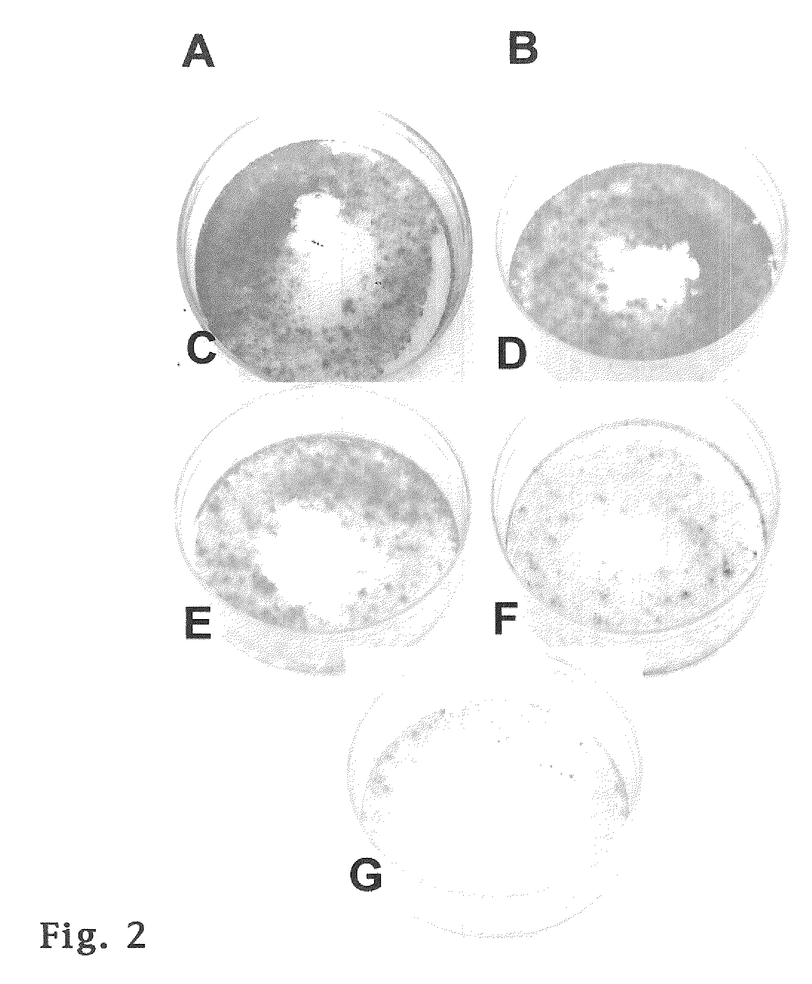
Inhibition of Cell Proliferation by Mitomycin C Incorporated Into P-Hema Hydrogels

[0061]In this Experiment 1 a device including a P-HEMA matrix was tested that releases low concentrations of mitomycin-C over a 3-week period using an in vitro model.

MATERIALS AND METHODS

[0062]Materials: Neutral buffered formalin, toluidine blue, hematoxylin and eosin solution, sodium dodecyl sulfate, mitomycin C (Streptomyces caespitosus), reagents for tissue culture (culture medium, trypsin, antibiotics) and reagents for hydrogel synthesis (2-hydroxyethyl methacrylate, N, N′-methylene-bisacrylamide, N,N,N′,N′-tetramethylethylenediamine, and ammonium persulfate) were purchased from Sigma / Aldrich (St. Louis, Mo.). Tissue culture dishes (60 mm diameter) were from Corning-Costar (Corning, N.Y.). COS-1 cells were obtained from the American Type Culture Collection (Manassas, Va.). Early passage human conjunctival fibroblasts were established in 1994 from a biopsy sample of a 43 year old white male. The pri...

experiment 2

SUMMARY OF EXPERIMENT 2

[0081]In order to test the hypothesis that a slow release drug delivery system using P(HEMA) loaded with Mitomycin C would decrease the fibrosis, a glaucoma drainage device was created in the laboratory that consisted of a 13 mm 10 mm P(HEMA) plate loaded with MMC (2 mg / dry weight of the polymer) attached to a silicone tube. This plate and tube were implanted into the rabbit eye (using standard surgical techniques). The results demonstrated significant reduction in the fibrosis with a resulting avascular bleb formation at the end of 1 week. This experiment proved that the concept of slow release drug delivery system worked in the rabbit model.

DEVICES

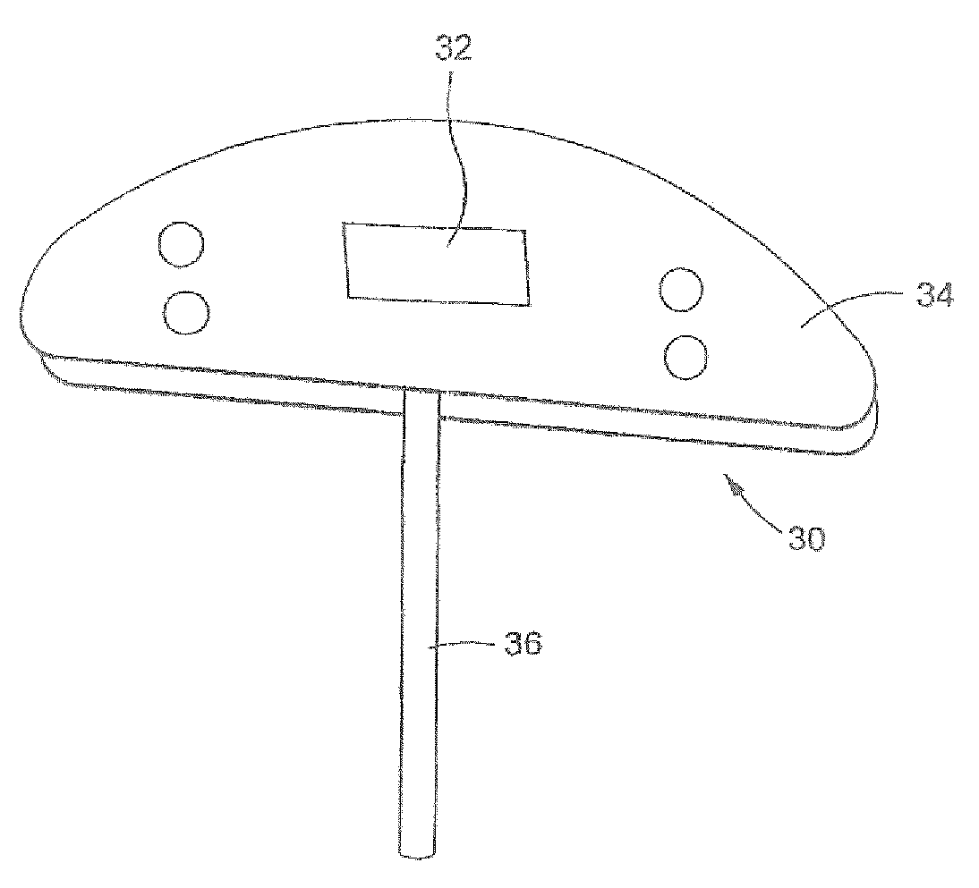
[0082]FIG. 7 schematically depicts one embodiment of my ophthalmological device identified by the numeral 10. The ophthalmological device 10 may be used to treat glaucoma and includes a distribution plate 12 carrying an antifibrotic agent and a tube 14 attached to the plate 12 near an inner end 14b so fluid exiting...

experiment 3

[0090]Incorporation of the p(HEMA) Drug Delivery System into a Commercial GDD

[0091]The next set of experiments concentrated on the preparation and standardization of p(HEMA) disks loaded with a lower concentration of MMC that could be attached to a commercially available glaucoma drainage device, such as the Ahmed Glaucoma Valve (New World Inc., Rancho Cucamonga, Calif.). Semicircular disks of p(HEMA) (5 mm×6 mm) with incorporated MMC (0. 173 mg MMC / g dry gel) were prepared and then attached to the lower half of a commercial Ahmed valve plate (model FP-7), as shown in FIG. 14C, modified for implantation. The arrow shows the p(HEMA) polymer with incorporated MMC. The incorporated MMC imparts the pink color to the polymer. The tube through which aqueous fluid drains is being held by forceps in this photograph FIG. 14C. The release of MMC from this device was measured using a syringe pump to inject sterile water through the tubing of the device. The injection rate was kept constant to ...


PUM


Abstract

An ophthalmological device comprises an implant member carrying an antifibrotic agent that is released slowly over a prolonged period when inserted into a wound in an eye produced by surgery. The member is placed in the vicinity of a wound created during the surgery to inhibit inflammatory cell proliferation, thereby preventing the growth of fibrous tissue after the eye surgery.

Description

RELATED PATENT APPLICATIONS & INCORPORATION BY REFERENCE[0001]This application is a continuation-in-part application of International Application No. PCT / U.S. 2007 / 012253, entitled “DEVICE FOR DELIVERY OF ANTIFIBROTIC AGENTS & METHOD,” filed May 23, 2007, which claims the benefit under 35 USC 119(e) of U. S. Provisional Patent Application No. 60 / 808,446, entitled “DEVICE FOR DELIVERY OF ANTIFIBROTIC AGENTS & METHOD,” filed May 25, 2006. These related applications are incorporated herein by reference and made a part of this application, and any and all U. S. patents, U. S. patent applications, and other documents, hard copy or electronic, cited or referred to in this application are incorporated herein by reference and made a part of this application.DEFINITIONS[0002]The words “comprising, ”“having, ”“containing,” and “including,” and other forms thereof, are intended to be equivalent in meaning and be open ended in that an item or items following any one of these words is not meant ...

Claims


Application Information

Patent Timeline
IPC IPC(8): A61F9/00A61M1/00A61F2/14
CPCA61F9/0017
Inventor AYYALA, RAMESH S.
Owner ELUTIMED LLC
Why Eureka
Try Eureka