Therapeutic Agent for Corneal Diseases

a technology of endothelial cells and therapeutic agents, which is applied in the direction of drug compositions, organic chemistry, genetic material ingredients, etc., can solve the problems of bullous keratopathy and the inability to maintain the transparency of the cornea, and achieve the effects of maintaining transparency, constant water content, and little ability to prolifera

Inactive Publication Date: 2009-06-25
KANSAI TLO KK
View PDF3 Cites 26 Cited by
  • Summary
  • Abstract
  • Description
  • Claims
  • Application Information

AI Technical Summary

Benefits of technology

[0046]Corneal endothelial cells are a monolayer cell layer present on the rear side of the cornea, and play an important role in maintaining transparency by maintaining a constant water content in the cornea with a pump function and a barrier function. Once the corneal endothelial cells are impaired and drop off due to an invasive intrusion, such as those occurring in external injury, dystrophy or intraocular surgery, corneal endothelium dysfunction occurs causing strong edema and opacity in the cornea because corneal endothelial cells in primates such as human beings and monkeys have little ability to proliferate in vivo. Such a pathological state is referred to as bullous keratopathy, where the patient develops a severe visual impairment. A full thickness cornea transplant is currently performed for advanced bullous keratopathy, but its long term results are worse than those for other corneal diseases, and the development of better therapeutic methods is desired. Also in eye banks in Japan, the donated corneas required for such cornea transplants are always in short supply, and patients with bullous keratopathy requiring such a transplant are currently forced to wait for a long time. Under such a social and medical context, the present inventors developed a new method for treating corneal endothelial diseases.
[0047]According to the present invention, it is possible to proliferate the corneal endothelial cells and effectively treat a disease or a disorder caused by a reduction in corneal endothelial cells.
[0048]For example, corneal endothelial cells can be proliferated to restore or regenerate the corneal endothelial tissue leading to the healing of bullous keratopathy by administering at least one nucleic acid molecule inhibiting the expression of a connexin 43 gene to an anterior chamber of an eye in a patient with bullous keratopathy whose cornea has low transparency.
[0049]Even when the corneal endothelial tissue becomes thin due to various causes, e.g., an external injury to the eye, an intraocular infection, an increase of intraocular pressure due to glaucoma or insufficient oxygen due to contact lenses, and thus surgery requiring an intraocular manipulation (intraocular surgery) or laser therapy cannot be given, the intraocular surgery can be allowed by administering at least one nucleic acid molecule inhibiting the expression of the connexin 43 gene to cause the corneal endothelial cells to proliferate and thicken the corneal endothelial tissue.
[0050]Intraocular surgery herein includes surgery requiring intraocular manipulation and laser therapy. Such intraocular surgery includes, but is not limited to, surgeries for cataract, glaucoma and strabismus, retinal detachment operations and vitreous surgery.
[0051]In the present invention, the “disease or disorder due to a reduction in corneal endothelial cells” includes not only bullous keratopathy but also states where because of reduced corneal endothelial cells, the risk of developing bullous keratopathy due to intraocular surgery or laser therapy is increased and such intraocular surgery or laser therapy cannot be performed.

Problems solved by technology

However, when damage above a certain level is inflicted upon the corneal endothelium to thereby reduce the thickness of the corneal endothelium, the transparency of the cornea cannot be maintained resulting in bullous keratopathy.

Method used


Image

  • Therapeutic Agent for Corneal Diseases
  • Therapeutic Agent for Corneal Diseases
  • Therapeutic Agent for Corneal Diseases

Examples


example 1

Subjects and Methods

1) Production of Corneal Wound-Healing Model Using Rats

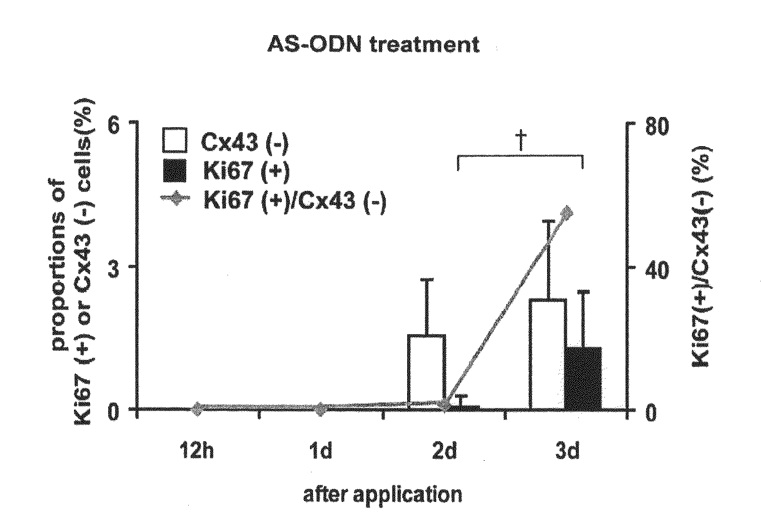
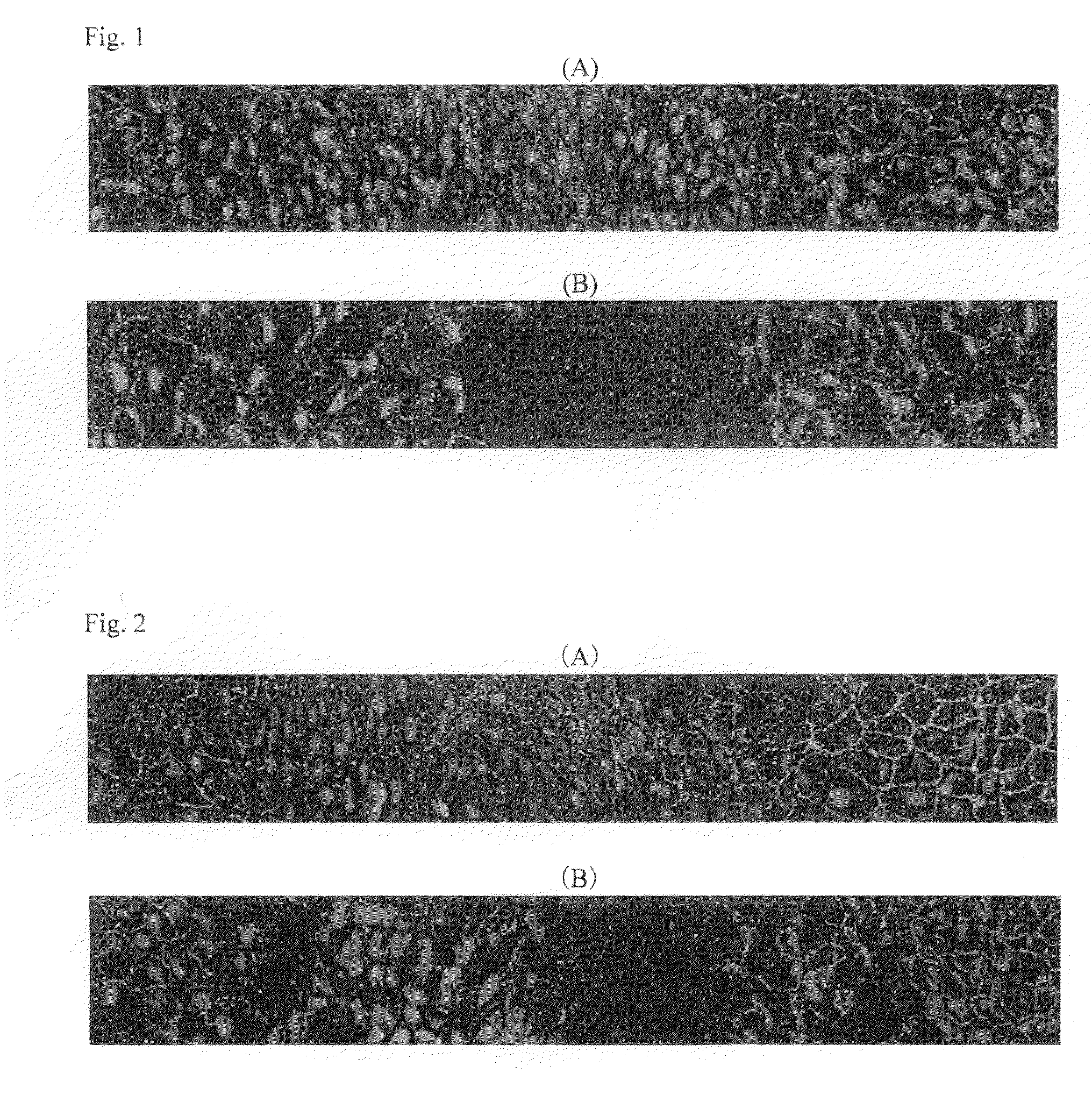
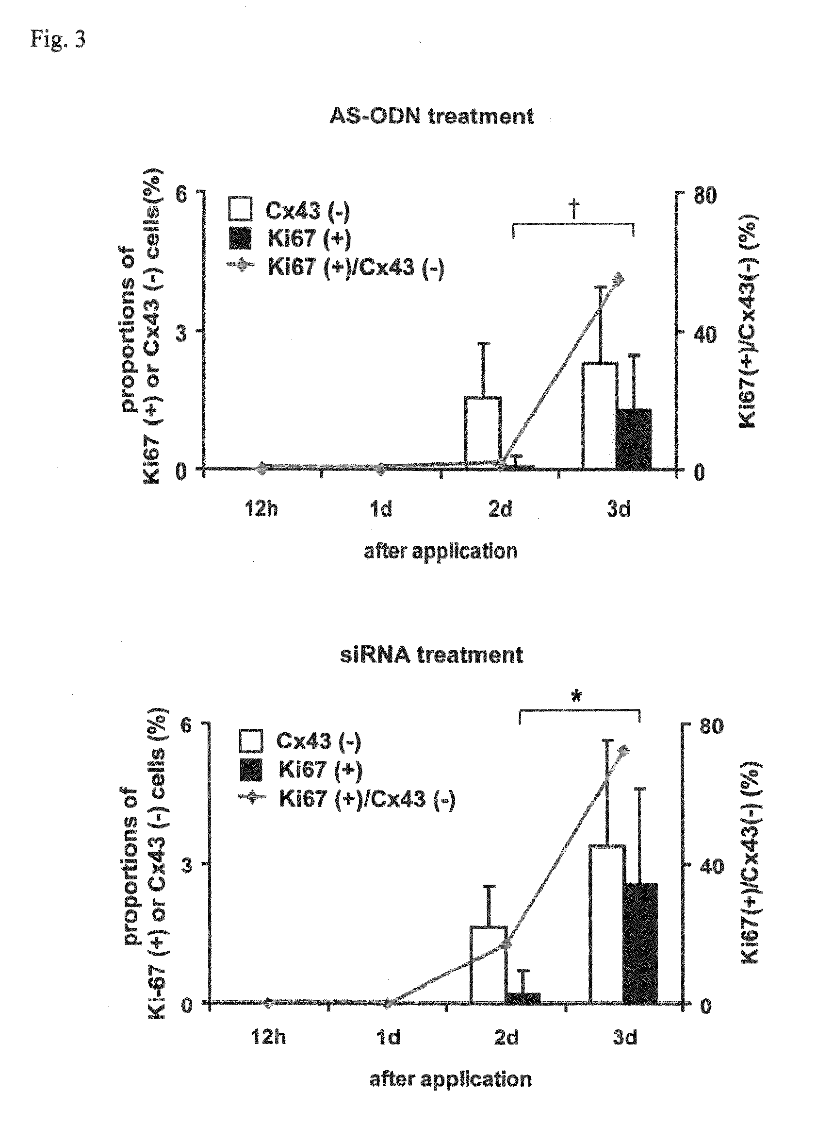
[0070]Wistar strain rats (male, 8 weeks of age) were used. After each rat was deeply anesthetized with pentobarbital, a needle of 30G (Nipro Medical Industries Co., Ltd.) was inserted from a limbus into an anterior chamber to take up 20 μL of anterior chamber fluid. The corneal endothelium was mildly scratched with the needle from the side of the anterior chamber to make a wound. Subsequently, in order to examine the effects of antisense oligonucleotides and RNAi for connexin 43 (Cx43) on corneal wound healing, 20 μL of 40 μM AS ODN or siRNA was injected into the anterior chamber using another needle inserted through the same incision (when the total amount of liquid in the anterior chamber is supposed to be 40 μL, the final concentration of AS ODN and siRNA in the liquid in the anterior chamber was 20 μM). After one or three days, the eye ball was removed and corneoscleral sections (diameter of 6 to 7 mm) we...

example 2

Effect of hCx43-siRNA on Regeneration of Monkey Corneal Endothelium

[0084]The effect of hCx43-siRNA on the proliferation of endothelial cells was observed by administering the following hCx43-siRNA to a cultured endothelial cell sheet obtained from the cornea of a crab-eating macaque and counting the number of cells in a DNA synthesis phase.

hCx43-siRNASense:5′-CAA UUC UUC UUG CCG CAA TT-3′(SEQ ID NO: 7)Antisense:5′-UUG CGG CAA GAA GAA UUG TT-3′(SEQ ID NO: 8)

[0085]As a result, the number of BrdU-positive cells was clearly increased in the hCx43-siRNA administration groups (#2-4) compared with the hCx43-siRNA non-administration group (#1), demonstrating that hCx43-siRNA (Cx43-siRNA of human and monkey have the same sequence) promotes the regeneration of monkey corneal endothelial cells.

TABLE 2AdministrationBrdU-positiveof hCx43-siRNAcells#1−19#2+29#3+40#4+30

[0086]From the results of Examples 1 and 2, it has been elucidated that the nucleic acid molecule (antisense oligonucleotides, siR...

example 3

In Vivo Effects of hCx43-siRNA on Regeneration of Monkey Corneal Endothelium

[0087]Using a corneal endothelium disorder model from crab-eating macaques, the corneal endothelial cells of which have a poor proliferative ability in vivo similar to human beings, it is possible to evaluate the effect of administering siRNA that selectively inhibits the expression of human connexin 43 into the anterior chamber on corneal endothelium wound healing.

1. Preparation of a Connexin 43-inhibiting Drug

[0088]The following siRNA (hCx43-siRNA) was used.

Sense:5′-CAA UUC UUC UUG CCG CAA TT-3′Antisense:5′-UUG CGG CAA GAA GAA UUG TT-3′

2. Preparation of Corneal Endothelium Disorder Models from Crab-Eating Macaques and Administration of Connexin 43-Inhibiting Drugs

1) General anesthesia: General anesthesia by intramuscular injection of ketamine hydrochloride and xylazine hydrochloride was given, and then the animal was carried from a cage to a medical table. On the medical table, inhalation anesthesia using ...


PUM

PropertyMeasurementUnit
diameteraaaaaaaaaa
temperatureaaaaaaaaaa

Abstract

The present invention relates to a treatment agent for a disease or a disorder caused by a reduction in corneal endothelial cells, comprising as an active component at least one nucleic acid molecule inhibiting the expression of a connexin 43 gene.

Description

TECHNICAL FIELD[0001]The present invention relates to the proliferation of corneal endothelial cells or the regeneration of corneal endothelium, and particularly relates to a treatment agent for treating a disease or a disorder caused by a reduction in corneal endothelial cells, the use thereof for such treatment, a pharmaceutical composition for such treatment, a method for treating the disease or the disorder, a treatment agent and a treatment method allowing an intraocular surgery in a patient having reduced corneal endothelial cells, and siRNA for human connexin 43 (Cx43).BACKGROUND ART[0002]The cornea is a transparent tissue having the role of an optical lens, and has corneal endothelial tissue as its innermost layer.[0003]Corneal endothelial cells are indispensable for maintaining the transparency of the cornea. When a human corneal endothelial cell is once impaired, it is not regenerated and a impaired portion is covered by the migration of the remaining surrounding endotheli...

Claims


Application Information

Patent Timeline
Patent Type & Authority Applications(United States)
IPC IPC(8): A61K31/7105C07H21/02
CPCC12N2310/14C12N15/1138A61P27/02A61P43/00
Inventor TAKAMATSU, TETSURODAI, PINGKINOSHITA, SHIGERU
Owner KANSAI TLO KK
Why Eureka
Try Eureka