
Systems and methods for ex vivo lung care

Active Publication Date: 2009-08-06
TRANSMEDICS
View PDF101 Cites 47 Cited by
  • Summary
  • Abstract
  • Description
  • Claims
  • Application Information

AI Technical Summary

Benefits of technology

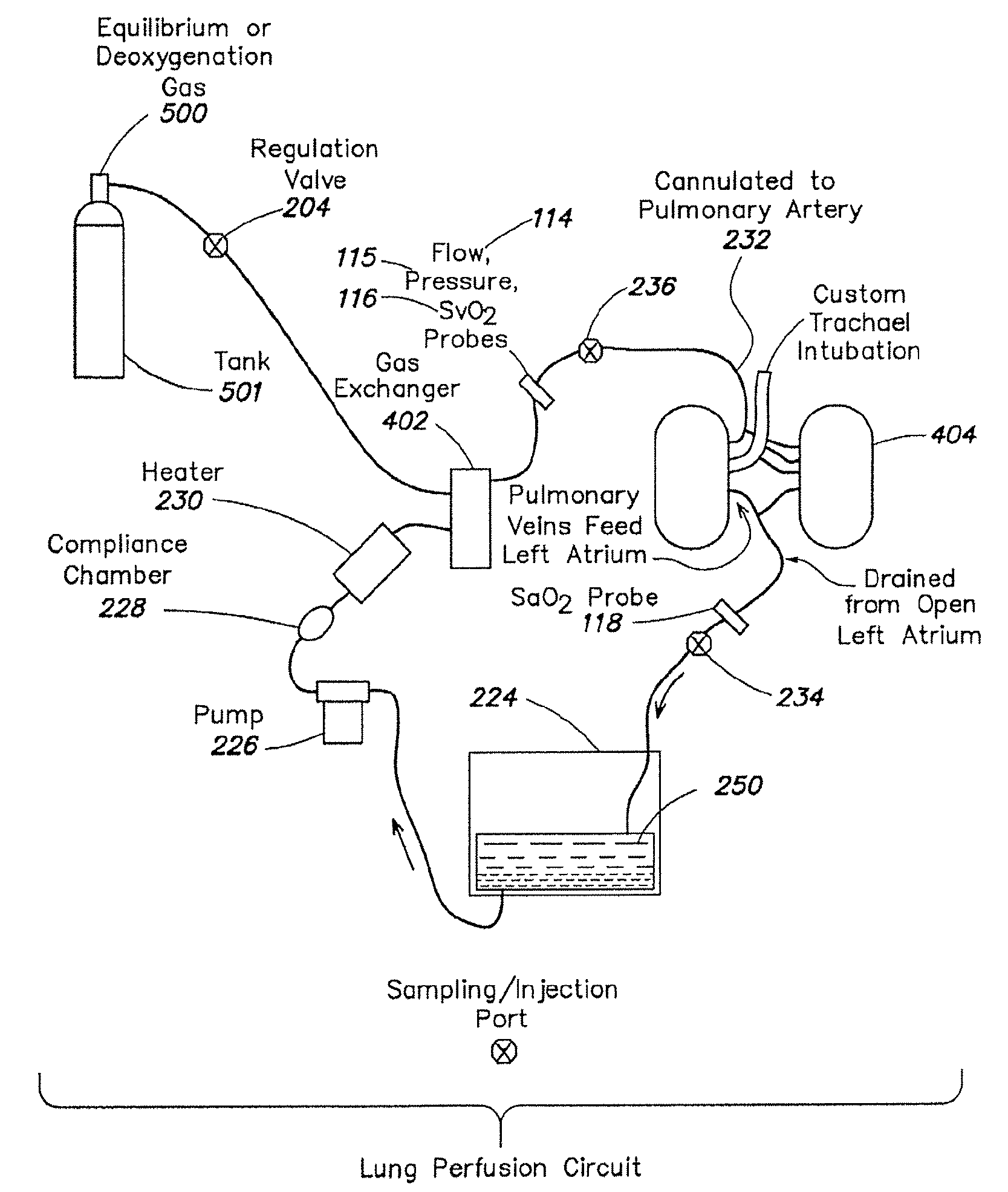
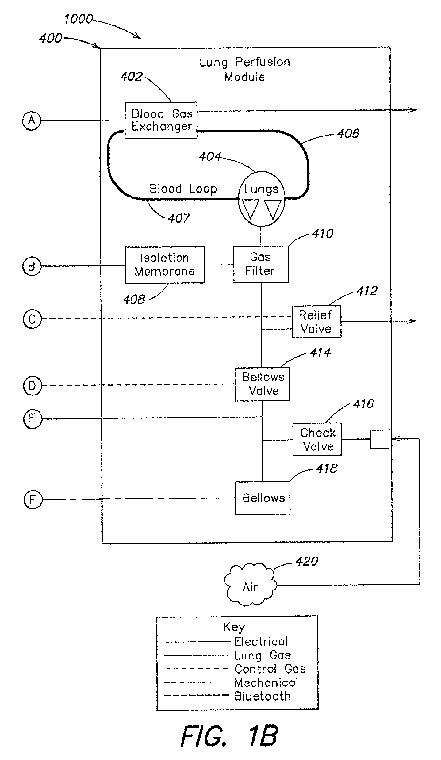
[0020]The predetermined composition of the ventilation gas includes about 12% oxygen and about 5.5% carbon dioxide. The hemoglobin saturation level of the perfusion fluid entering the lung reaches an equilibrium level of about 90-95% and a hemoglobin saturation level of the perfusion fluid leaving the lung reaches an equilibrium level of about 90-95%. The oxygen content of the perfusion fluid entering the lung is lower than physiologic levels, and the oxygen content of perfusion fluid leaving the lung is higher than physiologic levels. The following parameters are used in certain embodiments: the additional flow of ventilation gas is about 400-600 mL per minute; the captive volume is about 400-1200 mL; the minimum gas pressure of the captive volume is about 4-8 cm of H2O; and the maximum pressure of the ventilation gas is about 12-22 cm of H2O. Excess ventilation gas is vented through a relief valve in communication with the captive volume. The variable volume chamber is a bellows; compressing the bellows causes the flow of ventilation gas into the lung. The pulmonary artery interface includes a pulmonary artery cannula, a portion of the pulmonary artery cannula being inserted into a pulmonary artery of the lung. The perfusion fluid to flows away from the lung through an exposed left atrial cuff of the lung, or through a sealed or semi-sealed connection between the left atrial cuff and a left atrial cannula. The tracheal interface includes a tracheal cannula, a portion of the tracheal cannula being inserted into a trachea of the lung. The method includes measuring a first level of oxygen content in the perfusion fluid flowing into the lung and a second level of oxygen content in the perfusion fluid flowing out of the lung. The oxygen measurement involves measuring at least one of a level of oxygen saturation of hemoglobin in the perfusion fluid and a partial pressure of oxygen in the perfusion fluid flowing into the lung and flowing out of the lung. The perfusion fluid includes a blood product, and can deliver therapeutics to the lung. The gas exchange in the lung between the ventilation gas and the perfusion fluid causes the level of one or more gases, such as oxygen and carbon dioxide, in the perfusion fluid to reach equilibrium values. The lung may be preserved for a period of about 3-24 hours when maintained with the equilibrium levels of gas.

Problems solved by technology

These techniques utilize a variety of solutions, none of which sufficiently protect the organ from damage resulting from ischemia.
Such injuries are particularly undesirable when an organ is intended to be transplanted from a donor into a recipient.
This is of crucial importance when harvesting lungs because lungs are easily compromised even before harvesting within the donor's body.
Current transplantation techniques rely mainly on matching donor and recipient blood types, which by itself is a relatively unreliable indicator of whether or not the organ will be rejected by the recipient.
A more preferred test for organ compatibility is a Human Leukocyte Antigen (HLA) matching test, but current cold ischemic organ preservation approaches preclude the use of this test, which can often require 12 hours or more to complete.
These relatively brief time periods limit the number of recipients who can be reached from a given donor site, thereby restricting the recipient pool for a harvested organ.
Even within the time limits, the organs may nevertheless be significantly damaged.
A significant issue is that there may not be any observable indication of the damage.
Because of this, less-than-optimal organs may be transplanted, resulting in post-transplant organ dysfunction or other injuries.

Method used


Image

  • Systems and methods for ex vivo lung care
  • Systems and methods for ex vivo lung care
  • Systems and methods for ex vivo lung care

Examples


Embodiment Construction

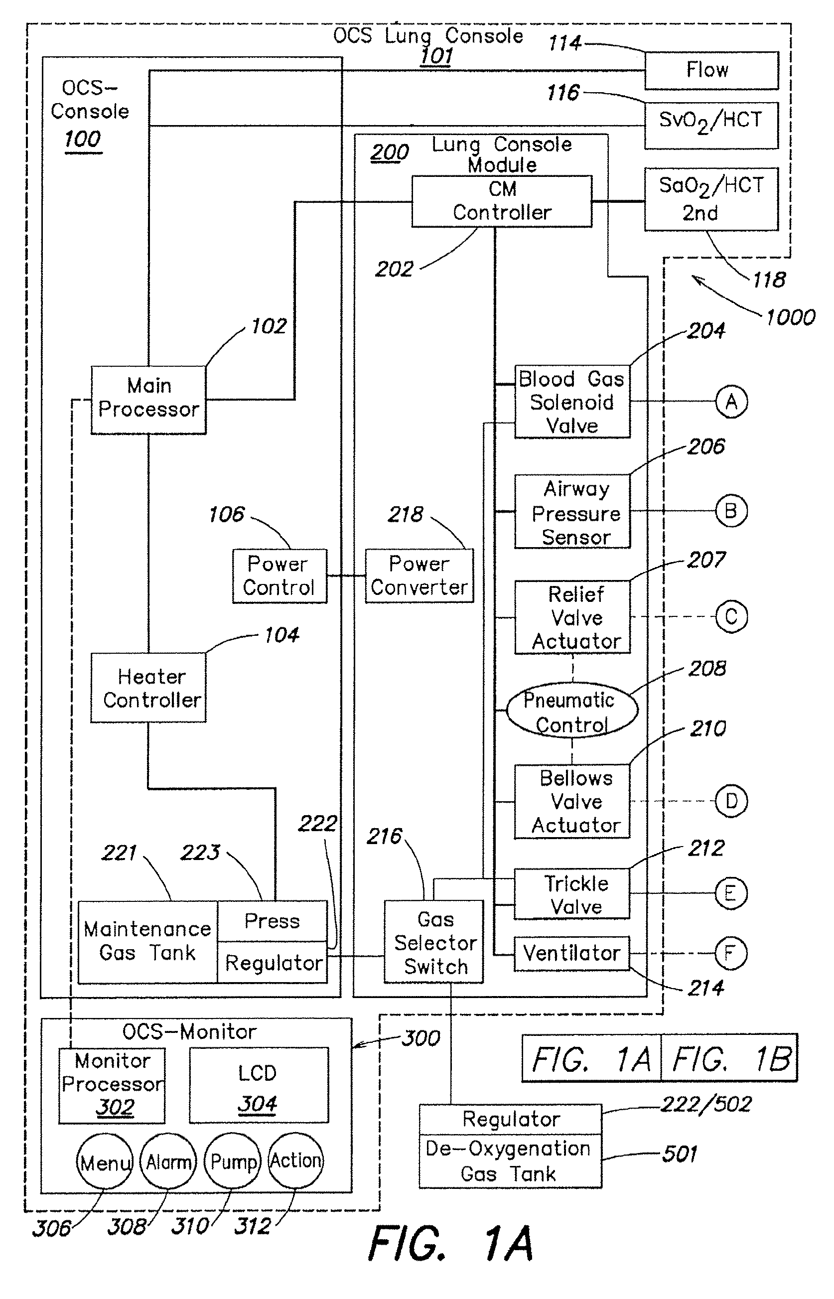
[0057]As described above in summary, the described embodiment generally provides improved approaches to ex vivo lung care, particularly in an ex vivo portable environment. The organ care system maintains a lung in an equilibrium state by circulating a perfusion fluid through the lung's vascular system, while causing the lung to rebreath a specially formulated gas having about half the oxygen of air. The perfusion fluid circulates by entering the pulmonary artery (PA) via a cannula inserted into the PA. After passing through the lung, the perfusion fluid exits the lung from an open, uncannulated left atrium (LA) where it drains into a reservoir. A pump draws the fluid out of the reservoir, passes it through a heater and a gas exchanger, and back into the cannulated PA. In the described embodiment, the perfusion fluid is derived from donor blood. In alternative embodiments, the perfusion fluid is blood-product based, synthetic blood substitute based, a mixture of blood product and blo...


PUM


Abstract

Methods and systems of maintaining, evaluating, and providing therapy to a lung ex vivo. The methods and systems involve positioning the lung in an ex vivo perfusion circuit; circulating a perfusion fluid through the lung, the fluid entering the lung through a pulmonary artery interface and leaving the lung through a left atrial interface; and ventilating the lung by flowing a ventilation gas through a tracheal interface. Maintaining the lung for extended periods involves causing the lung to rebreath a captive volume of air, and reaching an equilibrium state between the perfusion fluid and the ventilation gas. Evaluating the gas exchange capability of the lung involves deoxygenating the perfusion fluid and measuring a time taken to reoxygenate the perfusion fluid by ventilating the lung with an oxygenation gas.

Description

REFERENCE TO RELATED APPLICATIONS[0001]This application claims priority to and the benefit of U.S. Provisional Application Ser. No. 61 / 024,976, filed on Jan. 31, 2008, the entire contents of which are incorporated herein by reference.FIELD OF THE INVENTION[0002]The invention generally relates to systems, methods, and devices for ex vivo organ care. More particularly, in various embodiments, the invention relates to a portable device for caring, assessing, and applying therapeutic measures to a lung or a pair of lungs ex vivo at physiologic or near-physiologic conditions.BACKGROUND OF THE INVENTION[0003]Current organ preservation techniques typically involve hypothermic storage of the organ in a chemical preservation solution on ice. These techniques utilize a variety of solutions, none of which sufficiently protect the organ from damage resulting from ischemia. Such injuries are particularly undesirable when an organ is intended to be transplanted from a donor into a recipient.[0004...

Claims


Application Information

Patent Timeline
IPC IPC(8): C12Q1/02C12M1/00
CPCA01N1/0247A01N1/02A01N1/021
Inventor FISHMAN, ROBERTHAVENER, ROBERTFATTAH, IHAB ABDELABDELAZIM, ANASNEWELL, SCOTTBISHOP, TOMKHAYAL, TAMERKYI, STANLEYTAYLOR, RONHARRIOTT, DOUGDE REMER, MATTHEWMURRAY, PAULSULLIVAN, JOHNANDERSON, MARKBRINGHAM, RICHARDVAN DRIEL, MICHAELHASSANEIN, WALEED
Owner TRANSMEDICS
Learn More