Method of assessing tissue viability using near-infrared spectroscopy

a tissue viability and near-infrared technology, applied in the field of tissue viability assessment using near-infrared spectroscopy, can solve the problems of irreversible tissue damage, necrosis and tissue loss, and clinical signs which arise as a result of poor blood perfusion along the flap

Inactive Publication Date: 2009-08-27
NAT RES COUNCIL OF CANADA
View PDF2 Cites 14 Cited by
  • Summary
  • Abstract
  • Description
  • Claims
  • Application Information

AI Technical Summary

Benefits of technology

[0031]The probe may be located at least 1 mm away from the tissue portion. That is, the probe does not have to be mounted or attached to the damaged tissue to collect spectrum data.

Problems solved by technology

However, the clinical signs which arise as a consequence of poor blood perfusion along the flap become evident only after several hours of compromised perfusion.
Prolonged and severe deprivation of oxygen and other nutrients to the tissues results in irreversible tissue damage leading to necrosis and loss of tissue.
It was soon realized that the use of pharmacologic agents gave inconsistent results which were difficult to interpret.
Also, fluorescein dye or other labelled tracer measurements of blood flow were necessarily invasive procedures and the invasiveness of the procedure coupled with prolonged washout times limited the frequency with which these methods could be applied to the site of interest.
However, it only measured the blood flow within a small volume of tissue, and microvascular heterogeneity rendered the method prone to errors: motion of the probe during measurement and poor probe placement reproducibility both led to poor reproducibility.
In addition, the recognition of arteriovenous shunting, where blood flow bypasses the capillary bed resulting in non-nutrient flow, reduced the value of a strictly blood flow-based investigation.
Blood flow methods primarily address the issue of oxygen delivery, but provide little information on cellular utilization.
Magnetic resonance spectroscopy can provide information on the metabolic status of skin flaps at a cellular level (Cheung, 1994), but the extended measurement times, high costs, and limited portability render the method clinically impractical.
Clearly, this device is limited in that it provides information only on the oxygenation level of the blood.
However, this apparatus is arranged to measure changes and trends in metabolism and not tissue viability.
However, as with fluorescein dye and other vital dyes discussed above, this method is invasive and may harm the compromised tissue or the surrounding tissue.
Furthermore, the device is used to determine the depth of the burn in the tissue, not the viability of the tissue.
Clearly, this method is too invasive for use with damaged tissue, as it requires both the insertion of a probe below the surface of the tissue and injection of one or more metabolic substrates into the tissue.
However, the method is limited in that the device must be mounted onto the surface of the tissue being examined and, as a result, is prone to interference if the probe is removed and subsequently replaced on the suspect tissue.

Method used


Image

  • Method of assessing tissue viability using near-infrared spectroscopy
  • Method of assessing tissue viability using near-infrared spectroscopy
  • Method of assessing tissue viability using near-infrared spectroscopy

Examples


example i

Principles of Optical Near-Infrared Tissue Spectroscopy

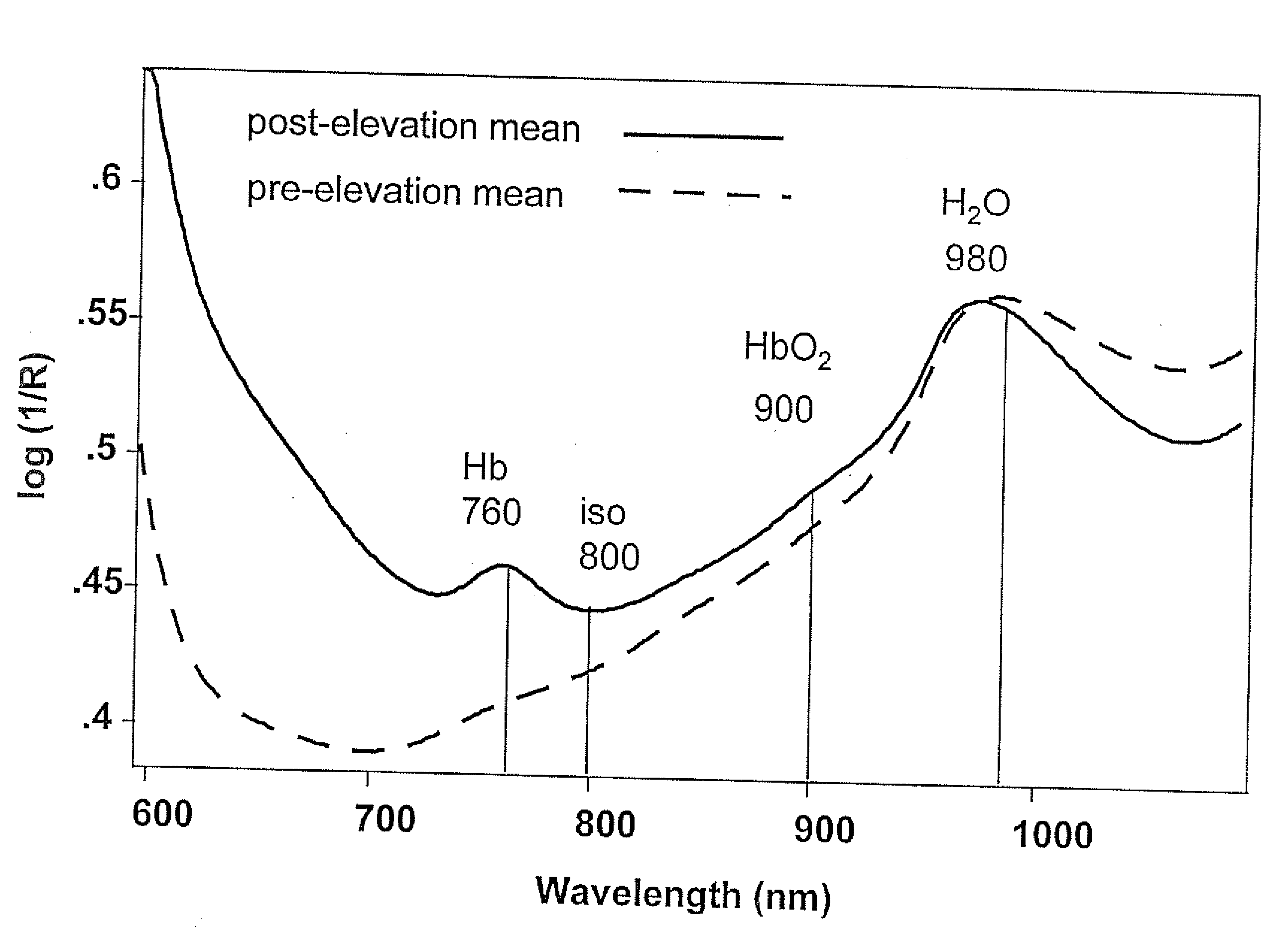
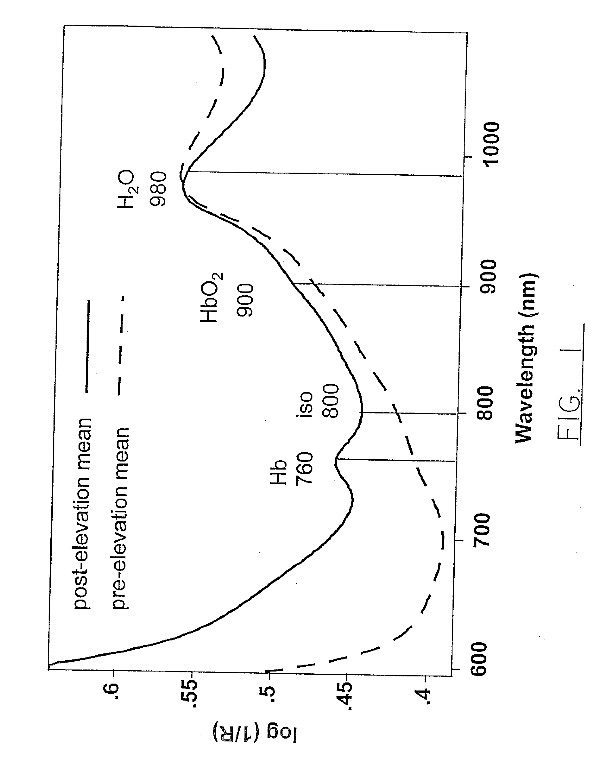
[0063]Infrared spectroscopy deals with the interaction of IR radiation with matter. The IR region of the electromagnetic spectrum is generally considered to lie in the wavelength range from 700-250,000 nm and is further subdivided into three subregions: The near-IR region, (from 700-2500 nm), the mid-IR region (from 2500-50000 nm) and the far-IR region (beyond 50000 nm). Visible and near-IR spectra in the 600 to 1100 nm range are presented in this study. The spectroscopy of tissues involves measuring the attenuation of the light intensity by the tissue relative to the incident light intensity.

[0064]Herein, the relative change and distribution of the levels of oxyhemoglobin (HbO2), and deoxyhemoglobin (Hb) in tissue, following flap elevation is examined and these data are used to predict tissue viability. The near-IR and visible absorption spectra of Hb, HbO2 and water are well understood and the differential absorption by these ...

example ii

Anaesthesia and Surgical Procedures

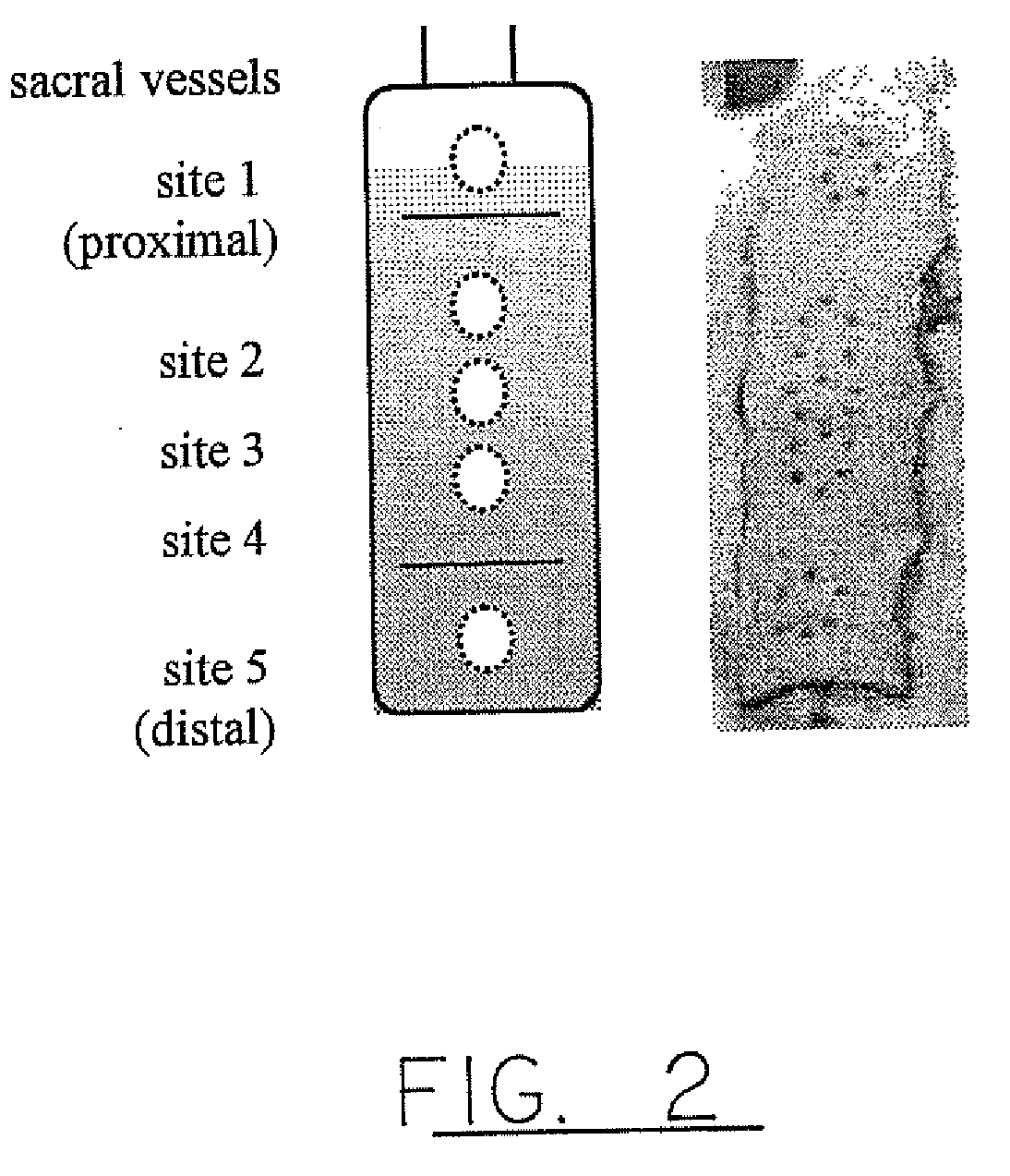
[0070]3×10 cm reverse McFarlane flaps raised on the dorsa of 9 Sprague Dawley rats weighing between 380-410 grams were used. The rats were acclimatized for a minimum period of 2 weeks prior, and the individual surgeries were performed over a three month period. All the procedures were done under 1.2-2% isoflurane inhalational anaesthesia. Five days prior to surgery, rats were shaved and a depilatory agent (Nair™) was applied on the rat dorsum. At that time, reproducible anatomic points for the near-IR spectroscopy measurements were marked on the skin, as shown in FIG. 2.

[0071]Surgery was performed 3 days (72 h) after pre-elevation measurements, which were to be used as control values in the analyses. Prior to surgery, the flap location was defined and marked based on the location of the sacral vessels, following which pre-elevation images and spectra were acquired. Twenty minutes prior to surgery, the rats were premedicated with 0.05 mg / kg of atrop...

example iii

Near-Infrared Measurements

[0073]Near infrared radiance images of 256×256 pixels were collected at 650, 700, 760, 800, 850, 900 and 980 nm using a Photometrics Series 200 CCD camera fitted with a Nikon Macro AF60 lens using 10 nm bandpass (FWHH) Lyot type filters (OCLI, Santa Rosa, Calif.). Visible and near-IR spectra were collected with a NIRSystems 6500 (Perstop, Silver Springs, Mass.) spectrometer using a custom bifurcated fiber optic bundle (CeramOptic, Enfield, Conn.). A 99% Spectralon® reflectance standard (LabSphere Inc.) was used as a reference to convert raw data into reflectance spectra. Each reflectance spectrum consisted of 32 co-added scans collected between 600-1100 nm at 10 nm resolution.

[0074]Pilot studies indicated that approximately the distal half of the flap failed while the half proximal to the vascular pedicle remained viable. Five dorsal monitoring sites were located along the length of the flap, as shown in FIG. 2. In order to have reproducible anatomical poin...


PUM


Abstract

Prolonged and severe tissue hypoxia results in tissue necrosis in pedicled flaps. We demonstrate the potential of near-infrared spectroscopy for predicting viability of compromised tissue portions. This approach clearly identifies tissue regions with low oxygen supply, and also the severity of this challenge, in a rapid and non-invasive manner, with a high degree of reproducibility. Tissues remaining below a certain hemoglobin oxygen saturation threshold (oxygen saturation index <1) for prolonged periods (>6 h) became increasingly dehydrated, eventually becoming visibly necrotic. Tissues above this threshold (oxygen saturation index >1), despite being significantly hypoxic relative to the pre-elevation saturation values, remained viable over the 72 h post-elevation monitoring period. The magnitude of the drop in tissue oxygen saturation, as observed immediately following surgery, correlated with the final clinical outcome of the flap tissue. These results indicate the potential of near infrared spectroscopy and imaging to monitor tissue oxygenation status and assess tissue viability following reconstructive surgery. Early, nonsubjective detection of poor tissue oxygenation following surgery increases the likelihood that intervention aimed at saving the tissue will be successful.

Description

PRIOR APPLICATION INFORMATION[0001]The instant application is a divisional application of U.S. Ser. No. 10 / 319,838, filed Dec. 16, 2002 which was a divisional application U.S. Ser. No. 09 / 402,348, filed Feb. 17, 2000 now U.S. Pat. No. 6,587,701 (Jul. 1, 2003) which was a 371 of PCT CA98 / 00292, filed Apr. 2, 1998 (expired), which claimed priority on U.S. Ser. No. 60 / 043,398, filed Apr. 3, 1997 (expired).SUMMARY OF THE INVENTION[0002]The present invention relates generally to a method of determining tissue viability. More specifically, the present invention relates to a non-invasive method of determining tissue viability using visible and near-infrared spectroscopy.BACKGROUND OF THE INVENTION[0003]In surgery, the ability to determine whether or not a tissue will survive is of paramount importance. This is particularly true in plastic surgery where efforts to develop a reliable method to predict skin flap viability are on-going. It is therefore not surprising that efforts to develop a ...

Claims


Application Information

Patent Timeline
Patent Type & Authority Applications(United States)
IPC IPC(8): A61B5/1455A61B5/00A61B5/026
CPCA61B5/0261A61B5/7264A61B5/413A61B5/14551
Inventor STRANC, MIROSLAW F.SOWA, MICHAEL G. R.MANTSCH, HENRY H.
Owner NAT RES COUNCIL OF CANADA
Why Eureka
Try Eureka