Adhesive cartilage implant

a cartilage implant and adhesive technology, applied in the field of medical technology, can solve the problems of limited regenerative capacity compared to other tissues, pain, cartilage damage, etc., and achieve the effect of decreasing the temperature of the polymer

Inactive Publication Date: 2010-07-01
HOWMEDICA OSTEONICS CORP
View PDF31 Cites 3 Cited by
  • Summary
  • Abstract
  • Description
  • Claims
  • Application Information

AI Technical Summary

Benefits of technology

[0012]In one embodiment, a method of producing a cartilage implant may include coating a substrate with a thermo-sensitive polymer, seeding the polymer with a cell suspension comprising cells that may differentiate into chrondocyte-like cells, wherein the seeding may be conducted at a first temperature, cu

Problems solved by technology

Loss of or damage to cartilage can lead to painful conditions such as osteoarthritis.
Damage to cartilage can be caused by traumatic injury, disease and/or age.
Since cartilage lacks nerves and blood vessels, it has very limited regenerative capabilities compared to other tissues.
Consequently, the healing of damaged joint cartilage results in a fibrocartilaginous repair tissue that lacks the structure and biomechanical properties of

Method used


Image

  • Adhesive cartilage implant
  • Adhesive cartilage implant
  • Adhesive cartilage implant

Examples


example 1

[0059]In this example, an in vitro study was performed to determine the appropriate cell seeding density in thermo-sensitive polymer coated 96-well culture plates.

[0060]Human mesenchymal stem cells (hMSCs) were expanded in DMEM / 10% FBS / 1% PS at 37° C. (a physiological temperature) up to passage 3. Cells were trypsinized, washed, and resuspended in DMEM / 10% FBS / 1% PS. Cells were then seeded in the 96-well culture plate coated with thermo-sensitive polymer at the following densities: 1×10⁴ cells / well, 2×10⁴ cells / well, 5×10⁴ cells / well, 1×10⁵ cells / well, and 2×10⁵ cells / well. Following a 24-hour culture at 37° C., the medium was changed to serum-free DMEM supplemented with TGF-β3. Macroscopic examination of cell morphology was conducted daily for 14 days. On the termination day, the culture plate was transferred to 4° C. (a nonphysiological temperature) to release the cell construct from the well.

[0061]The results of the study illustrated that within 24 hours of plating, cells became ...

example 2

[0063]In this example, the three-dimensional cell constructs formed in thermo-sensitive polymer coated 96-well culture plates were assessed.

[0064]As discussed in Example 1, hMSCs were seeded to form a three-dimensional configuration or construct in a 96-well plate coated with thermo-sensitive polymer at the density of 2×10⁴ cells per well and induced to form cartilage in serum-free DMEM medium supplemented with TGF-β3 for 21 days. Cell constructs were released from the culture plate by incubating at 4° C. for a few minutes. Then the cell constructs were embedded in 1% agarose and subjected to histological processing. Tissue sections of 5 μm were prepared for the following stains: H&E, Safranin O, Alcian Blue (pH 1.0), and integrin α5β1 antibodies.

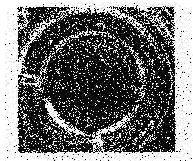
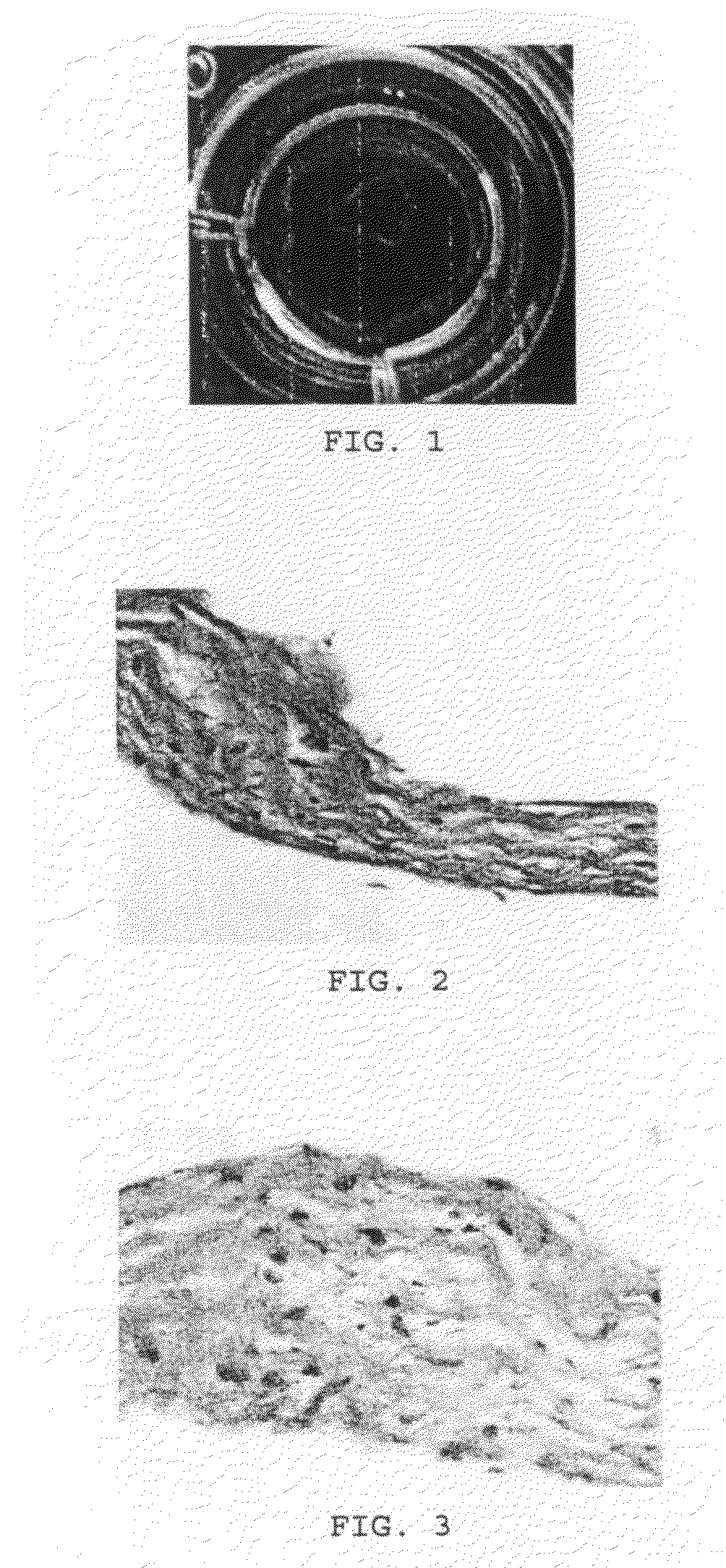
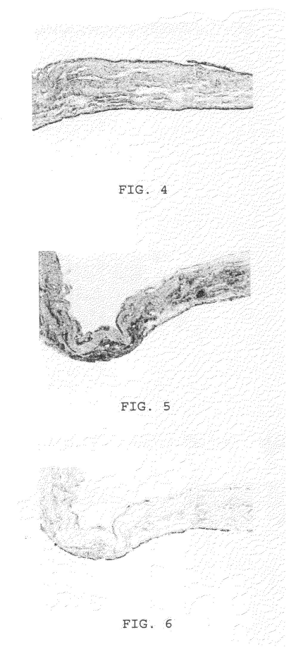
[0065]The cell construct remained attached to the plate over 21 days of culture. Once released from the plate, the construct maintained its circular shape (FIG. 1). Upon study of the cross sections, the construct appeared to consist of mult...


PUM

PropertyMeasurementUnit
Temperatureaaaaaaaaaa

Abstract

A method of producing a cartilage implant which may include coating a substrate with a thermo-sensitive polymer, seeding the polymer with a cell suspension comprising cells that may differentiate into chrondocyte-like cells, wherein the seeding may be conducted at a first temperature, culturing the cell suspension at an increased temperature, whereby cells in the suspension may adhere to the polymer and differentiate into chrondrocyte-like cells, which may form a cartilagenous implant, and decreasing the temperature of the polymer which may allow detachment of the implant from the polymer. The present invention may also include an implant based on this method. The invention may further include a cartilage implant for use in a patient in need thereof, which may include a random three-dimensional configuration of differentiated cells, which may have a natural adhesion surface on at least a portion of the outer surface of the three-dimensional configuration.

Description

FIELD OF THE INVENTION[0001]The present invention relates to the field of medical technology and is generally directed to a cartilage implant and methods of making cartilage implants.BACKGROUND OF THE INVENTION[0002]Cartilage is an avascular connective tissue made up of collagen and / or elastin fibers, and chondrocytes, all of which are embedded in a matrix. There are three main types of cartilage: elastic, fibrocartilage, and hyaline. Elastic cartilage is found in the outer ear and the epiglottis. Fibrocartilage is found between the bones of the spinal column, hips and pelvis. Hyaline cartilage can be found on the ends of bones which form joints, on the ends of the ribs, on the end of the nose, on the stiff rings around the windpipe, and supporting the larynx. Articular cartilage is a specialized type of hyaline cartilage which covers the surface of joints and provides a durable low friction surface that distributes mechanical forces and protects the joint's underlying bone.[0003]Di...

Claims


Application Information

Patent Timeline
IPC IPC(8): A61F2/00C12N5/06A61K35/12
CPCA61F2/30756A61F2/3094A61F2002/30762A61L27/227A61L27/34A61L27/3817A61L27/3852A61L27/3895A61L2430/06
Inventor SONG, LIN
Owner HOWMEDICA OSTEONICS CORP
Why Eureka
Try Eureka