Agent for promoting corneal endothelial cell adhesion

Active Publication Date: 2010-08-19
SENJU PHARMA CO LTD +1
View PDF11 Cites 19 Cited by
  • Summary
  • Abstract
  • Description
  • Claims
  • Application Information

AI Technical Summary

Benefits of technology

[0009]The agent for promoting adhesion of a corneal endothelial cell of the present invention contains a Rho kinase inhibitor as an active ingredient. The agent for promoting adhesion of a corneal endothelial cell of the present invention promotes adhesion of the corneal endothelial cell and enables formation of a corneal endothelial cell layer having good cell morphology and high cell density. Therefore, it is useful as a therapeutic agent or prophylactic agent for a disease accompanied by corneal endothelial dysfunction, such as bullous keratopathy and corneal endotheliitis. The agent for promoting adhesion of a corneal endothelial cell of the present invention is useful as an agent for protecting corneal endothelium in the treatment or prophylaxis of a disease accompanied by a corneal endothelial dysfunction. Furthermore, the agent for promoting adhesion of a corneal endothelial cell of the present invention can be utilized as an agent for protecting corneal endothelium in the treatment or prophylaxis of corneal endothelial dysfunction associated with an intraocular surgery such as cataract surgery, vitreous surgery and the like, corneal endothelial dysfunction caused by increased intraocular pressure (particularly glaucomatous attack), or corneal endothelial dysfunction caused by insufficient oxygen due to contact lenses. Since the culture medium or corneal preservation solution of the present invention contains a Rho kinase inhibitor, corneal endothelial cells can be cultured, maintained or preserved fine, and stable supply, maintenance or preservation of a corneal endothelial preparation is enabled.

Problems solved by technology

Once damaged, the cells lose an ability to regenerate.
However, donation of cornea is insufficient in Japan and, although there are about 5500 waiting patients of corneal transplantation, about 2700 corneal transplants are performed annually in Japan.
However, as for corneal endothelial cells, particularly those derived from human, since the number of corneal donors is limited and in vitro culture is difficult, production of cultured cells in the number necessary for transplantation requires time and cost.
However, it has a practical problem of drastically reduced cell number, since an operation to disperse the cells in a culture step easily causes cell death.

Method used


Image

  • Agent for promoting corneal endothelial cell adhesion
  • Agent for promoting corneal endothelial cell adhesion
  • Agent for promoting corneal endothelial cell adhesion

Examples


example 1

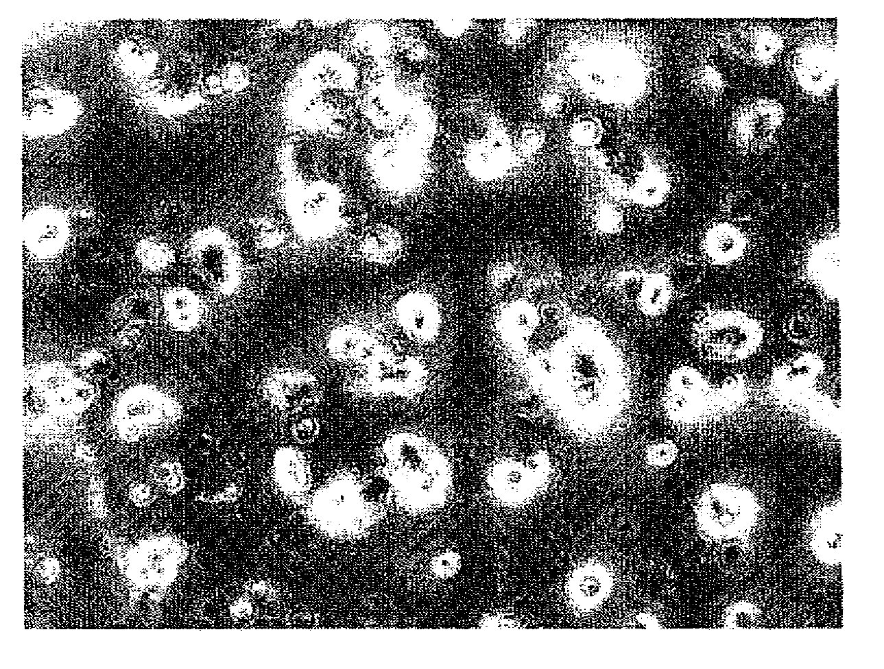
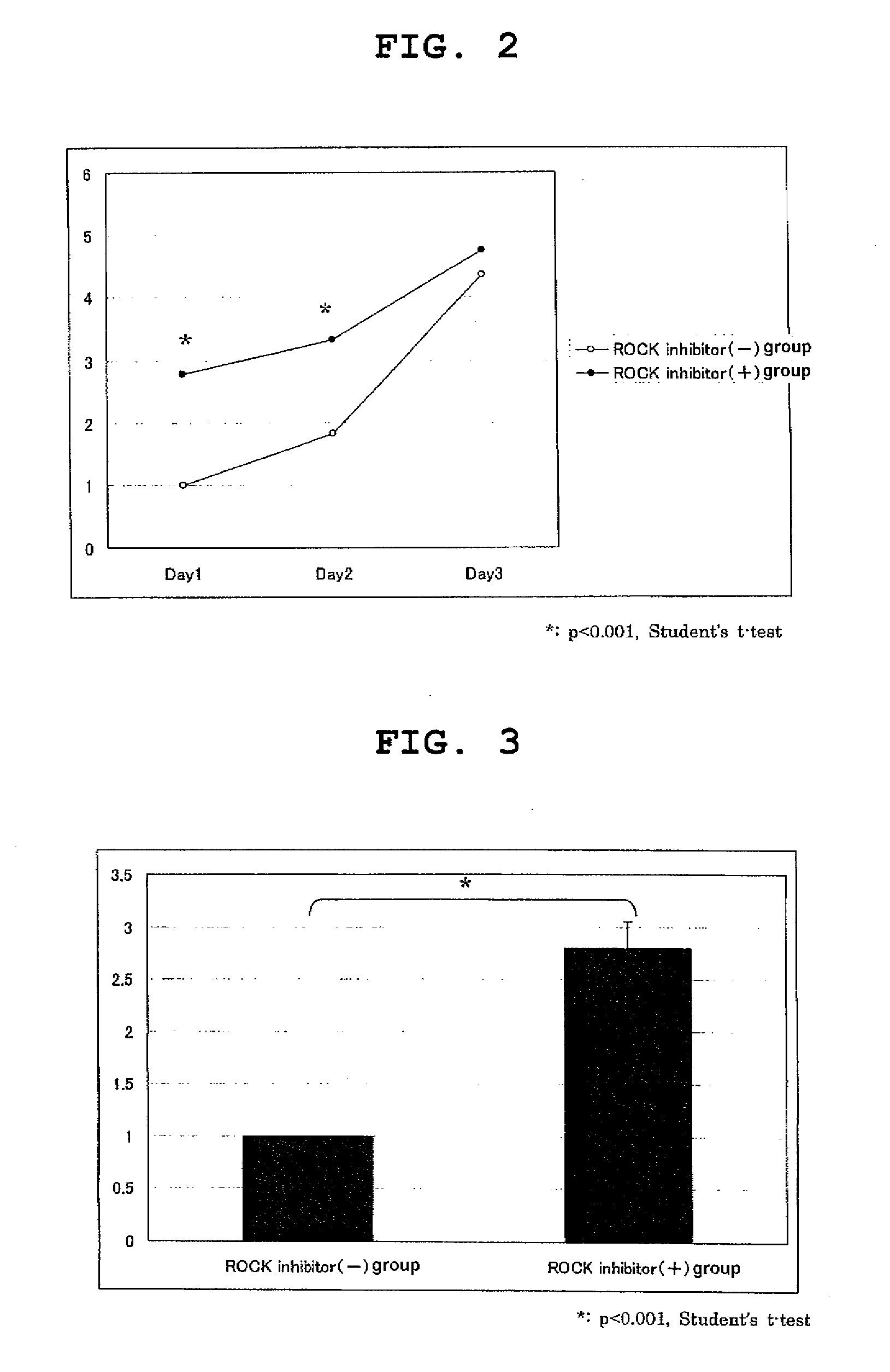
Study of Influence of ROCK Inhibitor on Culture of Rabbit Corneal Endothelial Cell

[0065]From a rabbit corneal tissue isolated immediately after euthanasia, Descemet's membrane with corneal endothelial cells attached thereto was separated. The Descemet's membrane was incubated together with Dispase II (1.2 U / ml, Roche Applied Science) under the conditions of 37° C., 5% CO2 for 45 min, and the corneal endothelial cells were mechanically separated by a pipetting operation. The separated corneal endothelial cells were centrifuged, the cells of a ROCK inhibitor(+) group were stirred in a medium for corneal endothelium added with Y-27632 (10 μM) and the cells of a ROCK inhibitor(−) group were stirred in a medium for corneal endothelium without addition of Y-27632, to the same concentration, and the cells were seeded on a 96 well plate at a density of about 2000 cells per well. As a medium for corneal endothelium, a culture medium (DMEM, Gibco Invitrogen) added with 1% fetal bovine serum a...

example 2

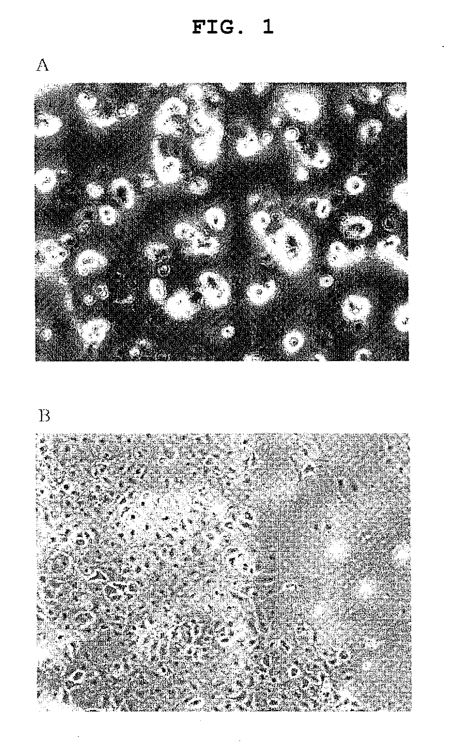
Study of Influence of Y-27632 on Culture of Monkey Corneal Endothelial Cell

[0067]From the corneal tissue isolated from Macaca fascicularis euthanized for other purpose, Descemet's membrane with corneal endothelial cells attached thereto was separated. For the ROCK inhibitor(+) group, the Descemet's membrane was placed in a medium for corneal endothelium added with Y-27632 (10 μM) and incubated under the conditions of 37° C., 5% CO2 for 10 min. For the ROCK inhibitor(−) group, the membrane was placed in a medium for corneal endothelium without addition of Y-27632 and incubated for 10 min under the same conditions. As the medium for corneal endothelium, the same cell culture medium as in Example 1 was used. The Descemet's membrane after the incubation was incubated together with Dispase II (1.2 U / ml, Roche Applied Science) under the conditions of 37° C., 5% CO2 for 45 min, and the corneal endothelial cells were mechanically separated by a pipetting operation. The separated corneal end...

example 3

Study of Influence of Y-27632 on Culture of Human Corneal Endothelial Cell

[0069]Of human corneal tissues obtained from the US eye bank, the central part (diameter 7 mm) had been used for corneal transplantation and the remaining surrounding corneal tissue was used. The Descemet's membrane with corneal endothelial cells adhered thereto was separated. For the ROCK inhibitor(+) group, the Descemet's membrane was placed in a medium for corneal endothelium added with Y-27632 (10 μM) and incubated under the conditions of 37° C., 5% CO2 for 10 min. For the ROCK inhibitor(−) group, the membrane was placed in a medium for corneal endothelium without addition of Y-27632 and incubated for 10 min under the same conditions. As the medium for corneal endothelium, the same culture medium as in Example 1 was used. The Descemet's membrane after the incubation was incubated together with Dispase II (1.2 U / ml, Roche Applied Science) under the conditions of 37° C., 5% CO2 for 45 min, and the corneal en...


PUM

PropertyMeasurementUnit
Adhesion strengthaaaaaaaaaa

Abstract

The invention provides an agent for promoting adhesion of a corneal endothelial cell, containing a Rho kinase inhibitor, as well as a culture medium for a corneal endothelial cell, a solution for preservation of cornea, and a method of producing a corneal endothelial preparation, which includes culturing the corneal endothelial cell using the aforementioned culture medium.

Description

TECHNICAL FIELD[0001]The present invention relates to an agent for promoting corneal endothelial cell adhesion, which is used for adhesion, maintenance or preservation of corneal endothelial cells.BACKGROUND ART[0002]Visual information is recognized when the light that entered from the cornea (transparent tissue at the forefront of the eyeball) reaches the retina to excite retinal nerve cells, and the developed electric signals are transmitted to the cerebral visual cortex via the optic nerve. To have good eyesight, the cornea needs to be transparent. The transparency of the cornea is maintained when the water content is kept at a constant level by the pumping function and barrier function of the corneal endothelial cells.[0003]The density of the corneal endothelial cells of human is about 3000 cells per 1 mm2 at birth. Once damaged, the cells lose an ability to regenerate. In endothelial corneal dystrophy and bullous keratopathy resulting from functional disorder of corneal endothe...

Claims


Application Information

Patent Timeline
IPC IPC(8): C12N5/071A01N1/02A61K35/44A61K31/4409A61K31/496A61K45/00A61P27/02A61P43/00
CPCA61K9/0048A61K31/4409A61K31/496C12N2501/70C07D401/12C12N5/0621C07D213/81A61K31/551A61P27/02A61P43/00
Inventor KOIZUMI, NORIKOKINOSHITAUENO, MORIO
Owner SENJU PHARMA CO LTD
Why Eureka
Try Eureka