Electroporative flow cytometry

a flow cytometry and electroporation technology, applied in the field of electroporation, flow cytometry, flow cytometry, protein translocation, cell biomechanics, can solve the problems of complex image analysis algorithm, intrinsic insensitivity of conventional flow cytometry, and low throughput. , to achieve the effect of reducing the number of cells/second, and improving the accuracy of flow cytometry

Inactive Publication Date: 2010-09-02
PURDUE RES FOUND INC
View PDF17 Cites 13 Cited by
  • Summary
  • Abstract
  • Description
  • Claims
  • Application Information

AI Technical Summary

Benefits of technology

[0012]Provided is a novel tool, referred to as electroporative flow cytometry, to detect the translocation of cellular proteins. Electroporation occurs when cells experience an electrical field with the intensity beyond a certain threshold. During electroporation the electrical field opens up pores in the cell membrane. Such pores allow the release of intracellular materials into the surrounding solution (Rols and Teissie, 1990, Biophys. J. 58: 1089-1098; Wang and Lu, 2006, Chem. Commun. 3528-3530; Wang and Lu, 2006, Anal. Chem. 78: 5158-5164). It is possible to differentiate a cell population with translocation from one without it with the information collected from individual cells of the entire population. This technique allows detection of protein translocation at the single cell level. Due to the frequent involvement of kinase translocations in disease processes such as oncogenesis, the methods of the present invention have utility in kinase-related drug discovery and tumor diagnosis and staging.

Problems solved by technology

However, conventional flow cytometry is intrinsically insensitive to the subcellular location of the probed protein.
However, the algorithm of quantification based on image analysis is complex and lacks robustness—the throughput is typically less than 100 cells / second, compared to about 104 cells / second for flow cytometry (Pozarowski et al., 2005, In: Cell Imaging Techniques: Methods and Protocols, Taatjes and Mossman, eds., Humana Press, Totowa, N.J., Vol. 319, pp 165-192).
This technique requires additional steps to isolate nuclei from living cells and the sample may potentially be contaminated by other organelles.
Although interesting for applications to mechanistic studies, the rather complex molecular biology makes this technique impractical for clinical applications.
In spite of the variety of the techniques demonstrated, most of the above techniques are only capable of studying a low number of single cells due to their low throughput.
The data generated by these techniques are not necessarily representative of a large cell population.
This problem becomes particularly serious when a heterogeneous cell population consisting of different cell types (e.g. tissue samples derived from animals and patients) is involved.
The low throughput issue may hinder the wide application of biomechanical assays as effective tools for cancer diagnosis and staging.

Method used


Image

  • Electroporative flow cytometry
  • Electroporative flow cytometry
  • Electroporative flow cytometry

Examples


example 1

Electroporative Flow Cytometry for Detection of Protein Translocation

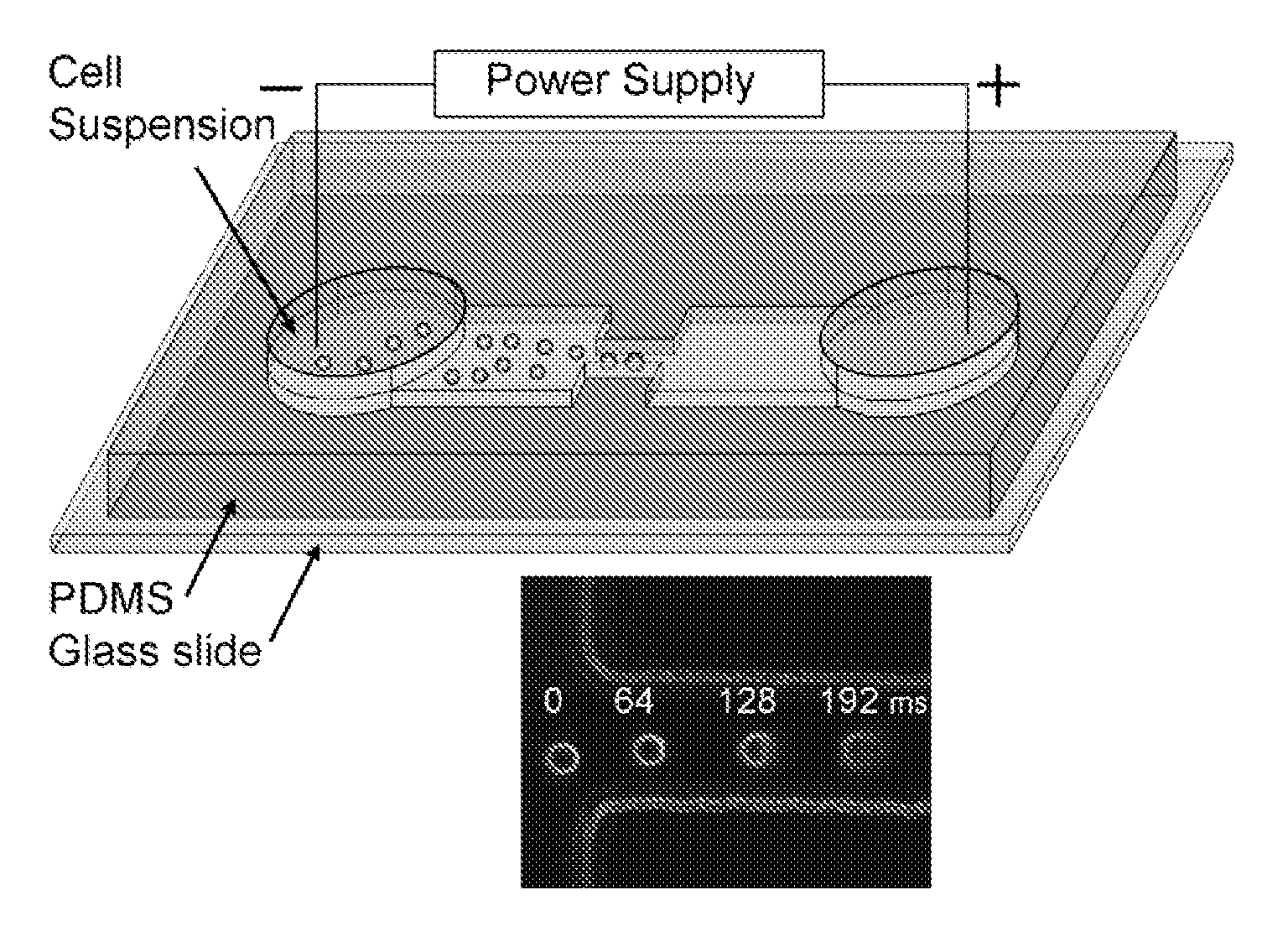
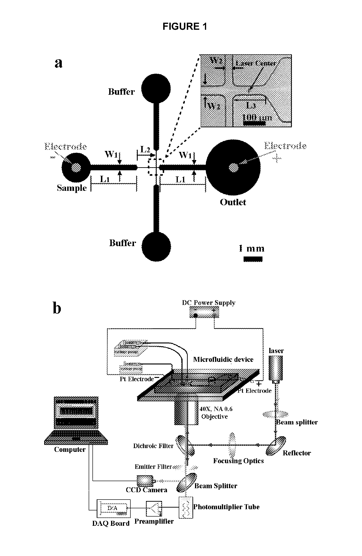
[0057]Microchip fabrication. Microfluidic EFC devices were fabricated based on PDMS using standard soft lithography method described before (Duffy et al., 1998, Anal. Chem. 70: 4974-4984; Wang and Lu, 2006, Anal. Chem. 78: 5158-5164). The microscale patterns were first created using computer-aided design software (FreeHand MX, Macromedia, San Francisco, Calif.) and then printed out on high-resolution (5080 dpi) transparencies. The transparencies were used as photomasks in photolithography on a negative photoresist (SU-8 2010, MicroChem. Corp., Newton, Mass.). The thickness of the photoresist and hence the depth of the channels was around 33 μm (measured by a Sloan Dektak3 ST profilometer). The pattern of channels in the photomask was replicated in SU-8 after exposure and development. The microfluidic channels were molded by casting a layer (approximately 5 mm) of PDMS prepolymer mixture (General Electric Silicones ...

example 2

Microfluidic Electroporative Flow Cytometry for Studying Single Cell Biomechanics

[0092]Cell culture. MCF-7 and MCF-10A cell lines were obtained from American Type Culture Collection (ATCC, Manassas, Va.) and cultured according to recommended protocols. Briefly, MCF-10A cell line was cultured in DMEM F-12 supplemented with Horse Serum (5.6%), EGF (20 ng / ml), Insulin (10 μg / ml), antibiotics (1%) and Hydrocortisone (0.5 μg / ml). The MCF-7 cell line was cultured in DMEM supplemented with FBS (10%), Penicillin / streptomycin (1%) and L-glutamine (2 mM). The TPA treated MCF-7 cell line was generated by treating MCF-7 cells with 100 nM 12-O-tetradecanoylphorbol-13-acetate (TPA) for 18 hr before experiments. In order to detach cells from culture flasks, cells were treated with 0.1% Trypsin / EDTA, washed and resuspended using the electroporation buffer (10 mM Na2HPO4, mM NaH2PO4, and 250 mM sucrose). For the treatment of MCF-7 cells with colchicine, MCF-7 cells were incubated in the culture medi...


PUM

PropertyMeasurementUnit
timeaaaaaaaaaa
width W2aaaaaaaaaa
width W2aaaaaaaaaa

Abstract

Novel devices and methods are provided, which include electroporative flow cytometry, where electroporation is combined with flow cytometry. The devices and methods can be used for the detection a variety of cellular features, including protein translocation, and for monitoring biomechanics at single cell level. Using the novel devices methods it is possible to observe the release of proteins such as intracellular kinases out of the cells during electroporation. Using the novel devices methods it is possible to study cytoskeleton dynamics and deformability at a single cell level, and to correlate these to diseases such as cancers.

Description

CROSS-REFERENCE TO RELATED APPLICATIONS[0001]This invention claims priority to U.S. Provisional Patent Application Ser. No. 60 / 967,407, filed Sep. 4, 2007, which is herein incorporated by reference.GOVERNMENT INTERESTS[0002]This invention was made with United States government support from the National Cancer Institute, grant number CA37372, and by the National Science Foundation, grant number CBET-0747105. The United States government has certain rights in this invention.FIELD OF THE INVENTION[0003]This invention relates to the fields of fluidic devices, electroporation, flow cytometry, protein translocation, and cell biomechanics.BACKGROUND[0004]Translocation of a protein between different subcellular compartments is a common event during signal transduction in living cells. Integrated signaling cascades often lead to the relocalization of protein constituents such as translocations between the cytoplasm and the plasma membrane or nucleus. Such events can be essential for the acti...

Claims


Application Information

Patent Timeline
Patent Type & Authority Applications(United States)
IPC IPC(8): C12Q1/02C12M1/34
CPCG01N15/147G01N33/574G01N33/5005G01N15/1484
Inventor LU, CHANGWANG, JUNBAO, NINGGEAHLEN, ROBERT L.
Owner PURDUE RES FOUND INC
Why Eureka
Try Eureka