Method for Dynamic Prior Image Constrained Image Reconstruction

a prior image and image reconstruction technology, applied in the field of medical imaging, can solve the problems of data gaps and overlap between different sectors, and the uniform distribution of the highly undersampled data set for each cardiac phase, and achieve the effects of increasing the signal-to-noise ratio (snr), increasing the snr, and increasing the temporal resolution

Active Publication Date: 2010-12-09
WISCONSIN ALUMNI RES FOUND
View PDF33 Cites 33 Cited by
  • Summary
  • Abstract
  • Description
  • Claims
  • Application Information

AI Technical Summary

Benefits of technology

[0016]The present invention provides an image reconstruction method applicable to a number of different imaging modalities including x-ray computed tomography (“CT”), x-ray C-arm imaging, magnetic resonance imaging (“MRI”), positron emission tomography (“PET”), and single photon emission computed tomography (“SPECT”). More specifically, the present invention provides an image reconstruction method that combines the merits of accurate reconstruction with a gain in signal-to-noise ratio (“SNR”) by incorporating an appropriate prior image of the subject into the image reconstruction process. In addition to the increased SNR, the method of the present invention provides an increase in temporal resolution. Moreover, for a given desired image and a given number of projections, the method of the present invention provides accurate image reconstruction where previous image reconstruction methods, such as compressed sensing (“CS”) fail.
[0017]One aspect of the present invention provides a method for producing a time series of images indicative of a higher temporal resolution than the temporal resolution at which the corresponding image data was acquired. For example, the present invention provides a method for high temporal resolution cardiac imaging. For cardiac CT, a slow gantry rotation data acquisition is employed. More specifically, high temporal resolution is achievable that significantly improves the diagnostic accuracy of coronary CT angiography and measurement of cardiac function. Moreover, by using a slow gantry rotation during a single breath-hold, problems with view angle synchronization are significantly reduced. Increased temporal resolution is also achievable in MR imaging applications. The increase in temporal resolution is achieved while mitigating undesired image artifacts by incorporating image data not associated with a current time frame into a data consistency condition during the reconstruction of the image frame corresponding to the current time frame. The expanded image frame data set that is formed in this manner includes, for example, 5-15 percent of data acquired that is not associated with the current time frame.
[0019]Another aspect of the present invention provides a fundamentally different form of segmented acquisition, where individual projections are segmented into to different cardiac phases, without the need to synchronize the gantry angle, or other data acquisitions, with the appropriate cardiac phase. In this way, excellent image quality can be achieved with the benefits of improved temporal resolution of segmented acquisitions. For example, in conventional multi-detector CT (“MDCT”), an irregular heart beat causes data gaps and overlap between different sectors. However, when practicing the present invention, an irregular heart beat only leads to a non-uniform distribution of the highly undersampled data set for each cardiac phase. This is true without dependence on the particular imaging modality utilized. This non-uniformity has very little effect on the images reconstructed in accordance with the present invention.
[0020]Yet another aspect of the present invention provides an accurate image reconstruction in cardiac imaging without a constraint on the patient's heart rate. In general, the higher a patient's heart rate, the more acquired projections available for each cardiac phase. Thus, improved image quality is achievable using the image reconstruction method of the present invention. More notably, an immediate clinical significance is that beta blockers are not required to be prescribed to the patient practicing the method of the present invention.
[0021]Yet another aspect of the present invention provides that the same imaging system utilized during catheter-based interventional procedures can be employed to produce a time-resolved four-dimensional cardiac “roadmap.” This alleviates the need for tedious 3D-to-2D image registration during interventional procedures. This will be advantageous for hospitals in terms of imaging system space and resource allocation, in addition to more efficient patient throughput from a definitive diagnosis to a successful treatment.

Problems solved by technology

For example, in conventional multi-detector CT (“MDCT”), an irregular heart beat causes data gaps and overlap between different sectors.
However, when practicing the present invention, an irregular heart beat only leads to a non-uniform distribution of the highly undersampled data set for each cardiac phase.

Method used


Image

  • Method for Dynamic Prior Image Constrained Image Reconstruction
  • Method for Dynamic Prior Image Constrained Image Reconstruction
  • Method for Dynamic Prior Image Constrained Image Reconstruction

Examples


Embodiment Construction

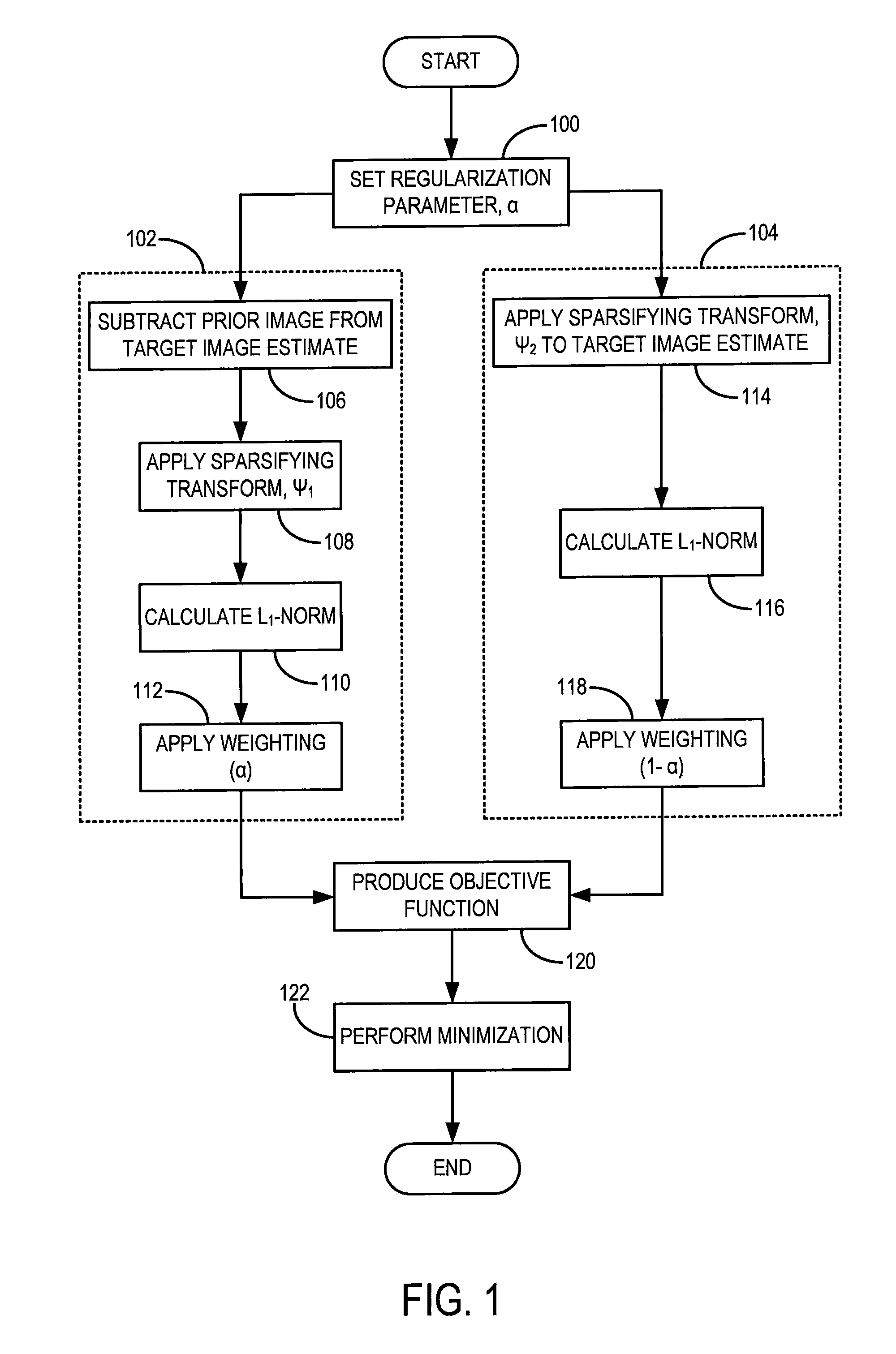
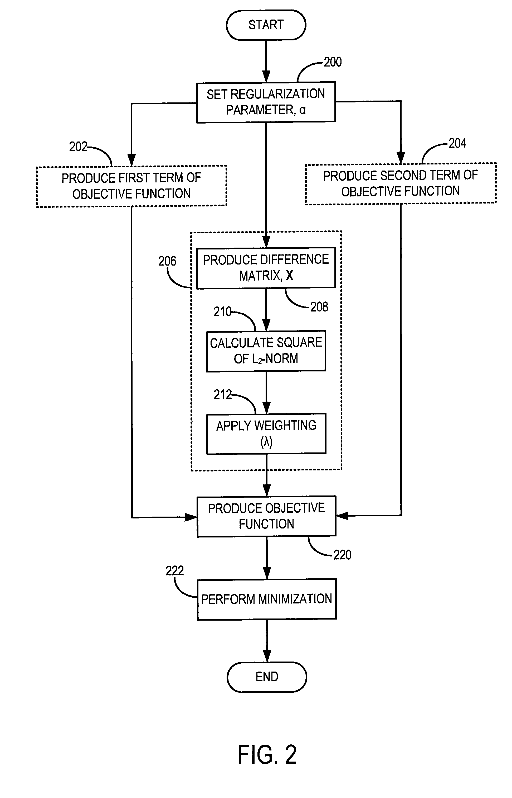
[0038]Generally speaking, the method of reconstructing an image from a set of data includes a series of numerical steps to estimate a desired image, I, from the measured data samples, Y. More specifically, the image reconstruction should fulfill the following consistency condition:

AI=Y   Eqn. (1);

[0039]where A is a system matrix. In general, the system matrix, A, can be viewed as a forward projection operator that relates the desired image, I, to the acquired data samples, Y. When dealing with computed tomography (“CT”) imaging, the system matrix can include a reprojection operation, while in magnetic resonance imaging (“MRI”), it can include a Fourier transform operation. The consistency condition of Eqn. (1), put in other words, states that when an image is faithfully reconstructed, the forward operation should substantially mimic the actual data acquisition procedure in order to generate a correct estimate of the measured projection data.

[0040]Turning now to the method of the pre...


PUM


Abstract

A method for reconstructing a high quality image from undersampled image data is provided. The image reconstruction method is applicable to a number of different imaging modalities. Specifically, the present invention provides an image reconstruction method that incorporates an appropriate prior image into the image reconstruction process. One aspect of the invention is to provide an image reconstruction method that produces a time series of desired images indicative of a higher temporal resolution than is ordinarily achievable with the imaging system, while mitigating undesired image artifacts. This is generally achieved by incorporating a limited amount of additional image data into the data consistency condition imposed during a prior image constrained image reconstruction. For example, cardiac phase images can be produced with high temporal resolution using a state-of-the-art multi-detector CT system with either fast gantry rotation speed or CT imaging system with a slow gantry rotation speed.

Description

STATEMENT REGARDING FEDERALLY SPONSORED RESEARCH[0001]This invention was made with United States government support awarded by the following agency: National Institutes of Health, NIH EB005712. The United States government has certain rights in this invention.CROSS-REFERENCE TO RELATED APPLICATIONS[0002]None.BACKGROUND OF THE INVENTION[0003]The field of the invention is medical imaging and particularly, methods for reconstructing images from acquired image data.[0004]In a computed tomography system, an x-ray source projects a fan-shaped beam which is collimated to lie within an x-y plane of a Cartesian coordinate system, termed the “image plane.” The x-ray beam passes through the object being imaged, such as a medical patient, and impinges upon an array of radiation detectors. The intensity of the transmitted radiation is dependent upon the attenuation of the x-ray beam by the object and each detector produces a separate electrical signal that is a measurement of the beam attenuatio...

Claims


Application Information

Patent Timeline
Patent Type & Authority Applications(United States)
IPC IPC(8): G06K9/00
CPCG06T11/006G06T2211/424G06T2211/412
Inventor CHEN, GUANG-HONGTANG, JIE
Owner WISCONSIN ALUMNI RES FOUND
Why Eureka
Try Eureka