
Dna Fragments, Primers, Kits, and Methods for Amplification the Detection and Identification of Clinically Relevant Candida Species

a technology of dna fragments and kits, applied in the field of dna fragments, primers, kits, and methods for identifying clinically relevant candida species, can solve the problems of difficult cultivation, limited clinical value of positive culture, and difficult laboratory diagnosis and treatment of fungal infections, so as to facilitate the extremely high degree of implementation in diagnostic laboratories, the effect of facilitating the application

Inactive Publication Date: 2010-12-09
UNIVERSITY OF MINHO
View PDF0 Cites 4 Cited by
  • Summary
  • Abstract
  • Description
  • Claims
  • Application Information

AI Technical Summary

Benefits of technology

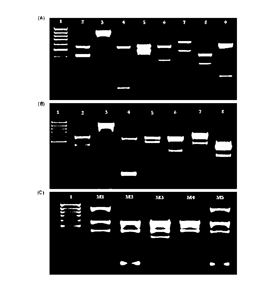
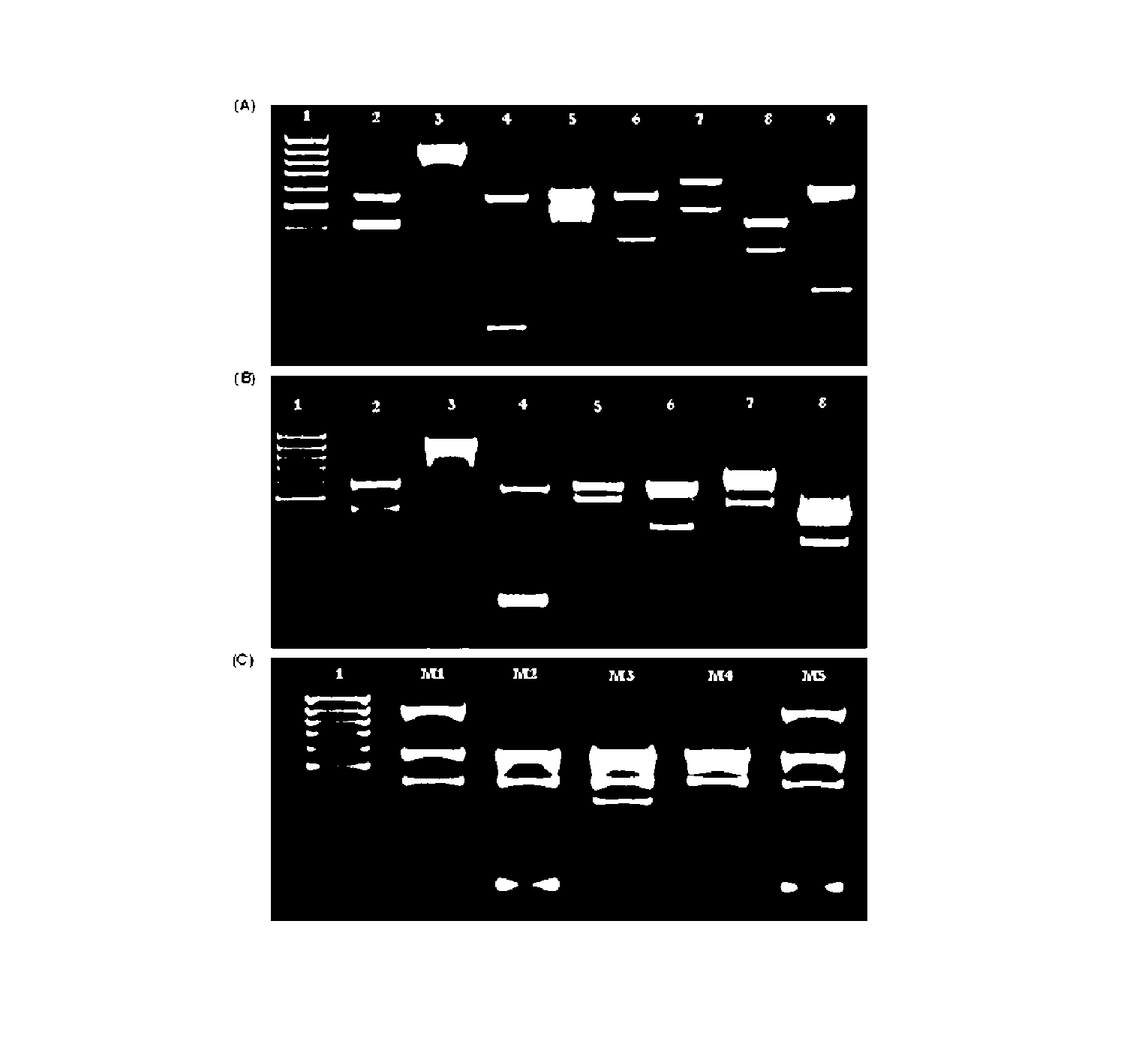
[0015]The simplicity and efficiency of this method are also shown by other characteristics displayed by the described invention. The designed strategy allows the use of whole cells directly in the PCR reaction, bypassing the time-consuming and laborious steps necessary for the DNA isolation and reducing the time required for the identification (FIG. 2B). Additionally, a PCR-based method has the possibility to detect both viable as well as dead cells, increasing many fold the target sequence to amplify, being advantageous when compared to cultural methods.
[0018]Also in economical terms, this strategy reveals extremely attractive, when taking into account that most of the expenses related to the components of the PCR mixture, not being necessary DNA probes or restriction enzymes of elevated cost. Finally, one the major advantages that this technology presents is the reproducibility of the technique when performed by different persons and using different machines, what per se facilitates extremely its implementation in diagnostic laboratories. Additionally, its execution requires just common equipment generally in use in most of the clinical laboratories and if not, easily implemented. Altogether, the features presented herein by this new technique together with the easiness of interpretation of the results point to a highly advantageous applicability concerning the identification of the etiological agent in Candida infections, both in the clinical practice as well as in epidemiological studies.

Problems solved by technology

The laboratory diagnosis and treatment of fungal infections are usually problematic and are still a great challenge both to clinicians as well as to microbiologists.
The fungi are difficult to cultivate from samples such as blood or urine and taking into account their ubiquity, a positive culture presents a limited clinical value due to the elevated probability of contamination.
The isolation of Candida from blood is a highly predictive fact of invasive infections, although the success rate of cultivation is inferior to 20% in patients with candidemia.
In addition, it is necessary to take into account the time necessary to cultivate and identify the yeast, a generally time-consuming procedure.
Nevertheless, the sensitivity of this methodology is very low (usually inferior to 50%), since the patients that are immunocompromised have difficulty to generate adequate immune responses.
The flaws, from both the cultural methods as well as those from the antibody detection, resulted in that the attention of researchers was focussed in tests that detected antigens or fungal metabolites in body fluids.
A major problem of this technique is the transient nature of the antigens in the serum, whereby the sensibility is generally much reduced for most of the antigen tests.
In addition, none of the afore-mentioned methods allow the identification of the fungus to the species level, necessary for the effective treatment of fungal infections.
Although the molecular methods available present an increased specificity, they are generally costly or compel sophisticated technology not readily available or not easily implementable in diagnostic laboratories.
In addition, conventional methods of identification do not allow distinguishing and identifying two or more Candida species present in multiple infections, a fact occurring with some frequency and that can affect the antifungal therapy.
Finally, most of the genotypical methods of identification are based on the use of purified DNA of the species to identify, which invariably leads to the need of implementing time-consuming methods of DNA isolation.

Method used


Image

  • Dna Fragments, Primers, Kits, and Methods for Amplification the Detection and Identification of Clinically Relevant Candida Species
  • Dna Fragments, Primers, Kits, and Methods for Amplification the Detection and Identification of Clinically Relevant Candida Species
  • Dna Fragments, Primers, Kits, and Methods for Amplification the Detection and Identification of Clinically Relevant Candida Species

Examples


example 1

DNA Isolation of Candida Cells in Culture

[0035]Candida cells were grown overnight in liquid YEPD medium at 26° C. with aeration on a mechanical stirrer (150 rpm). The cells were collected by centrifugation and the sediment was suspended in 200 μl of lysis buffer (2% Triton X-100, 1% SDS, 100 mM NaCl, 10 mM Tris-Hcl e 1 mM EDTA, pH 8.0). For cellular disrupture, 200 μl of glass beads with a 0.5 mm diameter and 200 μl of phenol / chloroform (1:1) were added and the tubes agitated during three intervals of 60s intercalated with periods of cooling on ice. After the removal of cellular debris by a centrifugation of 5 minutes at 18.000×g, the supernatant was collected and 1 ml of cold absolute alcohol was added before mixing by inversion. The tubes were centrifuged at 18.000×g during 3 minutes and the sediment was suspended in 400 μl of TE buffer (100 mM Tris-HCl, 1 mM EDTA, pH 8.0). A 5 minutes treatment with RNase A (1 mg / ml) at 37° C. was carried out before the addition of 10 μl of sodiu...

example 2

Multiplex PCR Amplification

[0036]Multiplex PCR amplification was performed in a 20 μl volume consisting of 0.8×PCR buffer [160 mM (NH4)2SO4, 670 mM Tris-HCl (pH 8.8)], 3.5 mM MgCl2, dNTPs mixture (200 μM each), primer mix (SEQ ID NO 1 and 2, 0.55 μM each; SEQ ID 3 and 6, 0.15 μM each; SEQ ID NO 4 and 7, 0.2 μM; SEQ ID NO 5, 0.3 μM; SEQ ID NO 8, 0.05 μM; SEQ ID NO 9, 0.4 μM), 1 U Taq polymerase DNA and 100 ng of genomic DNA, with the remaining volume consisting of sterilized water. To perform multiplex PCR amplification using whole cells, part of an isolated colony was directly suspended in the reaction tube. The reaction was carried out as usual in a thermal cycler Biometra Tpersonal (Whatman Biometra) under the following conditions: 40 cycles of 15 s at 94° C., 30 s at 55° C., and 45 s at 65° C., after an initial period of 10 minutes for denaturation and enzyme activation at 94° C. Negative control reactions were performed simultaneously with each test replacing the DNA by steriliz...

example 3

Detection and Identification of Candida in Peripheral Blood

[0037]In order to determine the detection limit of the method, human peripheral blood was seeded separately with cells from several Candida species, amongst which C. albicans, C. glabrata, C. krusei, C. parapsilosis and C. tropicalis, until concentration of 2.5×10⁵ CFU / ml was obtained. The cell number (CFU / ml) was estimated by hemacytometer counting and confirmed by plating serial dilutions of seeded blood with Candida onto solid medium Sabouraud plates and colony counting forming units after 2 days of incubation at 30° C. The seeded blood was then diluted several times with unseeded blood (concentrations in the range from 2.5×10⁵ to 1.25×10³ CFU / ml) and 200 μl of the diluted samples were exposed to DNA isolation, according to Example 4.


PUM

PropertyMeasurementUnit
Volumeaaaaaaaaaa

Abstract

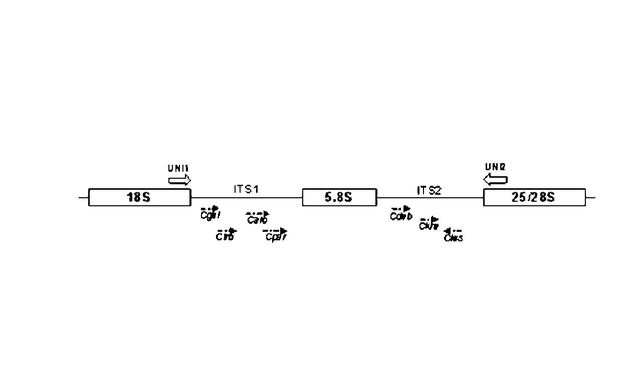
The present invention describes a novel molecular method based on the polymerase chain reaction (PCR) in a multiplex variant in order to detect and identify Candida species with clinical relevance, namely C. albicans, C. glabrata, C. krusei, C. parapsilosis, C. tropicalis, C. guilliermondii, C. lusitaniae e C. dubliniensis. The strategy uses the existence of sequences, whether conserved or variable, in fungal ribosomal genes and in the use of a combination of universal primers, specific for fungi, and internal primers, specific for each one of the Candida species (FIG. 1). In this sense, two fragments from the internal transcribed spacer (ITS) regions of the ribosomal RNA (rRNA) are amplified by multiplex PCR, allowing the easy identification of the Candida species in question. This methodology allows a rapid, effective and low-cost identification of Candida species with clinical relevance, bearing several advantages over the currently available diagnostic methods.

Description

SCOPE OF THE INVENTION[0001]The described invention is included in the detection and identification of clinically important fungi. More particularly, this inventions is related with the identification of Candida species with clinical relevance, namely C. albicans, C. glabrata, C. krusei, C. parapsilosis, C. tropicalis, C. guilliermondii, C. lusitaniae and C. dubliniensis. BACKGROUND OF THE INVENTION[0002]In the last years, changes in the epidemiological profile of infectious diseases have been observed characterized with the increase of fungal infections when compared to bacterial infections, clearly proven by a 400% increase in fungemias from 1990 to 2000 (Kao et al., Clin. Infect. Dis. 29:1164-70 (1999)), with the consequent elevated cost for the public health system (Edmond et al., Clin. Infect. Dis. 29:239-244 (1999)).[0003]Invasive fungal infections, namely candidemia, assume particular relevance in nosocomial infections (Jarvis et al., Clin. Infect. Dis. 20:1526-30 (1995); Fri...

Claims


Application Information

Patent Timeline
IPC IPC(8): C12Q1/68C07H21/00C12P19/34
CPCC12Q1/6895C12Q2600/16
Inventor RODRIGUES CARVALHO, AGOSTINHO ALBERICODOS SANTOS RODRIGUES, FERNANDO JOSE
Owner UNIVERSITY OF MINHO
Learn More