Imaging system with dynamic range maximization

a dynamic range and imaging system technology, applied in material analysis, instruments, computer peripheral equipment, etc., can solve the problems of large differences in fluorescence intensity and set the gain, and achieve the effect of rapid test, easy loading and processing

Active Publication Date: 2011-03-17
HURON TECH INT
View PDF4 Cites 38 Cited by
  • Summary
  • Abstract
  • Description
  • Claims
  • Application Information

AI Technical Summary

Benefits of technology

[0021]“Proper exposure” is defined as a gain setting such that in the output image file no (or only a small number of) pixels are saturated, and the dynamic range of the image data matches the dynamic range of the output image file (8 bits for an 8 bit file, 12 bits for a 12 bit file, etc.) and includes substantially the entire range of pixel amplitudes from the noise floor to the brightest pixel. The output image file may have a smaller dynamic range than that of the detection system, and that of the image file that is collected during scanning. This patent describes two methods of maximizing the dynamic range of data stored in the output image file—(1) accurately estimating the gain required to maximize the dynamic range of each detection channel when the dynamic range of the detection channel and the dynamic range of the output image data file are the same, and (2) using a dynamic range in the detection channel that is larger than that required in the final image data file and contracting the acquired data to utilize substantially the entire dynamic range of the final image data file. Where there are bright pixels that are not part of the required data set, such as from dust particles on the slide, or from position markers or areas not included in the required image, these pixels are not included in the calculation to maximize dynamic range. Using the entire dynamic range available is particularly important in fluorescence imaging, where variation in fluorescence intensity from one part of the image to another is often an important part of the data analysis. In addition, there is often a large difference in fluorescence intensity from one fluorophore to another, and it is very difficult (and important) to set the gain for each fluorophore to maintain the maximum dynamic range for each. This is especially true for fluorescence images of macroscopic specimens, since an image file that contains only 8 bits / pixel for each fluorophore is only half the size of an image with 12 bits / pixel (usually stored as 16-bit numbers) or 16 bits, and smaller file sizes can greatly reduce the time for image acquisition, storage, manipulation, analysis and transmission.
[0034]It is an object of this invention to provide a Raman imaging system for large specimens where a sparse pixel preview scan can be used to map changes in composition of the specimen at low resolution, and to direct the operator where to image small areas at high resolution.
[0036]It is an object of this invention to provide a method of acquiring fluorescence images in which the image data from each fluorophore substantially fills the dynamic range available in the final image file, by estimating the gain required to maximize the dynamic range for each fluorophore in a fluorescence image before scanning, using detection channels that have larger dynamic range than that required in the final image, and contracting the dynamic range of the acquired data to fill substantially the entire dynamic range of the output image data file for each fluorophore.
[0050]It is an object of this invention to provide a means and method for imaging macroscopic specimens whereby a sparse image of the specimen is calculated during scanning and stored as metadata with the output image file. NOTE: This sparse image file can be used to suggest, direct and test additional image processing and analysis operations to be applied to the large output image file. For example, if the output image file is a 10 GB file, a sparse image file containing 1 / 100 of the pixels in the original output image file is 100 MB in size, a file size that can easily and quickly be loaded into and processed by commercially-available image processing programs, and since it contains a fraction of the same pixels that are in the output image file, allows a rapid test to predict the outcome of some image processing algorithms that are planned for the output image file, allowing the operator to try different algorithms and settings without having to process the entire file. In addition, the sparse image file can be used by image storage and retrieval programs as a basis on which to calculate a smaller thumbnail (or the sparse image file, itself can be used as a thumbnail).

Problems solved by technology

In addition, there is often a large difference in fluorescence intensity from one fluorophore to another, and it is very difficult (and important) to set the gain for each fluorophore to maintain the maximum dynamic range for each.

Method used


Image

  • Imaging system with dynamic range maximization
  • Imaging system with dynamic range maximization
  • Imaging system with dynamic range maximization

Examples


first embodiment

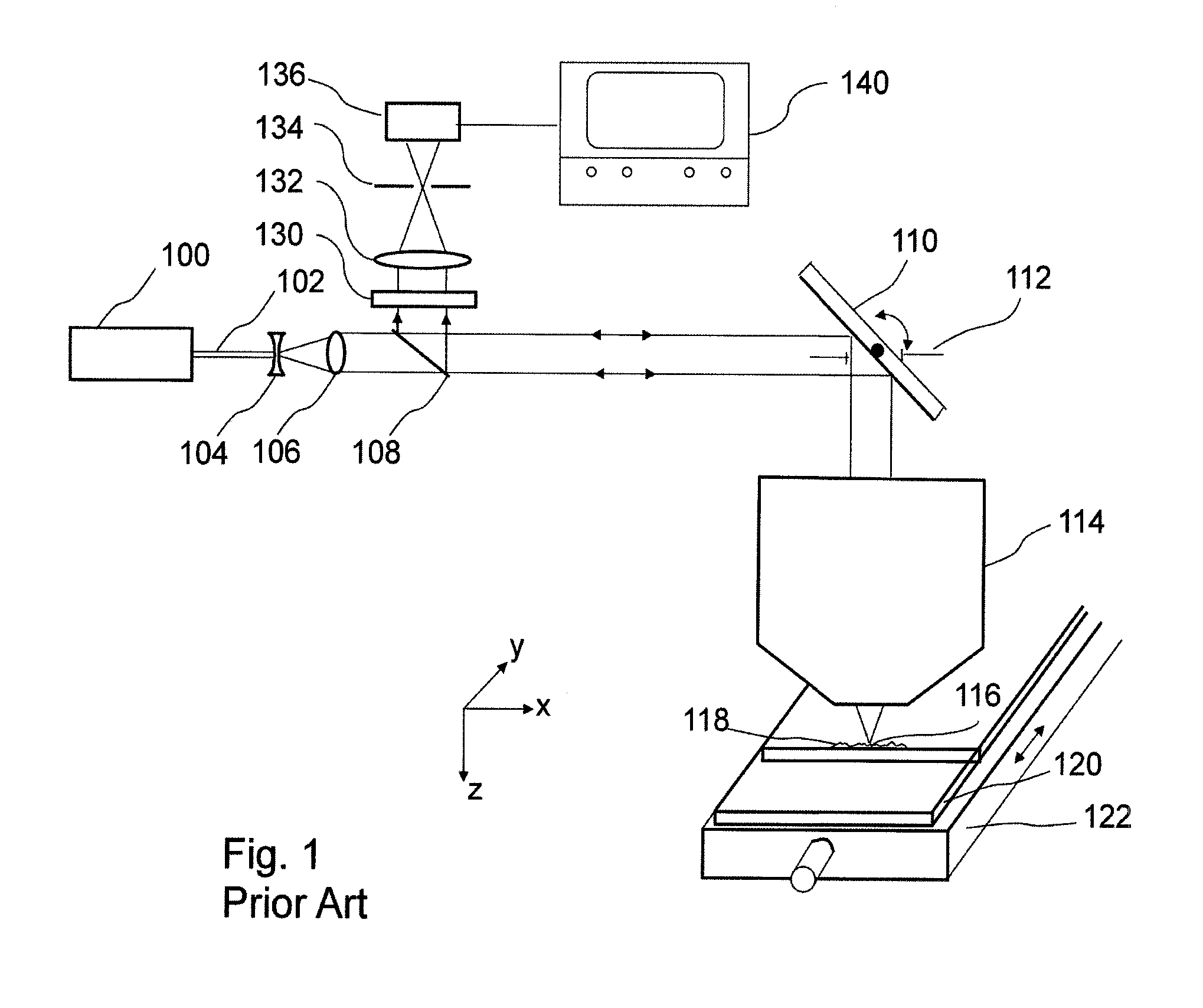
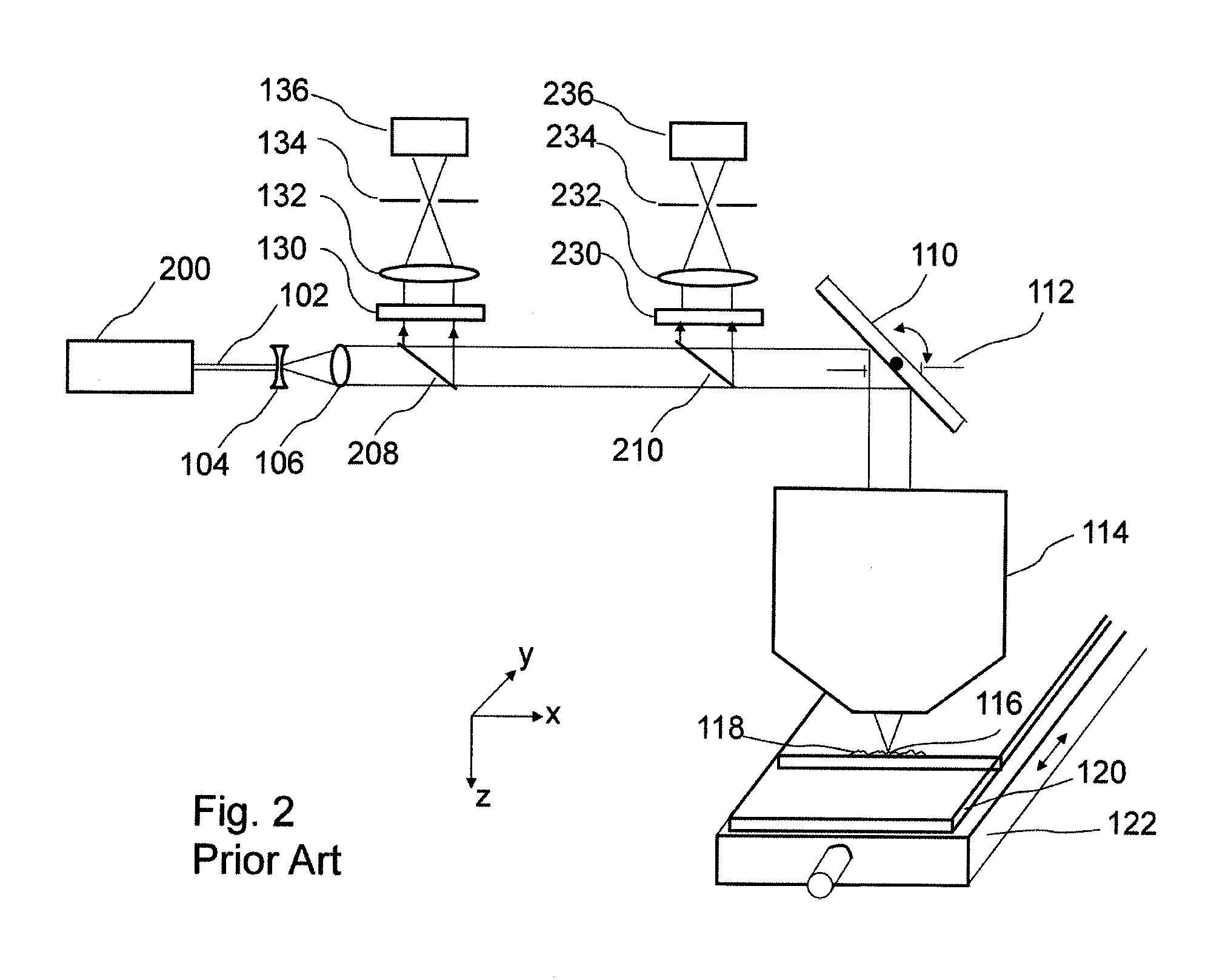
[0141]this invention is a method and macroscope that will:[0142]image the entire specimen rapidly in preview mode, where only a small fraction of pixels (a sparse image) are recorded across a field-of-view that includes the entire specimen, where those pixels have the same size and exposure as those same pixels would have in the final image if no changes were made in detection gain and offset before scanning,[0143]Calculate and display a histogram of the preview image,[0144]Increase (or reduce) the detection channel gain so that the brightest pixel in the preview image has a value less than the maximum pixel value for the dynamic range of the detection system,[0145]Adjust the preamplifier offset (if possible) to move the dimmest pixel in the preview image close to the 0 end of the histogram.

[0146]FIG. 24 shows the series of steps for imaging in which a histogram of the Sparse Preview Image (a separate histogram for each fluorophore or channel) is used to set gain and offset for each...

second embodiment

[0152]this invention is a microscope (or slide scanner) that has a larger dynamic range for measurement than the dynamic range required in the final image, and a method for contracting the dynamic range of the measured data to use all or substantially all of the dynamic range available in the final image file. (The image shown in FIG. 5 is a good example of an image where the gain and offset were incorrect, and only part of the dynamic range of the final image file was utilized.)

[0153]One example of such an instrument is a macroscope with a dynamic range of 12 bits per channel, where the dynamic range required in the final image file is 8 bits per fluorophore (each fluorophore is imaged using a separate channel). A histogram of hypothetical fluorescence image data from one 12-bit channel of the macroscope is shown at the top of FIG. 23, which shows a plot of the Number of Pixels (vertical axis) vs. Pixel Value (fluorescence intensity) on the horizontal axis. This plot shows the numb...

third embodiment

[0161]this invention is to provide a microscope (or slide scanner) and method for calculating, displaying and storing as metadata attached to the final image file a histogram of the pixel intensity data in that image file, where the histogram is calculated on-the-fly during scanning. A separate histogram is required for each fluorophore.

[0162]When using a 12-bit detector, 4096 memory locations are dedicated to storing the histogram. The TISSUEscope will be used as an example. During scan, data from each fluorophore scan line is transmitted from the TISSUEscope optics module to the computer controlling the scan. These scan lines usually contain from 10,000 to 40,000 data points (pixels) which are used to update the image display on the computer screen and are stored in a data file in RAM (for small image files) or on a hard drive or other storage device (for large image files). To calculate an image data histogram on-the-fly, during scan, the pixel value of each data point in that sc...


PUM


Abstract

A method of operating an instrument that is a macroscope, microscope, or slide scanner is provided where the instrument has a larger dynamic range for measurement than a dynamic range required in the final image of a specimen. In the method, data is measured from a specimen using the instrument, and the dynamic range of the measured data is contracted in the final image file during scanning.

Description

BACKGROUND OF THE INVENTION[0001]1. Field of the Invention[0002]This invention relates to the fields of confocal and non-confocal imaging of large microscope specimens with particular emphasis on scanning beam fluorescence and photoluminescence imaging systems, including multi-photon fluorescence, spectrally-resolved fluorescence, and second and third harmonic imaging. Applications include imaging tissue specimens, genetic microarrays, protein arrays, tissue arrays, cells and cell populations, biochips, arrays of biomolecules, detection of nanoparticles, photoluminescence imaging of semiconductor materials and devices, Raman imaging, and many others.[0003]2. Description of the Prior Art[0004]FIG. 1 shows one embodiment of a prior art confocal scanning laser macroscope, as described in U.S. Pat. No. 5,760,951. In this embodiment, the incoming collimated laser beam 102 from laser 100 passes through a beam expander (comprised of lens 104 and lens 106), and is expanded to match the diam...

Claims


Application Information

Patent Timeline
Patent Type & Authority Applications(United States)
IPC IPC(8): G06K9/00
CPCG01N21/6428G01N21/6458G01N2021/6419G02B21/367G01N2021/6441G02B21/0024G02B21/16G01N2021/6421
Inventor DIXON, ARTHUR E.
Owner HURON TECH INT
Why Eureka
Try Eureka