Nucleic acid nanotube liquid crystals and use for nmr structure determination of membrane proteins

a technology liquid crystals, applied in the field of nucleic acid nanotubes, can solve the problems of significant bottleneck in the direction of reradiation, measurement using nmr, structure determination of membrane proteins, and slow rate of membrane-protein structure determination,

Inactive Publication Date: 2011-04-21
PRESIDENT & FELLOWS OF HARVARD COLLEGE
View PDF0 Cites 8 Cited by
  • Summary
  • Abstract
  • Description
  • Claims
  • Application Information

AI Technical Summary

Benefits of technology

[0010]A further object of the invention is to provide nucleic acid nanotubes in which the average length of the nucleic acid nanotubes is given by the length of the single-stranded scaffold nucleic acid divided by the number of double stranded nucleic acid helices comprising each nanotube. In some embodiments the length of the helices comprising the nucleic acid nanotubes varies by no more than 20% of the average length of the helices, and in certain embodiments by no more than 10% of the average length of the helices. The length of the nucleic acid nanotubes is greater than about 50 nanometers, and about 400 nanometers. The nanotubes comprise at least 3 adjacent double-stranded helices. In certain embodiments the nanotubes consist of 5, 6, or 7 adjacent helices. In other embodiments, DNA heterodimer nanotubes 800 nanometers in length can be constructed from 400 nanometer monomers of two types, one type of monomer self-assembling with the second type of monomer.

Problems solved by technology

Structure determination of membrane proteins is an important challenge for biomedical science.
The slow rate of membrane-protein structure determination represents a significant bottleneck for both basic and applied bioscience discovery.
This bottleneck largely derives from difficulties in forming well-ordered three-dimensional crystals of membrane proteins.
In many of these cases, RDC's would enable full structure determination that otherwise would be impossible.
However, none of the existing liquid-crystalline media used to align water-soluble proteins are compatible with the detergents required to solubilize membrane proteins.
This increase in effective size is especially problematic for α-helical membrane proteins greater than 15 kD in size, where resonance peaks are closely spaced and become irresolvable with the fast coherence relaxation of slowly tumbling macromolecules.
However, none of these media are compatible with detergent-solubilized membrane proteins.
Despite such progress, full-scale structure determination of α-helical membrane proteins remains challenging and rare.
Due to the large fraction of methyl-bearing residues in membrane proteins and to the added molecular weight of detergent micelles, the low chemical-shift dispersion of α-helical proteins is obscured by resonance overlap and line broadening, making assignment of side-chain methyl resonances extremely difficult.
Without side-chain chemical shifts, it is impossible to obtain a sufficient number of long-range NOE-derived distance restraints for folding secondary segments into the correct tertiary structure.
However, none could be applied to membrane proteins due to incompatibility with the zwitterionic or anionic detergents typically used to solubilize membrane proteins for structural study.
However, dissolving protein-micelle complexes to high concentration in gels is notoriously difficult due to the inhomogeneous pore size of randomly cross-linked gel matrices.
Thus the measured RDC's are of limited accuracy.

Method used


Image

  • Nucleic acid nanotube liquid crystals and use for nmr structure determination of membrane proteins
  • Nucleic acid nanotube liquid crystals and use for nmr structure determination of membrane proteins
  • Nucleic acid nanotube liquid crystals and use for nmr structure determination of membrane proteins

Examples


example 1

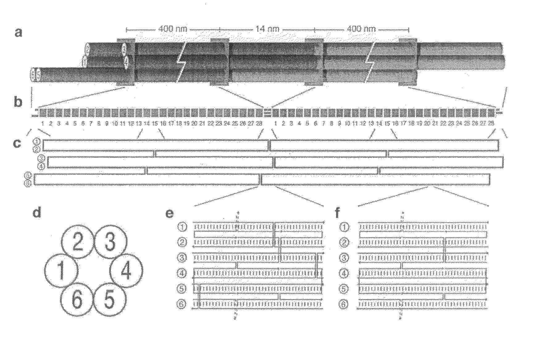
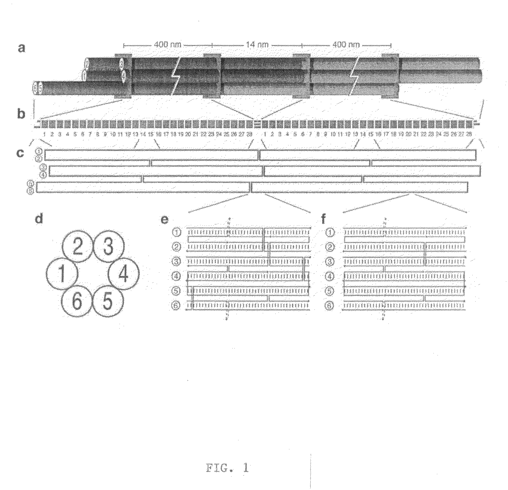
Preparation of DNA Nanotubes

[0087]M13 single stranded scaffold DNA (sequence shown in FIG. 12A-C) was obtained from phage produced from infected F+ bacteria grown in 2× YT media. Purified single-stranded DNA was extracted from the phage using a Qiagen Gigaprep ion-exchange column. Six-helix bundle DNA nanotubes were folded directly from the eluate of a Qiagen Gigaprep ion-exchange column, eluted at 50 mM Tris pH 8.5 (Fisher Scientific), 1.6 M NaCl (Fisher Scientific), 15% isopropanol. In the folding reaction, the buffer was diluted to 1 M NaCl, 9% isopropanol, along with 50 nM of the organic chemical buffer HEPES (4-2-hydroxyethyl-1-piperazineethanesulfonic acid) pH 7.5 (Sigma), 10 mM MgCl2 (Fisher Scientific). The scaffold concentration was at 6 nanomolar and the staple oligonucleotide (sequences shown in FIG. 13A-D) concentrations were at 36 nM each. The isopropanol did not interfere with the folding. Folding was performed by heating the suspension in 100 mL Pyrex bottles in 2 L b...

example 2

[0089]Recombinant M13 Bacteriophage Plasmid (p7308) Construction

[0090]Recombinant M13 filamentous bacteriophage was prepared by replacement of the BamHI-XbaI segment of Ml3mp18 by a polymerase chain reaction-generated 59 base pair (bp) fragment encoding a randomly-selected sequence (FIG. 14), flanked by positions −25 to +25 of the middle of the XbaI cut site(T̂CTAGA, or base 6258). A list of oligodeoxyribonucleotides that were used to construct the insert with flanking regions (109 bp total) is shown in FIG. 15. Double-stranded (replicative form) bacteriophage M13 DNA bearing the 59 base insert was prepared as described in Sambrook, J. & Russell, D. Molecular cloning: A Laboratory Manual (Cold Spring Harbor Laboratory Press, Cold Spring Harbor, 2001). The 59 bp insert was verified by a double restriction digest with BamHI and XbaI, followed by sequencing. The result was a modified bacteriophage M13 genome, 7308 bases in length. The full sequence is shown in FIG. 16.

example 3

Nanomole-Scale Production of M13 Bacteriophage Single-Stranded DNA

[0091]Recombinant M13 bacteriophage RF dsDNA was transformed into JM101 cells and grown overnight at 37° C. on an LB-agar plate (BD Diagnostics). A single, well-isolated plaque was used to inoculate 2 ml of 2× YT medium in a 14 mL sterile culture tube and agitated for 8 hours at 37° C. Bacterial cells were pelleted by centrifugation and phage was recovered from the supernatant by polyethylene glycol fractionation (incubation on ice for 30 minutes using a final concentration of 4% PEG8000, 0.5 M NaCl) followed by centrifugation. The phage was resuspended in 100 μL of 10 mM Tris.Cl pH 8.5 (Fisher Scientific) and labelled “pre-inoculation phage.”E Coli JM109 cells were grown overnight in 3 mL of 2× YT medium at 37° C. The 3 mL of JM109 culture was added to a 2 L flask containing 300 mL 2× YT medium supplemented with MgCl2 to 5 mM final concentration and incubated at 37° C. on a shaker at 300 rpm. When the bacterial cultu...


PUM

PropertyMeasurementUnit
lengthaaaaaaaaaa
lengthaaaaaaaaaa
lengthaaaaaaaaaa

Abstract

Compositions and methods for preparing nucleic acid nanotubes using DNA origami techniques are described, which provide for nanotubes of predictable and uniform length. The nucleic acid nanotubes thus formed are suitable as liquid crystal preparations enabling liquid-crystal NMR spectroscopy of proteins solubilized in detergent.

Description

RELATED APPLICATIONS[0001]This application claims the benefit of priority to U.S. Provisional Application Ser. No. 60 / 793,788, filed Apr. 21, 2006, and U.S. Provisional Application Ser. No. 60 / 904,266, filed Feb. 28, 2007.TECHNICAL FIELD[0002]The invention generally relates to nucleic acid nanotubes. More particularly the invention relates to compositions and methods for making nucleic acid nanotubes that are suitable for performing liquid-crystal NMR spectroscopy of detergent-solubilized membrane proteins.BACKGROUND[0003]Structure determination of membrane proteins is an important challenge for biomedical science. About thirty percent of expressed proteins span lipid bilayers, yet structures of only about one hundred membrane proteins have been resolved. Membrane proteins are encoded by 20-35% of genes but represent fewer than one percent of known protein structures to date. Knowledge of their structures will be enormously insightful for cell biology. Furthermore, membrane proteins...

Claims


Application Information

Patent Timeline
Patent Type & Authority Applications(United States)
IPC IPC(8): C07H21/04C07H1/00G01R33/44C09K19/04B82Y15/00B82Y40/00
CPCB82Y5/00Y10T436/24Y10T428/2975C12P19/34
Inventor SHIH, WILLIAM M.DOUGLAS, SHAWN M.CHOU, JAMES J.
Owner PRESIDENT & FELLOWS OF HARVARD COLLEGE
Why Eureka
Try Eureka