
Device and method of using superparamagnetic nanoparticles in treatment and removal of cells

a superparamagnetic nanoparticle and cell technology, applied in the field of devices and methods for using and removing magnetic nanoparticles, can solve the problems of high invasiveness, malignant cells will have broken free and be left, and the procedure is surgical in nature, so as to achieve the effect of removing nanoparticles from patients

Inactive Publication Date: 2011-04-28
GEORGIA TECH RES CORP
View PDF11 Cites 76 Cited by
  • Summary
  • Abstract
  • Description
  • Claims
  • Application Information

AI Technical Summary

Benefits of technology

[0009]It is a further object of the invention to provide i

Problems solved by technology

However, often during the surgical procedure, malignant cells will have broken free and be left in the peritoneal cavity.
Following surgical extraction of the tumor, chemotherapy is typically recommended to kill the residual malignant cells but chemotherapy is not completely effective.
Currently, most of the suggested procedures are surgical in nature and highly invasive.

Method used


Image

  • Device and method of using superparamagnetic nanoparticles in treatment and removal of cells
  • Device and method of using superparamagnetic nanoparticles in treatment and removal of cells
  • Device and method of using superparamagnetic nanoparticles in treatment and removal of cells

Examples


example 1

In Vitro Capture of Murine Cancer Cells with Superparamagnetic CoFe2O4 Nanoparticles Coated with Peptide

[0224]Preparation of CoFe2O4 Nanoparticles with a Biocompatible Polymer Coating and with YSA Peptide Conjugation

[0225]The superparamagnetic CoFe2O4 nanoparticles were synthesized with a micelle method, and the mean diameter was 8 nm with a size distribution of less than 15%. The detailed experimental procedures are reported in Scarberry, et al., “Magnetic Nanoparticle-Peptide Conjugates for in Vitro and in Vivo Targeting and Extraction of Cancer Cells”, J. Amer. Chem. Soc'y., 130 (31), 10258-10262 (2008).

[0226]The nanoparticles (200 mg) and polygalacturonic acid (600 mg, Alfa Aesar) were added into 80 mL of 5 M NaOH solution at ambient temperature. After sonication for 5 h with a Model 60 Sonic Dismembrator (Fisher Scientific), the coated nanoparticles were separated from the solution using a magnet. After being washed a few times with water, the coated nanoparticles were resuspen...

example 2

In Vitro Binding of Superparamagnetic CoFe2O4 Nanoparticles Coated with Peptide to Cells in Human Ascites Samples

[0258]Nanoparticle Synthesis

[0259]The superparamagnetic CoFe2O4 nanoparticles were synthesized with a microemulsion technique and the mean diameter was 8 nm with a size distribution of less than 15%. The detailed experimental procedures are reported in Scarberry, et al., “Magnetic Nanoparticle-Peptide Conjugates for in Vitro and in Vivo Targeting and Extraction of Cancer Cells”, J. Amer. Chem. Soc'y., 130 (31), 10258-10262 (2008).

[0260]Nanoparticle Coating and Peptide Conjugation

[0261]600 mg of CoFe2O4 nanoparticles were added to 300 mL of 5M NaOH and sonicated for 10 min (Model 60 Sonic Dismembrator (Fisher Scientific)—power setting of 19). 1800 mg of glucuronic acid was added to the solution and sonication continued for 1.5 hours. The product was magnetically separated using a 5000 gauss magnet, washed 3× in PBS and resuspended in 600 mL of distilled H2O, bringing the n...

example 3

Ovarian Cancer Study in Mice

[0311]An ovarian cancer survival study was conducted to evaluate whether the capture and removal of disseminated tumor cells could be employed as a curative measure to mitigate metastasis and thereby increase longevity. A murine ovarian cancer cell line (ID8 GFP VEGF) transfected with the gene for green fluorescent protein (GFP) and vascular endothelial growth factor (VEGF) expression was used for the study. VEGF expression can expedite tumor progression by stimulating angiogenesis and abating the immune response. The expression of GFP can be analyzed both qualitatively and quantitatively, providing a mechanism for tracking the dissemination of the malignant cells.

[0312]The mice used in the study were divided into two control groups and one experimental group. Each group received an intraperitoneal (I.P.) injection of 7 million IDS GFP VEGF cells. The first control group (Control A) contained 7 female C57BL / 6 mice (5-8 weeks old), which received no furthe...


PUM

PropertyMeasurementUnit
Densityaaaaaaaaaa
Temperatureaaaaaaaaaa
Flow rateaaaaaaaaaa

Abstract

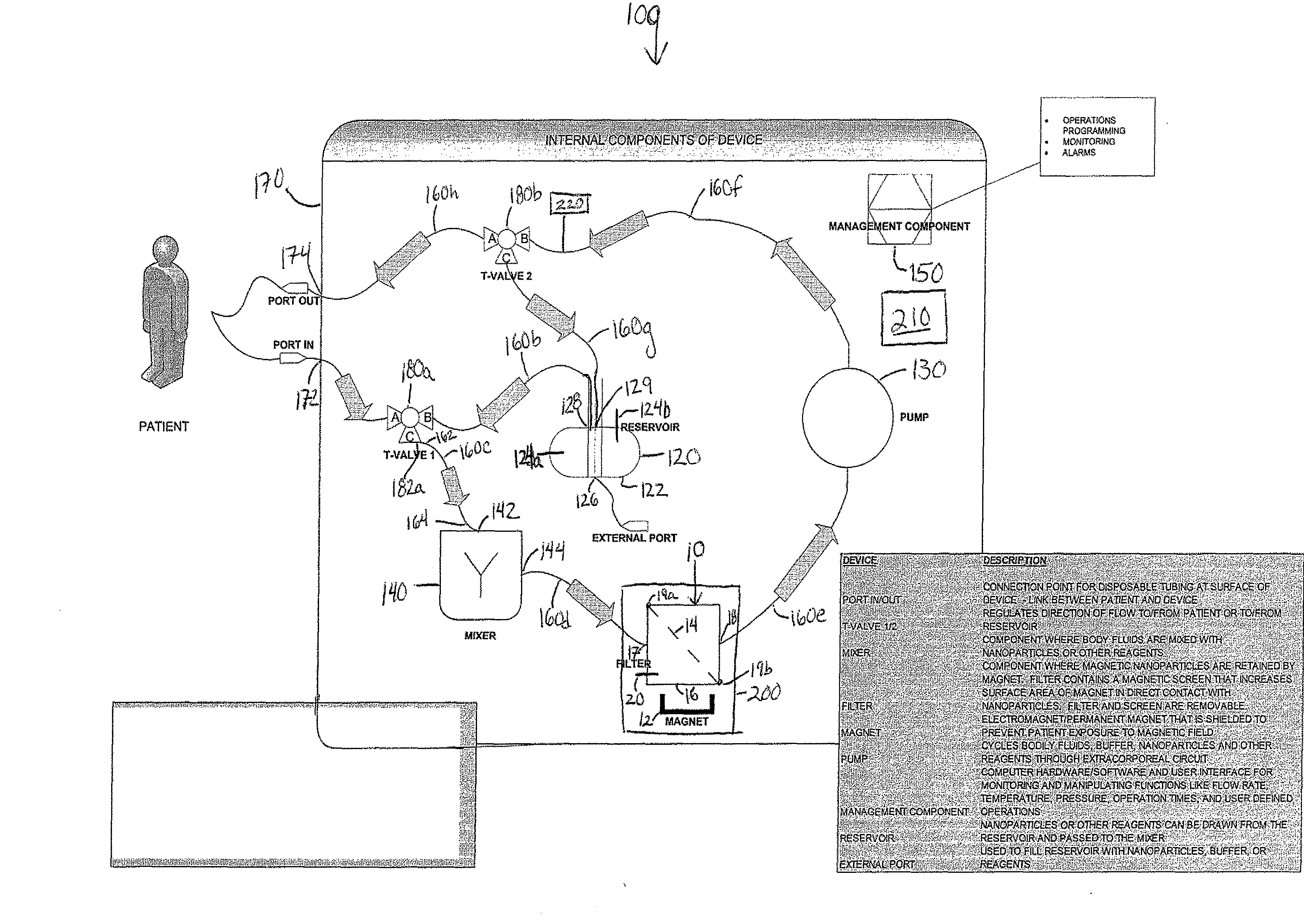
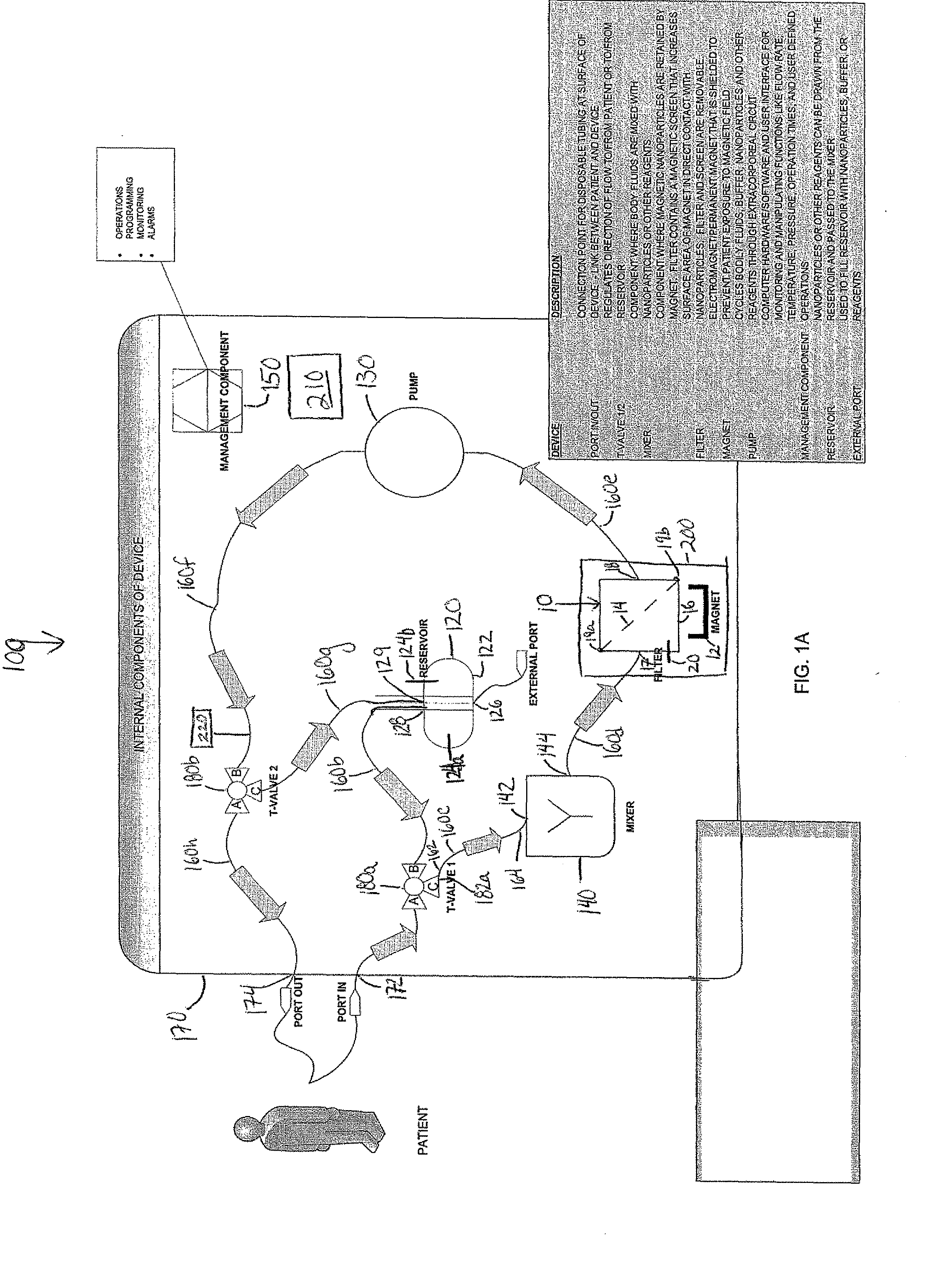
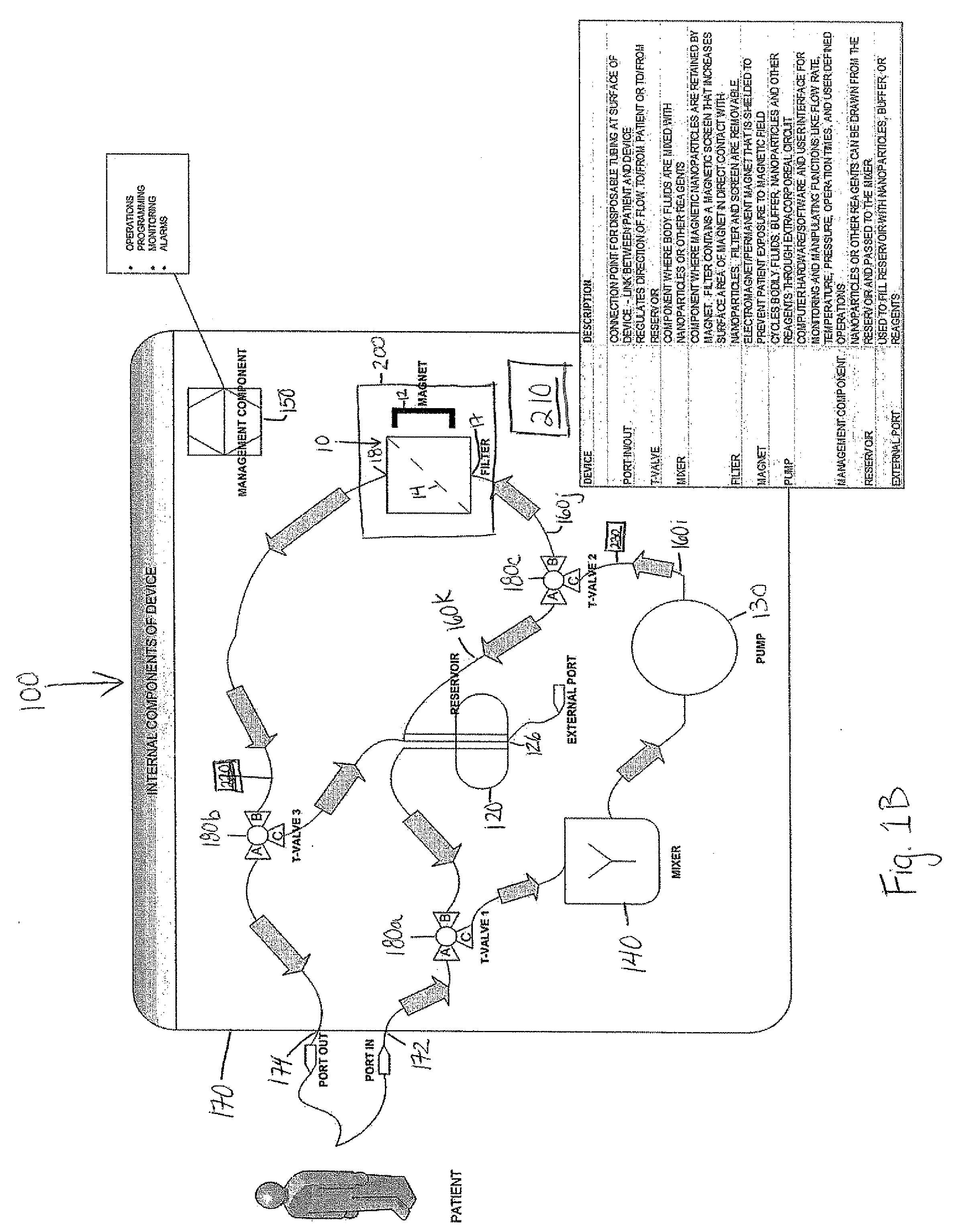
Methods and devices for selectively removing from a subject a target cell, pathogen, or virus expressing a binding partner on its surface are presented. In one embodiment, the device contains an excorporeal circuit, which includes, at least, a magnetic filter comprising a magnet and a removable, magnetizable substrate capable of capturing magnetic nanomaterials; and a pump in fluid communication with the magnetic filter, wherein the pump moves fluid through the excorporeal circuit. The magnet is capable of generating a magnetic field sufficient to capture magnetic nanomaterials in the magnetic field. In a preferred embodiment, the target cells are cancer cells and / or cells infected with pathogenic agents. The devices may be designed for extracorporeal or in vivo uses. Functionalized superparamagnetic nanoparticles are either mixed ex vivo with a biological fluid from the patient or injected into the patient. Then the biological fluid, which includes the nanoparticles is transported to the magnetic filter to remove any nanoparticles that are complexed to the target cells, pathogens, or virus, and any free nanoparticles. Optionally, the functionalized nanoparticles contain and deliver a therapeutic agent. In one embodiment, the therapeutic agent is released when the nanoparticle binds to the target cells, pathogens, or virus.

Description

CROSS-REFERENCE TO RELATED APPLICATION[0001]This application claims priority to U.S. Provisional application, U.S. Ser. No. 61 / 073,161, filed Jun. 17, 2008 and U.S. Provisional application, U.S. Ser. No. 61 / 073,973, filed Jun. 19, 2008.FIELD OF THE INVENTION[0002]This invention is in the field of devices and methods for using and removing magnetic nanoparticles, particularly for the treatment of cancer.BACKGROUND OF THE INVENTION[0003]Cancer of all types is the second leading cause of death in the United States. The morbidity of most cancers is often related to the metastatic dissemination of cells sloughing off the primary tumor. Although improved chemical and radiation therapies are resulting in less collateral damage to healthy cells, there remains a large market for targeted modalities. Long-term survival rates could be extended significantly if metastatic cells or the cells remaining after surgical excision of the tumor mass could be more effectively removed from the patient.[0...

Claims


Application Information

Patent Timeline
IPC IPC(8): A61M1/34A61F2/01B01D35/06B03C1/02B82Y5/00
CPCB03C1/01B03C2201/26B03C2201/18B03C1/288A61P31/04A61P31/12A61P35/00A61P35/02A61M1/34B03C1/02A61M1/3618A61M2205/75A61M2205/057A61M2202/20A61M2202/206
Inventor ZHANG, ZHONGJU JOHNSCARBERRY, KENNETH EDWARDDICKERSON, ERIN BETHMCDONALD, JOHN FRANCIS
Owner GEORGIA TECH RES CORP
Learn More