
Remote detection of substance delivery to cells

a technology of endocytosis and remote detection, which is applied in the field of endocytosis sensitive probe development, can solve the problems of poor encapsulation efficiency of paramagnetic ions, early results were hindered, liposome instability, etc., and achieve the effects of increasing the effect of proton relaxivity, increasing the effect of fluorescence, and low effect on proton relaxivity

Inactive Publication Date: 2011-05-05
SUTTER WEST BAY HOSPITALS +1
View PDF10 Cites 7 Cited by
  • Summary
  • Abstract
  • Description
  • Claims
  • Application Information

AI Technical Summary

Benefits of technology

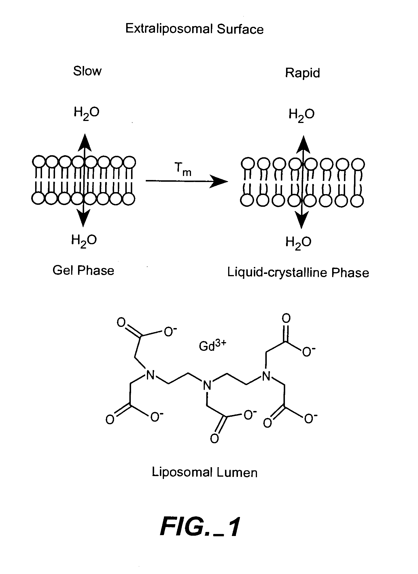
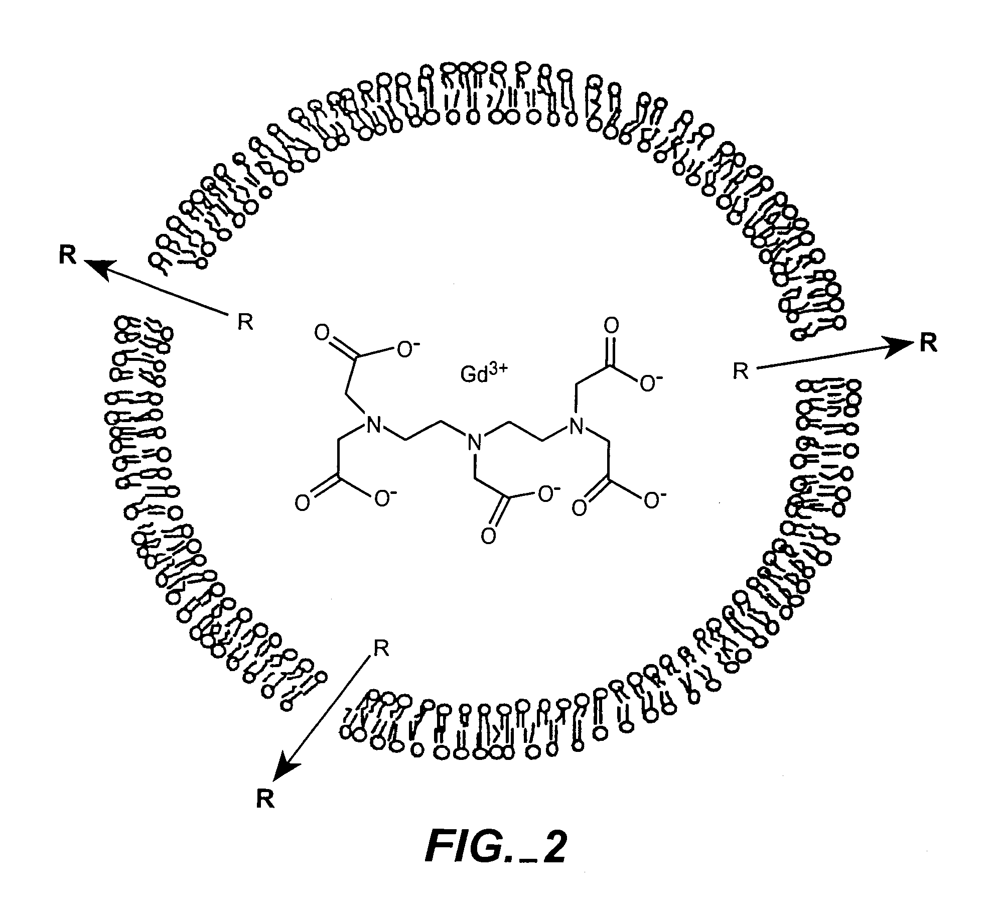
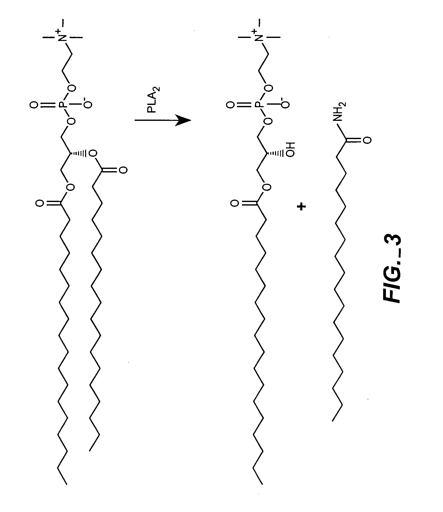
[0008]One embodiment of the invention provides a liposome for remote sensing of endocytosis for example, by pathological cells, useful in diagnosis of disease by non-invasive methods (MRI, radionuclide, intravital optical imaging). The liposome may comprise a lipopolymer conjugated to a detectable marker, for example, a paramagnetic metal (e.g. gadolinium) chelate, whose signal is modulated upon endocytosis of the liposome into a cell of interest. In a preferred embodiment, liposomes comprising gadolinium DTPA-BMA, gadolinium DTPA, or gadolinium HP-DO3A complexes encapsulated within poorly water-permeable membrane have low effect on proton relaxivity. Upon endocytosis, the liposome is degraded releasing the chelate and concomitantly increasing the effect on proton relaxivity which is remotely detected by MRI method. Alternatively, a liposome is made with encapsulated fluorescent marker in the presence of a fluorescent quencher, or under self-quenching concentrations. Endocytosis and consequent lysosomal degradation of the liposome causes the release of the marker which is accompanied by increase of fluorescence intravitally detected by laser imaging methods. The detection is made specific to a cell comprising a surface marker molecule by incorporation of a cell-internalizable ligand, such as an antibody, specific to the marker. To improve the endocytosis-specific signal modulation, the liposome is sensitized to intracellular conditions, such as decreased pH, for example, by incorporation of pH-sensitive lipopolymers.
[0011]In yet another embodiment, the invention provides improved liposomes co-encapsulating a therapeutic agent and a remotely detectable marker. The therapeutic agent and detectable marker are members of an ionically coupled pair, that is, if one is an anion, the other is a cation, and vice versa. A member of the pair is encapsulated in the liposome in a form providing for transmembrane ionic, chemical, or electrochemical gradient that enhances the encapsulation of the other member through a highly efficient “remote loading” principle. Optionally, two members form a stable ionic complex, or salt, that also improves the drug encapsulation stability and / or drug efficacy. An exemplary embodiment includes a liposome with encapsulated anionic MRI marker, Gd-DTPA, in the form of a diammonium salt. Upon removal of extraliposomal marker, transmembrane gradient of ammonium ion is created that affords highly efficient (e.g. >95%) co-loading of a cationic anticancer drug, such as doxorubicin, in the same liposome. The resulting dual-loaded liposome allows non-invasive monitoring of the therapeutic agent in a patient.

Problems solved by technology

Early results were hindered by obstacles such as poor encapsulation efficiency of paramagnetic ions, liposome instability, toxicity of certain encapsulated agents and poor relaxivity (Caride, et al., Magn Reson Imaging, 2: 107-112, 1984; Magin et al., Magn Reson Med, 3: 440-447, 1986; Navon et al., Magn Reson Med, 3: 876-880, 1986; Unger et al., Invest Radiol, 20: 693-700, 1985).
However, a limitation of using liposomes with increasingly smaller diameters is that the amount of encapsulated agent decreases compared to the amount of lipid in the bilayer.

Method used


Image

  • Remote detection of substance delivery to cells
  • Remote detection of substance delivery to cells
  • Remote detection of substance delivery to cells

Examples


example 1

Preparation of Caged Liposomal Carriers

[0061]Multiple formulations of low permeability Gd-liposomes were prepared. Multiple liposome formulations of Gd-chelates have been prepared. These liposomes were composed of distearoylphosphatidylcholine (DSPC), cholesterol, and N-poly(ethylene glycol)-distearoylphosphatidylethanolamine (PEG2000-DSPE) or mixtures of DSPC, dipalmitoylphosphatidylcholine (DPPC), and cholesterol (phospholipid:cholesterol (3:2). DPPC was added at small molar ratios to increase the rate of degradation upon reaching the endosomes. However, DPPC does have a lower phase transition than DSPC, and will likely increase the rate of diffusion of water molecules across the liposomal membrane. It remains to be seen to what extent this modification will have on the change in relaxivity upon encapsulation. This is currently being studied and will be compared to other liposomes of varying lipid composition in attempt to optimize the rate of lipid degradation and water permeabil...

example 2

Synthesis of bis-carboxymethyl-PEG600-mono(N,N-dioctadecyl)amide (DSA-PEG-COOH)

[0070]3.039 g of bis-carboxymethyl-poly(ethylene glycol) (M.w. 600; Aldrich p / n 40, 703-8) were dissolved in 20 mL of chloroform, and stirred with 250 mg of DCC at room temperature for 1 hour. The precipitated urea was filtered out using glass fiber filter, the filtrate was cooled down in an ice bath, mixed with 1 mL of anhydrous triethylamine, and 520 mg of dioctadecylamine (Fluka p / n 42358). The reaction mixture was brought to 40-45° C. with stirring until all amine was dissolved, overlaid with argon, and stirred at room temperature overnight. The precipitated additional amount of DCU was filtered out, the filtrate was diluted with 20 mL of chloroform and washed consequently with 100 mL of water (3 times), 100 mL of saturated NaHCO3 (1 time), and 100 mL of water (3 times). Organic layer was dried over sodium sulfate, the solvents were removed on a rotary evaporator, and additionally at 1-2 mm Hg and 50°...

example 3

Synthesis of bis-carboxymethyl-PEG mono-(N,N-dioctadecyl)amide tetra(carbobenzoxy-L-lysine) conjugate (DSA-PEG-(Z-Lys)4) (FIG. 10)

[0071]55 mg of DSA-PEG-COOH in 0.55 mL of chloroform were combined with the solution of 7.9 mg of N-hydroxysuccinimide in 0.5 mL of chloroform. With stirring at room temperature, 13.1 mg of DCC in 0.3 mL chloroform were added dropwise to this solution, and stirred for 2 hours. The precipitate of dicyclohexylurea was separated by filtration through GF / c glass fiber filter. The filtrate was mixed with the solution of 50 mg of poly(N-carbobenzoxy-L-lysine) Mol. weight 1,000 (Sigma) in 0.5 mL of anhydrous dimethylformamide (DMF). The chloroform was removed in vacuum, and additional 1 mL of DMF was added to the residue. 0.042 ml of triethylamine was added to this solution, and stirred overnight under argon. The solvent was removed under vacuum (1-2 mm Hg) at 50° C., and the residue was treated with 4 M HBr in glacial acetic acid for removal of the carbobenzoxy...


PUM

PropertyMeasurementUnit
diameteraaaaaaaaaa
sizeaaaaaaaaaa
sizeaaaaaaaaaa

Abstract

The present invention provides for the development of endocytosis-sensitive probes, and a remote method for measuring cellular endocytosis. These probes are based on the reduced water permeability of a nanoparticle or liposomal delivery system, and inherent degradability or disruption of barrier integrity upon endocytosis. The invention also provides for liposomes having combined therapeutic and diagnostic utilities by co-encapsulating ionically coupled diagnostic and therapeutic agents, in one embodiment, by a method using anionic chelators to prepare electrochemical gradients for loading of amphipathic therapeutic bases into liposomes already encapsulating an imaging agent. The invention provides for imaging of therapeutic liposomes by inserting a lipopolymer anchored, remotely sensing reporter molecules into liposomal lipid layer. The invention allows for an integrated delivery system capable of imaging molecular fingerprints in diseased tissues, treatment, and treatment monitoring.

Description

CROSS-REFERENCE TO OTHER APPLICATIONS[0001]This application claims priority to U.S. Provisional Application No. 60 / 486,080 filed Jul. 9, 2003, and incorporates the entire contents of that provisional application herein by reference.STATEMENT REGARDING FEDERALLY SPONSORED RESEARCH[0002]The U.S. Government has a paid-up license in this invention and the right in limited circumstances to require the patent owner to license others on reasonable terms as provided for by the terms of contract number N01-CO-27176 awarded by the National Cancer Institute.INTRODUCTION[0003]1. Field of the Invention[0004]The present invention provides for the development of endocytosis-sensitive probes, and a remote method for measuring cellular endocytosis. The invention also provides for liposomes having combined therapeutic and diagnostic utilities. The invention additionally provides for imaging of therapeutic liposomes. Further, the invention allows for an integrated delivery system capable of imaging mo...

Claims


Application Information

Patent Timeline
Patent Type & Authority Applications(United States)
IPC IPC(8): A61K9/127G01N33/53A61B5/00A61B5/055A61K49/00A61K49/18
CPCA61B5/0059A61B5/055A61K9/1272A61K9/1273A61K47/10A61K47/14A61K51/1234A61K47/36A61K47/48561A61K47/48823A61K49/0084A61K49/126A61K49/1812A61K47/18A61K47/6849A61K47/6913A61P35/00
Inventor DRUMMOND, DARYL C.HONG, KEELUNGKIRPOTIN, DMITRI B.
Owner SUTTER WEST BAY HOSPITALS
Learn More