Methods for Measuring Changes in Optical Properties of Wound Tissue and Correlating Near Infrared Absorption (FNIR) and Diffuse Reflectance Spectroscopy Scattering (DRS) With Tissue Neovascularization and Collagen Concentration to Determine Whether Wound is Healing

a tissue optical property and measurement method technology, applied in the field of measuring changes in tissue optical properties, can solve problems such as incomplete information, misdiagnosis, and inability to change treatment, and achieve the effect of reducing scattering coefficien

Active Publication Date: 2011-05-26
DREXEL UNIV
View PDF2 Cites 38 Cited by
  • Summary
  • Abstract
  • Description
  • Claims
  • Application Information

AI Technical Summary

Benefits of technology

[0016]In part to take advantage of these observations, a method of collecting information regarding the healing state of a wound is provided. In an exemplary embodiment, the method includes illuminating wound tissue with light from a light source, measuring the amplitude and / or phase shift of the light as it propagates through the wound tissue, calculating an optical absorption coefficient and / or a reduced scattering coefficient using the measured values, and correlating collagen concentration in the wound tissue with the reduced scattering coefficient calculated from measured parameters and / or correlating blood vessel in-growth and / or ischemia in the wound tissue with the optical absorption coefficient using the measured values. Changes in collagen concentration over time may be determined from changes in the reduced scattering coefficient over time. Similarly, changes in blood vessel in-growth and / or ischemia over time may be determined from changes in the optical absorption coefficient over time. The light is preferably provided by a laser and transmitted at a near infrared wavelength such as of 650-870 nm. The light may also be transmitted at specific near infrared wavelengths such as 685 nm, 780 nm, 830 nm, and / or 950 nm. The light output by the laser may be modulated to produce a diffuse photon density wave (DPDW) in the wound tissue. On the other hand, the light may be used to differentiate an impaired wound (e.g., chronic wounds such as diabetic, pressure ulcer, venous ulcer, ubiquitous ulcer, and ischemic wounds) from a non-impaired wound (normally healing wound) by measuring changes in blood vessel in-growth and / or ischemia in the wound over time and correlating optical absorption coefficients obtained from the wound over time with blood vessel in-growth and / or ischemia seen histologically for impaired and control wounds. In embodiments, the method may include detecting pressure ulcers or venous ulcers in the wound from changes in the optical absorption coefficients over time.

Problems solved by technology

However, the effectiveness of each treatment is not the same in all patients, so rapid and accurate evaluation of healing progress in each individual is critical so that unsuccessful treatments can be discontinued and alternate treatments initiated as soon as possible.
The main limitation of traditional wound evaluations is that they can give information mostly from the surface of the wound.
Such surface characteristics of a wound do not take into account the health of the wound environment beneath the surface in the whole wound bed, and provide inadequate information regarding the wound healing status of a wound.
Therefore, misdiagnosis may occur or treatment may not be altered as early as possible, with direct implications on the quality and cost of care for chronic wounds.
However, it is unclear how this method would translate to chronic wounds that are very different in shape, size, and also have more ambiguous boundaries than acute wounds.
As with HFUS, the clinical utility of OCT as a wound monitoring methodology is uncertain due to the size and complexity of human chronic wounds.
These studies identified regions of increased blood flow within chronic wounds that may correlate to granulation tissue; however, changes in blood flow were not monitored over time.
The clinical utility of LDF and LDI for serial assessment of chronic wounds is limited due to low penetration depths (˜1-2 mm) and issues with light reflection caused by curvature of the feet and presence of moisture on the surface of the wound.
However, changes of the surface appearance due to bleeding and other reasons will significantly affect the capability of DRS to provide on its own information about the wound status and oxygenation.
Generally speaking, the determination of wound surface area is highly inaccurate and subjective.
Wound edges may be hard to determine because of complex wound geometry.
Width and depth measurements may vary from between observers during the same clinic session and are highly inaccurate between visits.
Ultrasound measurements and image analysis of digital photos provide more accurate information but are difficult to use in a busy clinical setting.
Moreover, it is established that wounds, burns and lesions need oxygen to heal and that ischemic conditions represent impaired healing environments.
These methods are highly subjective and more importantly cannot assess the probability of wound healing in impaired environments, such as in chronic wounds due to diabetes, venous ulcers, pressure ulcers, ubiquitous ulcers, and others.
Invasive monitoring based on biopsies could provide information about the physiology and biochemistry of healing but is invasive and impractical, while monitoring based on wound fluid is controversial due to debates over appropriate correlation of wound fluid composition to wound tissue.
In wound characterizations, the absence of significant depth penetration makes DRS data difficult to interpret.

Method used


Image

  • Methods for Measuring Changes in Optical Properties of Wound Tissue and Correlating Near Infrared Absorption (FNIR) and Diffuse Reflectance Spectroscopy Scattering (DRS) With Tissue Neovascularization and Collagen Concentration to Determine Whether Wound is Healing
  • Methods for Measuring Changes in Optical Properties of Wound Tissue and Correlating Near Infrared Absorption (FNIR) and Diffuse Reflectance Spectroscopy Scattering (DRS) With Tissue Neovascularization and Collagen Concentration to Determine Whether Wound is Healing
  • Methods for Measuring Changes in Optical Properties of Wound Tissue and Correlating Near Infrared Absorption (FNIR) and Diffuse Reflectance Spectroscopy Scattering (DRS) With Tissue Neovascularization and Collagen Concentration to Determine Whether Wound is Healing

Examples


Embodiment Construction

[0058]A detailed description of illustrative embodiments of the present invention will now be described with reference to FIGS. 1-36. Although this description provides a detailed example of possible implementations of the present invention, it should be noted that these details are intended to be exemplary and in no way delimit the scope of the invention.

Monitoring Surface of Wound to Collect Data Regarding Healing State of Wound

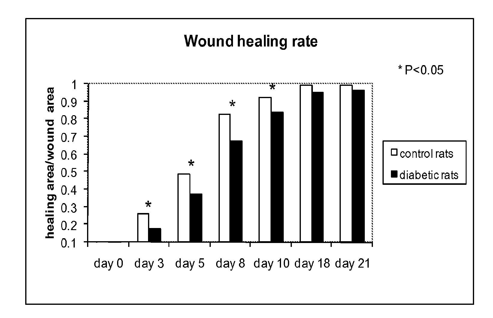
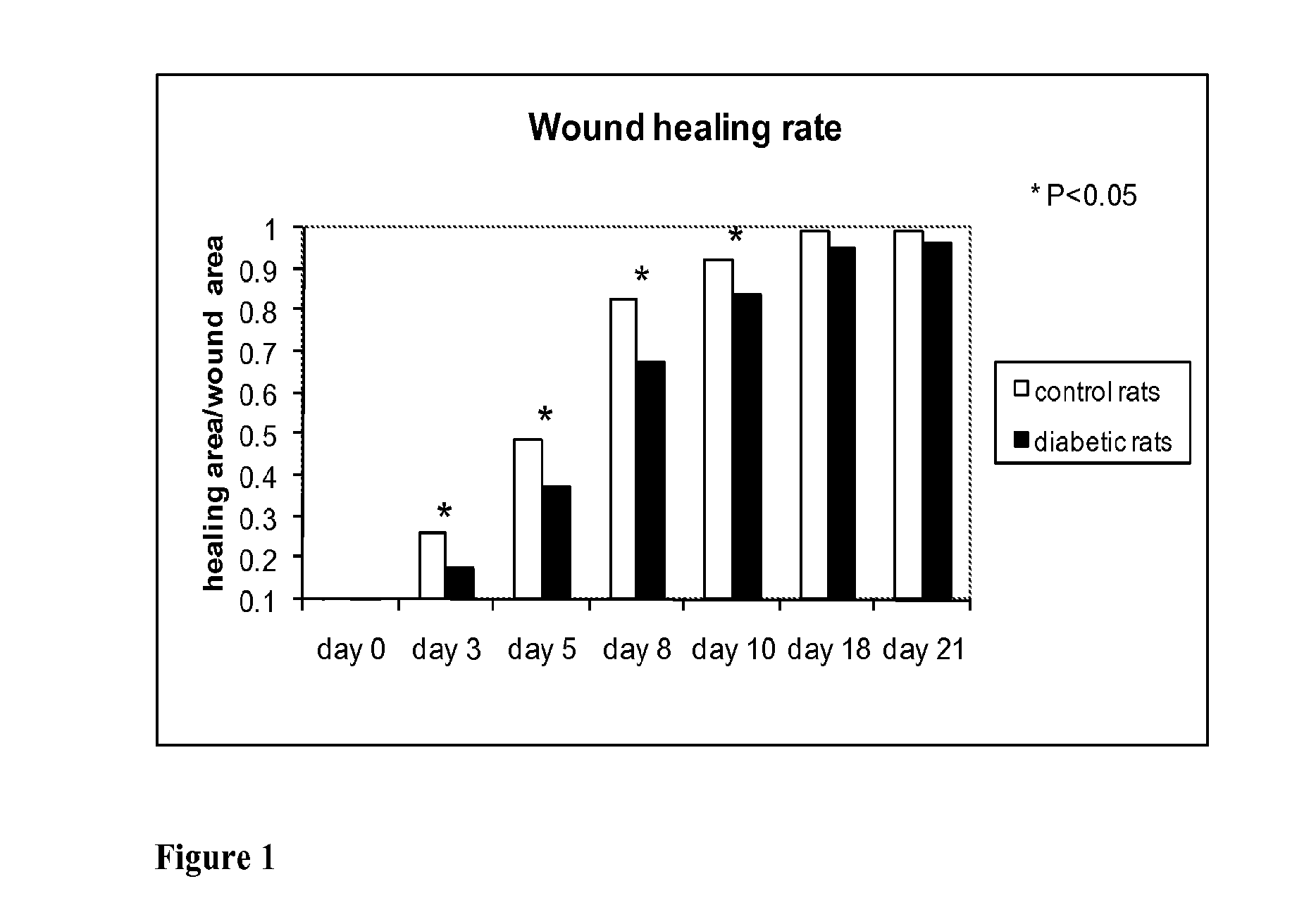
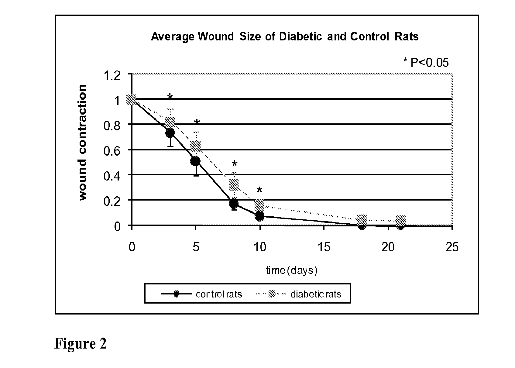
Material and Methods

[0059]A frequency domain diffuse optical tomography instrument developed by the School of Biomedical Engineering at Drexel University was used to non-invasively measure the optical properties of tissue at depths up to several millimeters. The instrument includes four laser diodes (685, 785, 830, and 950 nm) controlled by an optical switch, four avalanche photodiode detector channels, and a radio-frequency (RF) generator that modulates the laser output at a frequency of 70 MHz. The device measures the amplitude and phase shift of light as...


PUM


Abstract

Optical changes of tissue during wound healing measured by Near Infrared and Diffuse Reflectance Spectroscopy are shown to correlate with histologic changes. Near Infrared absorption coefficient correlated with blood vessel in-growth over time, while Diffuse Reflectance Spectroscopy (DRS) data correlated with collagen concentration. Changes of optical properties of wound tissue at greater depths are also quantified by Diffuse Photon Density Wave (DPDW) methodology at near infrared wavelengths. The diffusion equation for semi-infinite media is used to calculate the absorption and scattering coefficients based on measurements of phase and amplitude with a frequency domain or time domain device. An increase in the absorption and scattering coefficients and a decrease in blood saturation of the wounds compared to the non wounded sites was observed. The changes correlated with the healing stage of the wound. The methodologies used to collect information regarding the healing state of a wound may be used to clinically assess the efficacy of wound healing agents in a patient (e.g., a diabetic) and as a non-invasive method

Description

CROSS-REFERENCE TO RELATED APPLICATIONS[0001]The present application claims priority to U.S. Provisional Patent Application No. 61 / 046,640, filed Apr. 21, 2008, and to U.S. Provisional Patent Application No. 61 / 054,535, filed May 20, 2008. The contents of these patent applications are hereby incorporated by reference in their entireties.FIELD OF THE INVENTION[0002]The invention relates to methods for measuring changes in optical properties of tissue during acute wound healing and, more particularly, to the use of diffuse photon density wave (DPDW) methodology at near infrared frequencies to calculate the absorption and scattering coefficients of wound tissue based on measurements of phase and / or amplitude with a continuous wave, a frequency domain, or a time domain near infrared device. The invention also relates to determining whether a wound is healing by assessing tissue neovascularization and collagen concentration in a wound by correlating measurements made using near infrared ...

Claims


Application Information

Patent Timeline
Patent Type & Authority Applications(United States)
IPC IPC(8): A61B5/1455
CPCA61B5/0059A61B5/02007A61B5/445A61B5/1455G01N21/4738A61B5/0075A61B5/14546G01N21/359
Inventor PAPAZOGLOU, ELISABETH S.WEINGARTEN, MICHAEL S.ZUBKOV, LEONIDNEIDRAUER, MICHAEL T.ZHU, LINDAPOURREZAEI, KAMBIZ
Owner DREXEL UNIV
Why Eureka
Try Eureka