
Inhibitors of stim1 for the treatment of cardiovascular disorders

Inactive Publication Date: 2011-06-23
INST NAT DE LA SANTE & DE LA RECHERCHE MEDICALE (INSERM)
View PDF0 Cites 0 Cited by
  • Summary
  • Abstract
  • Description
  • Claims
  • Application Information

AI Technical Summary

Benefits of technology

[0018]The present invention provides methods and compositions (such as pharmaceutical compositions) for inhibiting the proliferation of smooth muscle cells, in particular arterial smooth muscle cells. The present invention also provides methods and compositions (such as pharmaceutical compositions) for treating and / or preventing vascular disorders such as atherosclerosis, post-angioplasty restenosis, pulmonary arterial hypertension and vein-graft disease. The present invention also provides methods and compositions (such as pharmaceutical compositions) for inhibiting the hypertrophic response of cardiomyocytes. The present invention also provides methods and compositions (such as pharmaceutical compositions) for treating and / or preventing cardiac hypertrophy cardiac arrhythmias, valvulopathies, diastolic dysfunction, chronic heart failure, ischemic heart failure, and myocarditis. The treatment may improve one or more symptoms of cardiac hypertrophy or heart failure, such as providing increased exercise capacity, increased blood ejection volume, left ventricular end diastolic pressure, left ventricular end systolic and diastolic dimensions, wall tension and wall thickness, quality of life, disease-related morbidity and mortality, reversal of progressive remodeling, improvement of ventricular dilation, increased cardiac output, relief of impaired pump performance, improvement in arrhythmia.
[0031]Both antisense oligonucleotides and ribozymes useful as inhibitors of STIM1 expression can be prepared by known methods. These include techniques for chemical synthesis such as, e.g., by solid phase phosphorothioate chemical synthesis. Alternatively, anti-sense RNA molecules can be generated by in vitro or in vivo transcription of DNA sequences encoding the RNA molecule. Such DNA sequences can be incorporated into a wide variety of vectors that incorporate suitable RNA polymerase promoters such as the T7 or SP6 polymerase promoters. Various modifications to the oligonucleotides of the invention can be introduced as a mean of increasing intracellular stability and half-life. Possible modifications include but are not limited to the addition of flanking sequences of ribonucleotides or deoxyribonucleotides to the 5′ and / or 3′ ends of the molecule, or the use of phosphorothioate or 2′-O-methyl rather than phosphodiesterase linkages within the oligonucleotide backbone.
[0034]Preferred viruses for certain applications are the adenoviruses and adeno-associated (AAV) viruses, which are double-stranded DNA viruses that have already been approved for human use in gene therapy. Actually 12 different AAV serotypes (AAV1 to 12) are known, each with different tissue tropisms (Wu, Z Mol Ther 2006; 14:316-27). Recombinant AAV are derived from the dependent parvovirus AAV2 (Choi, V W J Virol 2005; 79:6801-07). The adeno-associated virus type 1 to 12 can be engineered to be replication deficient and is capable of infecting a wide range of cell types and species (Wu, Z Mol Ther 2006; 14:316-27). It further has advantages such as, heat and lipid solvent stability; high transduction frequencies in cells of diverse lineages, including hemopoietic cells; and lack of superinfection inhibition thus allowing multiple series of transductions. Reportedly, the adeno-associated virus can integrate into human cellular DNA in a site-specific manner, thereby minimizing the possibility of insertional mutagenesis and variability of inserted gene expression characteristic of retroviral infection. In addition, wild-type adeno-associated virus infections have been followed in tissue culture for greater than 100 passages in the absence of selective pressure, implying that the adeno-associated virus genomic integration is a relatively stable event. The adeno-associated virus can also function in an extrachromosomal fashion and most recombinant adenovirus are extrachromosomal.

Problems solved by technology

However, it is now considered that such remodelling following disease-inducing stimuli is maladaptive and contributes to heart failure progression and favour arrhythmia and sudden death.

Method used


Image

  • Inhibitors of stim1 for the treatment of cardiovascular disorders
  • Inhibitors of stim1 for the treatment of cardiovascular disorders
  • Inhibitors of stim1 for the treatment of cardiovascular disorders

Examples


example 1

STIM1 and Vascular Smooth Muscle Cell (VSMC) Proliferation

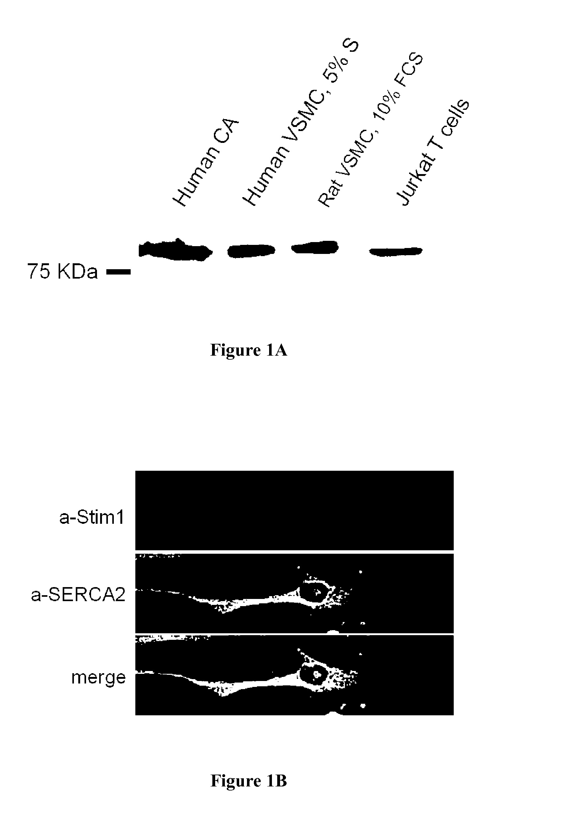
[0077]STIM1 is expressed in vascular smooth muscle cells: Immunofluorescence analysis of balloon-injured rat carotid arteries (a well-characterized model of SMC proliferation) revealed that STIM 1 was expressed in the media as well as in highly proliferative SMC in the neointima. The expected 90 kDa protein (the same molecular weight than the protein observed in human Jurkat T cell) was present in both vascular smooth muscle cells isolated from human coronary artery (hCASMC) and in rat aorta smooth muscle cells (FIG. 1A). Confocal immunofluorescence analysis in isolated vascular smooth muscle cells revealed a predominant endoplasmic reticulum distribution of STIM1, which was similar to the one of SERCA2, an endoplasmic reticulum marker (FIG. 1B).

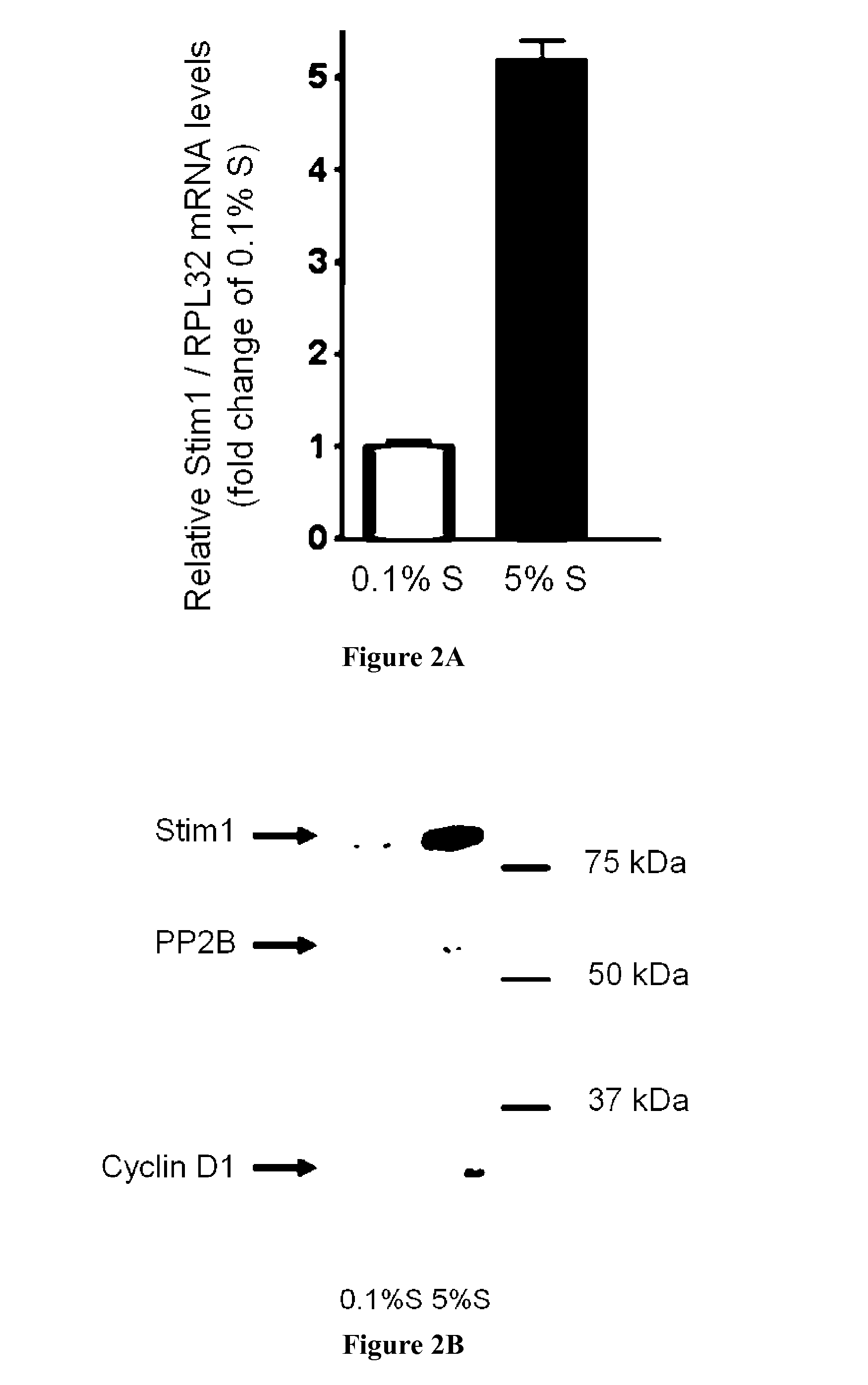
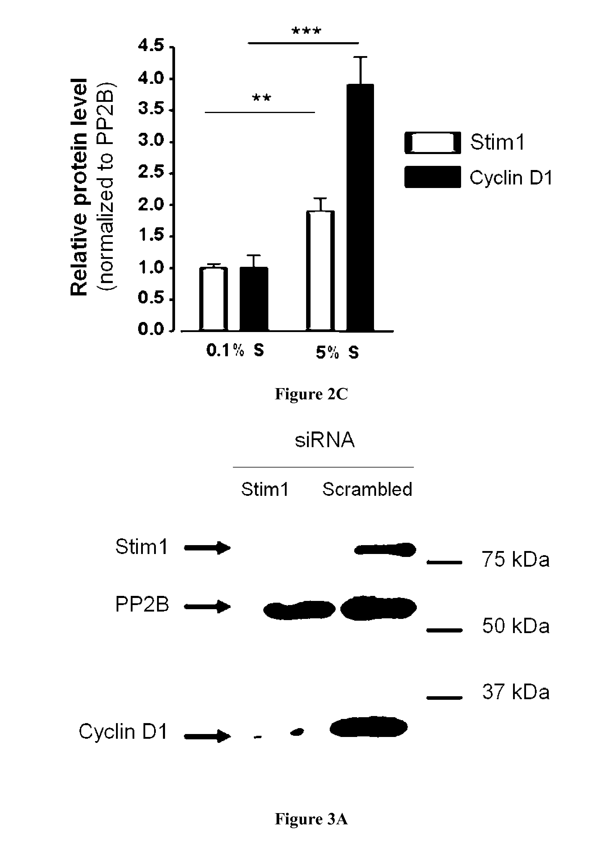
[0078]STIM1 is upregulated in proliferative VSMC: Relative expression level of STIM1 mRNA was obtained by quantitative Real Time PCR in quiescent (0.1% supplement mix, S, cultured hCA...

example 2

STIM1 and Cardiomyocyte Hypertrophy

[0087]STIM1 mRNA was detected by PCR in the human heart, in both atria and ventricles (FIG. 7A). The protein was also detected in human and rat ventricles (FIG. 7B). STIM1 protein was detected by Western-blot and immunofluorescence in isolated adult or neonatal cardiomyocytes (FIG. 7B). Stim1 expression persist for at least 7 days in culture (FIG. 7B).

[0088]To determine the expression of STIM1 in pathological growth, we used a model of pressure overload induced by abdominal aortic banding (AAB) in the rat. As shown in FIG. 8A the heart weight / to body weight ratio was increased in the AAB without increase in total body weight reflecting pathological cardiac growth. This pathological cardiac growth was confirmed by an increased ANF and MCIP mRNA levels, two markers of cardiac hypertrophy (FIG. 8B). Interestingly, STIM1 mRNA level detected by qRT-PCR was also significantly increased (p=0.08) (FIG. 8B). The expression in STIM1 expression was confirmed ...


PUM


Abstract

The invention relates to inhibitors of STIM1 for the treatment and / or the prevention of cardiac disorders such as atherosclerosis, post-angioplasty restenosis, pulmonary arterial hypertension, vein-graft disease, cardiac hypertrophy, cardiac arrhythmias, valvulopathies, diastolic dysfunction, chronic heart failure, ischemic heart failure, and myocarditis.

Description

FIELD OF THE INVENTION[0001]The invention relates to inhibitors of Stromal Interaction Molecule 1 (STIM1) for the treatment and / or the prevention of cardiac disorders, such as cardiac hypertrophy and heart failure and for vascular disorders such as atherosclerosis, post-angioplasty restenosis, pulmonary arterial hypertension and vein-graft disease. The present invention concerns gene regulation and cellular physiology in cardiomyocytes and smooth muscle cells.BACKGROUND OF THE INVENTION[0002]Cellular proliferation and growth are two mechanisms leading to cardiovascular remodelling commonly observed in vascular and cardiac muscular cells in response to diverse pathological stimuli. Excessive smooth muscle cells proliferation is a fundamental process that contributes to the injury response in major arterial vessels. Such process is involved in numerous vascular disorders including atherosclerosis, post-angioplasty restenosis, pulmonary arterial hypertension and vein-graft disease (Dza...

Claims


Application Information

Patent Timeline
IPC IPC(8): A61K39/395A61K31/713A61K31/7088A61P9/00C12N15/113
CPCC12N15/1138C12N2310/53C12N2310/14C12N2310/111A61P9/00
Inventor HULOT, JEAN-SEBASTIENLOMPRE, ANNE-MARIE
Owner INST NAT DE LA SANTE & DE LA RECHERCHE MEDICALE (INSERM)
Learn More