Shiga toxin b-subunit/chemotherapeutics conjugates

Inactive Publication Date: 2011-06-23
INSTITUT CURIE +1
4 Cites 12 Cited by

AI-Extracted Technical Summary

Problems solved by technology

The clinical use of chemotherapeutic agents against malignant tumors is successful in many cases but also has several limitations (B. A. Chabner and T. G. Roberts, Nature Rev.
In particular, anti-cancer drugs often do not affect tumor cells selectively over healthy cells, which leads to high toxicity and side effects (M. V. Blagosklonny, Trends Pharmacol. Sci., 2005, 26: 77-81).
The lack of selectivity and resulting adverse toxicity limit the dose of drug that can be administered to a patient, and therefore the therapeutic potential of certain anti-cancer drugs.
Lack of selectivity is only one, albeit major, obstacle hindering the optimization of tumor drug effectiveness.
Another limitation of certain chemotherapeutics is their intrinsic low solubility in water.
In addition, parenteral administration of these hydrophobic agents is associated with some proble...

Benefits of technology

[0019]In yet another aspect, the present invention provides a method for increasing the selectivity of a chemotherapeutic agent, the method comprising a step of: covalently attaching the chemotherapeutic agent to a Shiga toxin B-subunit moiety, or a functional equivalent thereof, through a linker to form a conjugate, wherein the link...

Abstract

The present invention relates to the use of a Shiga toxin B-subunit moiety as carrier for therapeutic agents, for example, anti-cancer agents such as anti-cancer agents that require intracellular uptake to exert their anti-cancer effects. In particular, the present invention provides conjugates comprising a Shiga toxin moiety covalently linked to an anti-cancer agent through a self-immolative spacer, and methods of using such conjugates to increase cellular uptake and/or specificity for cancer cells of the anti-cancer drug. Also provided are methods of treatment involving administration of such conjugates, and pharmaceutical compositions and kits useful for carrying out such methods of treatment.

Application Domain

Technology Topic

Anti cancer drugsShiga bacillus Dysentery +4

Image

  • Shiga toxin b-subunit/chemotherapeutics conjugates
  • Shiga toxin b-subunit/chemotherapeutics conjugates
  • Shiga toxin b-subunit/chemotherapeutics conjugates

Examples

  • Experimental program(4)

Example

Example 1
Preparation and Stability of STxB/SN-38 and STxB/Biotin Conjugates
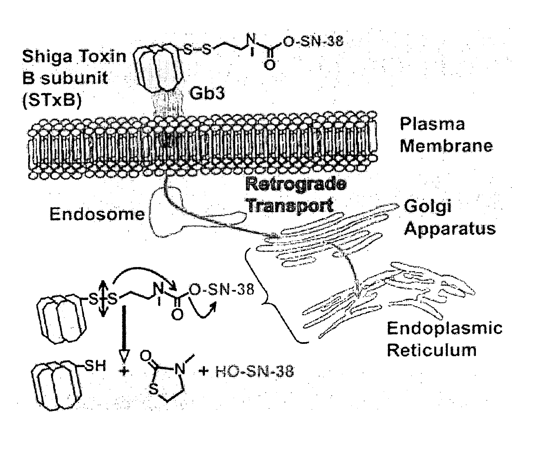
[0147]Preparation. Two prodrugs were designed and prepared based on SN-38 (compound 1), the active principle of CPT11 (Campto), which is used in the treatment of colorectal carcinoma (E. Van Cutsem et al., Eur. J. Cancer, 1999, 35: 54). SN-38 belongs to the class of camptothecin derivatives, which are cytotoxic by inhibition of topoisomerase I, and is one of the most efficient compounds in this family (B. Gatto et al., Curr. Pharm. Des., 1999, 5: 195). For coupling SN-38 to the Shiga toxin moiety, an STxB variant with a thiol functionality, termed STxB-Cys, was used that was specifically designed for site-directed chemical cross-linking in the laboratory of the present Applicants (PCT Publication No. WO 02/060937; and M. Amessou et al., Current Protocols in Cell Biology, J. Bonifacino et al. (Eds.), Wiley, Hoboken, 2006, chap. 15.10).
[0148]The phenolic position of SN-38 was chosen to build self-immolative spacers that include disulfide bonds. After cleavage of these bonds and release of a free thiol function, the free phenol is released without any other external reactant. To this end, two different spacers that exhibit variable stabilities in biological systems were envisioned. One of the spacers comprises an aromatic ring; the other an aliphatic chain.
[0149]The two prodrugs (compounds 2 and 3) and the cleavage reactions they respectively undergo are depicted in FIG. 3. Two variants were synthesized for each spacer arm: one (a) with SN-38 (1), and the other (b) with biotin derivative 4. The latter compound allowed to circumvent the fact that release of SN-38 from compounds 2a and 3a could not be monitored in vivo because of lack of sensitivity. The biotin group was derivatized with a phenol spacer to obtain a similar susceptibility to cleavage as that with the phenol function of the SN-38. The compounds were obtained according to FIG. 4.
[0150]For the synthesis of compounds 3a and 3b, commercial amino alcohol 5 was first monoprotected as a tert-butoxycarbonyl (Boc) derivative. The hydroxyl group was converted to bromide with CBr4 and then substituted by a thioacetate. The thiol function of 8 was activated as a pyridine disulfide 9. Liberation of the free amine was performed in acidic medium and the formed chlorhydrate 10 was kept as a salt because the free amine was unstable. Compound 10 was then reacted with phosgene and triethylamine to give the stable carbamoyl chloride 11. The phenol (SN-38 or biotin derivative) was first coupled in the presence of a stoichiometric amount of 4-dimethylaminopyridine (DMPA) to this bifunctional intermediate 11. Finally, STxB-Cys was reacted with carbamate 12 under basic conditions (pH 9).
[0151]The substitution levels of the coupling products were determined as 5 SN-38 or biotin molecules per STxB pentamer, using mass spectrometric analysis and fluorimetric dosage.
[0152]Stability. As a first step towards evaluation of the biological activity of compounds 2 and 3, the stability of the biotin versions was tested in different media. Compound 2b turned out to be readily activated even in the absence of cells, thus precluding its use in vivo. In contrast, compound 3b was completely stable over extended periods of up to 48 hours at 37° C. in all media, including pure fetal calf serum. Prodrug 3a was also stable in pure fetal calf serum, as shown by fluorimetric measurements.

Example

Example 2
In vitro Activity of STxB/SN-38 Conjugate 3
[0153]Compound 3 was chosen for an in-depth characterization on HT-29 colorectal carcinoma cells. ELISA analysis with 3b demonstrated that cleavage became detectable in the 6-24-h time interval, and was essentially complete at 48 hours (FIG. 5).
[0154]The same results were obtained using HeLa cells Immunofluorescence analysis was used to demonstrate that cleavage occurred intracellularly (FIG. 6). Consistent with ELISA data, no cleavage could be detected after short times of internalization (45 minutes), in which STxB (red) and biotin (green) co-localized with the Golgi marker Rab6 (blue). After 48 hours, STxB (red) could still be detected in the Golgi region (blue). However, the biotin signal was largely gone, which strongly suggests that reduction of the disulfide bond occurred with membranes of the biosynthetic/secretory pathway.
[0155]Having established that biotin model compound 3b is activated in HT-29 cells, the cytotoxic effect of corresponding prodrug 3a was determined As shown in FIG. 7, an IC50 value of 300 nM (in SN-38 equivalents) was observed on Gb3-expressing HT-29 cells. To establish specificity, two Gb3 negative-control situations were tested: HT-29 cells that were treated with glycosylceramide synthase inhibitor 1-phenyl-2-palmitoylamino-3-morpholino-1-propanol (PPMP) (A. Abe et al., J; Biochem., 1992, 111: 191), or spontaneously Gb3-negative Chinese hamster ovary (CHO) cells. In both cases, the cells were not sensitive at all to incubation with prodrug 3a, which was used in the same concentration range for all experiments with 3a (FIG. 7). Furthermore, the non-derivatized STxB-SH was shown to have no measurable cytotoxicity on Gb3-expressing HT-29 cells under the experimental conditions used (FIG. 7).
[0156]Importantly, neither nonvectorized SN-38 (IC50: 30 nM) nor its prodrug CPT-11 used in clinics (IC50: 70 μM) had a cytotoxic effect on HT-29 cells that were dependent on Gb3 expression (FIG. 7), thus further establishing the selectivity of compound 3a for Gb3-expressing tumor cells.
[0157]In summary, the present Applicants have identified a novel tumor-delivery approach based on retrograde prodrug targeting to membranes of the biosynthetic/secretory pathway, by using STxB. The disulfide linkage of prodrug 3a is slowly released, most likely in the endoplasmic reticulum whose function in cellular redox homeostasis is well-recognized (A. Gorlach et al., Antioxid. Redox Signaling, 2006, 8: 1391). Retrograde delivery will place the site of drug release close to the nucleus, where the molecular target of hydrophobic SN-38 resides.

Example

Example 3
In Vivo Activity of STxB/SN-38 Conjugate 3
[0158]Compound 3 was then investigated for its activity in vivo.
[0159]Protocol. Seventeen (17) APC1638N mice of 6 months of age were injected 3 times intravenously at day (D)=1, 8, and 15 with 100 μg of STxB-SN38. As a control, mice were injected with STxB (n=6) at the same molar dose. At D=28 after the first injection, the mice were sacrificed, and their intestine was analyzed first macroscopically on autopsy preparations for the presence of periampular tumors. The same preparations were then also treated for pathological examinations.
[0160]Statistical Analysis. The presence of periampular tumors in STxB-SN38 treated and control mice was determined by macroscopical observation and pathological analysis. Table 1 presents experimental results obtained and expected results.
TABLE 1 Numbers of periampular tumors per total number of mice that were analyzed. All mice Conditions Experimental results Expected results’ STxB-SN38 9/17 (53%) FT1, FT3* 17/32 (53%)1 FT1 Control 6/6 (93%) FT2, FT3 78/85 (92%)2 FT2 Control mice were injected with STxB. 1Expected results are deduced from in vivo tumor accumulation studies using fluorophore-labeled STxB. The underlying rational is that only tumors should be amenable to treatment by STxB-based therapy if they accumulate STxB in vivo. This is roughly only the case for 50% of them, due to heterogeneity in receptor expression (K. P. Jansen et al., Cancer Res., 2006, 66: 7230-7236). 2Expected results are deduced from historical analysis of the presence of periampular tumors in mice of the same genetic background. *Fisher Test: FT1 = 1; FT2 = 1; FT3 = 0.05.
[0161]This experiment clearly established that STxB-SN38 injected mice were protected from tumor growth, within the limits by which STxB has access to these tumors, as established by in vivo bioaccumulation experiments (K. P. Jansen et al., Cancer Res., 2006, 66: 7230-7236). In contrast, periampular tumors developed in control mice, irrespective of the injection of STxB alone or CPT11, the clinical prodrug version of SN38.
[0162]Pathology. Samples from STxB-SN38 injected mice (FIG. 8) or CPT11 injected control mice (FIG. 9) were prepared for H&E staining, and analyzed under the microscope. In STxB-SN38 injected mice, a strong inflammatory reaction was observed in the periampular region of mice in which no or only residual tumors could be detected. This strong inflammation could be interpreted as a “footprint” of the therapeutic response. In contrast, in CPT11 treated mice, high-grade adenomas and carcinomas could be detected. The inflammatory response was weak, except in the case of carcinoma.

PUM

PropertyMeasurementUnit
Molar density1.0E-9mmol / cm ** 3
Molar density1.0E-4mmol / cm ** 3
Composition
tensileMPa
Particle sizePa
strength10

Description & Claims & Application Information

We can also present the details of the Description, Claims and Application information to help users get a comprehensive understanding of the technical details of the patent, such as background art, summary of invention, brief description of drawings, description of embodiments, and other original content. On the other hand, users can also determine the specific scope of protection of the technology through the list of claims; as well as understand the changes in the life cycle of the technology with the presentation of the patent timeline. Login to view more.

Similar technology patents

Conjugation of mcr1 ligand with cytotoxic drugs for treating skin cancer

PendingUS20220143196A1Increase selectivityPharmaceutical delivery mechanismMelanocyte-stimulating hormoneDrug conjugationDrug resistance
Owner:THE ARIZONA BOARD OF REGENTS ON BEHALF OF THE UNIV OF ARIZONA

Dry-etching method

Owner:TOKYO ELECTRON LTD

Classification and recommendation of technical efficacy words

  • Increase selectivity
  • Increase specificity

Method for etching a layer on a silicon semiconductor substrate

InactiveUS20100203739A1Increase selectivitySemiconductor/solid-state device manufacturingMicrostructural device manufactureEtchingSemiconductor
Owner:ROBERT BOSCH GMBH

Catheter systems

ActiveUS20120059255A1Increase selectivityImprove accuracyElectrotherapyMedical devicesTemperature sensingChange color
Owner:ST JUDE MEDICAL ATRIAL FIBRILLATION DIV

Highly conductive ordered ion exchange membranes

InactiveUS20050238937A1Increase conductivityIncrease selectivityIon-exchanger regenerationFinal product manufactureIon-exchange membranesSolvent
Owner:BEN GURION UNIVERSITY OF THE NEGEV

Fluorescent in situ RT-PCR

InactiveUS7186507B2Increase specificityRapid detectionSugar derivativesMicrobiological testing/measurementBackground fluorescenceBiology
Owner:INDIANA UNIV RES & TECH CORP

Method for prediction of cardiac disease

InactiveUS20060155200A1Increase specificityMinimise numberElectrocardiographySensorsNatriuretic peptideLeft ventricular size
Owner:INVERNESS MEDICAL SWITZERLAND GMBH
Why Eureka
Try Eureka