

Peptides that target dorsal root ganglion neurons
a ganglion and dorsal root technology, applied in the field of neuronal biology, cell biology, molecular biology, medicine, can solve the problems of wildtype ad delivery not working, and achieve the effect of high-efficiency drg neuron targeting and impaired ability to transdu
- Summary
- Abstract
- Description
- Claims
- Application Information
AI Technical Summary
Benefits of technology
Problems solved by technology
Method used
Image

Examples
example 1
Isolation of DRG Neuron-Binding Peptides
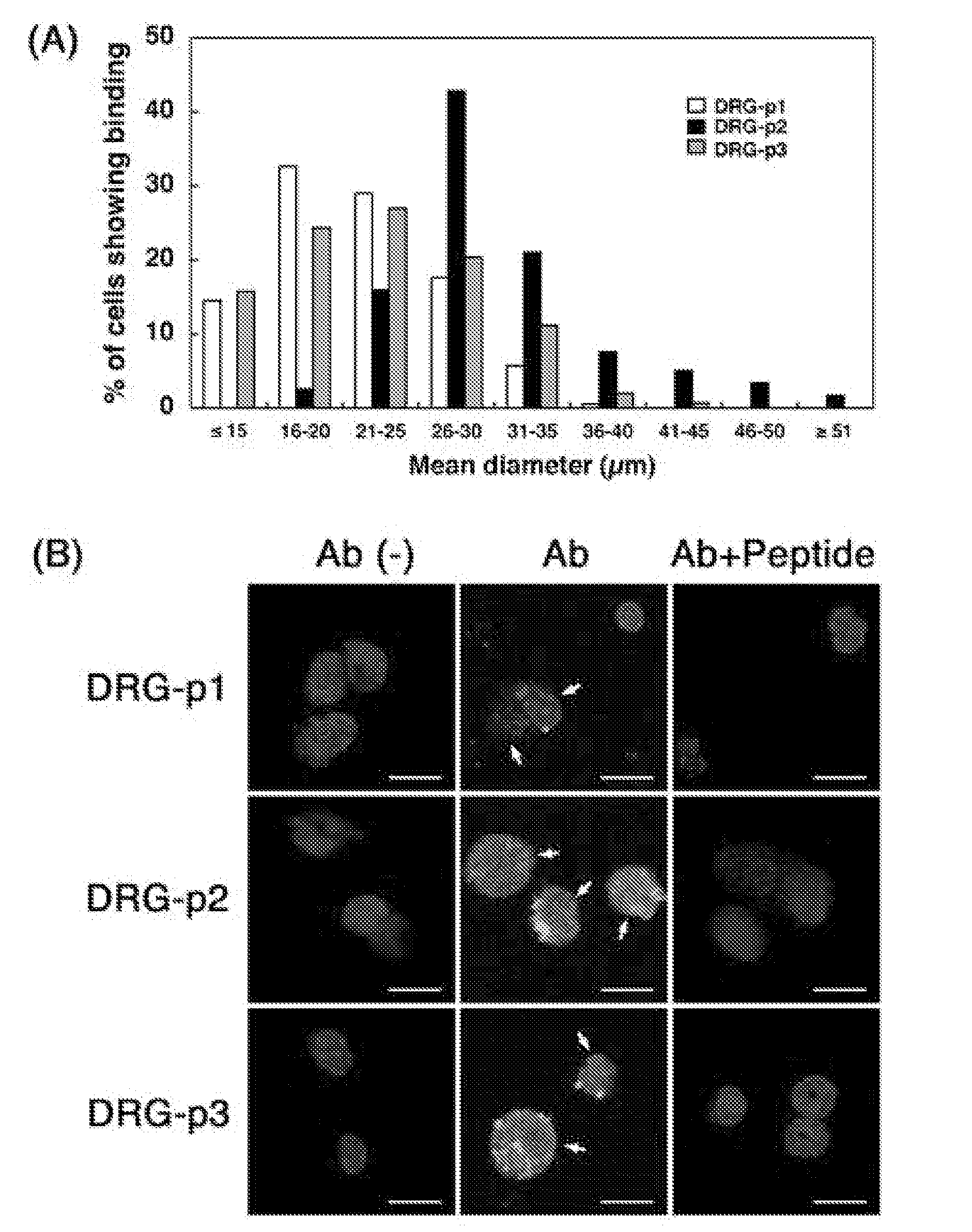
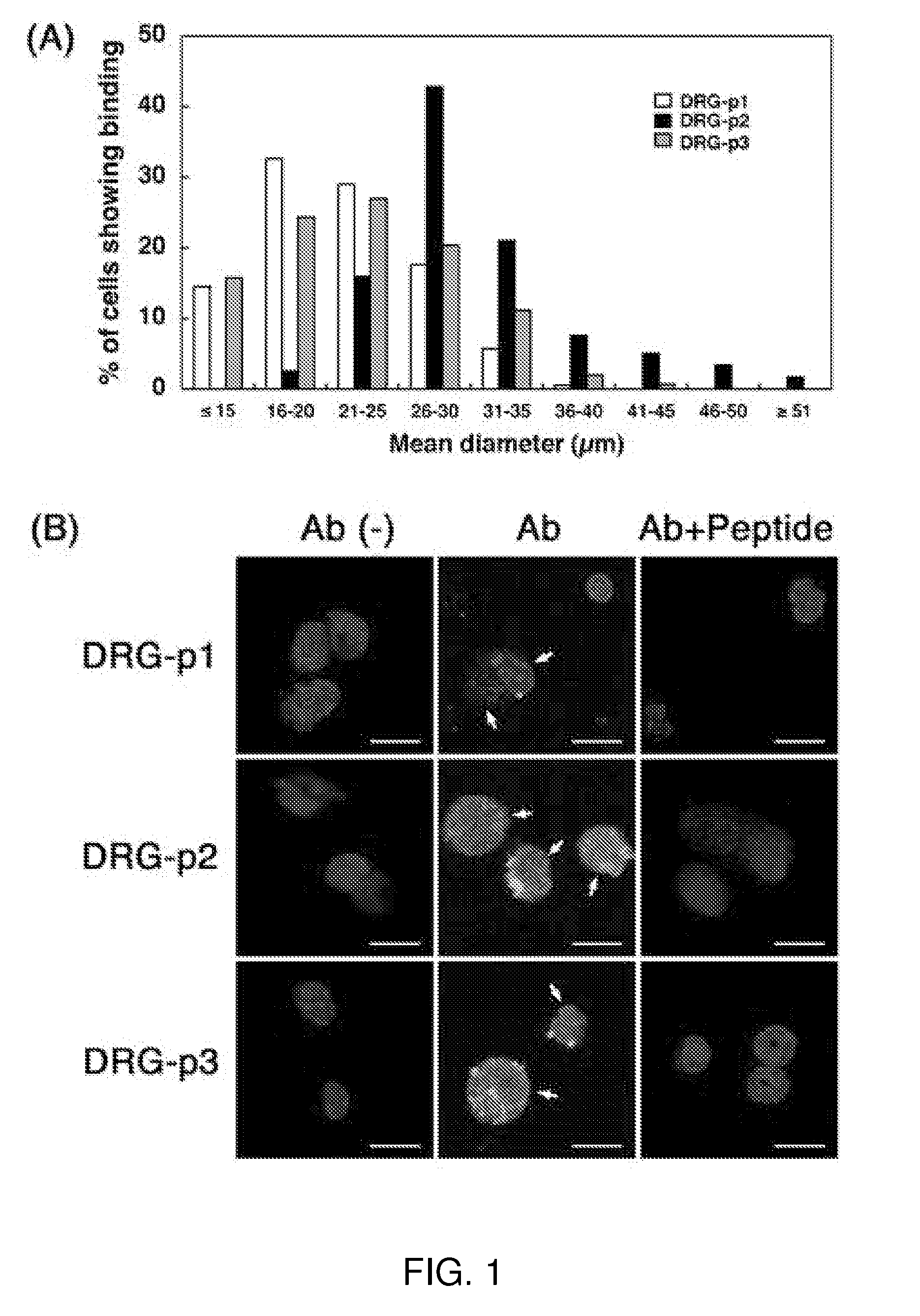
[0090]Adult C57BL / 6 mice aged 8-12 weeks were used in all experiments, and were housed in an animal room with a 12-h light and 12-h dark cycle in an illumination-controlled facility. All experiments were conducted with the approval of the Research Center for Animal Life Science at Shiga University of Medical Science. A phage library expressing random 7-mer peptides fused to a minor coat protein (pIII) of the M13 phage, at a complexity of about 1.3×10⁹ independent sequences, was purchased from New England BioLabs (The Ph.D.-C7C Phage display Peptide Library kit, Beverly, Mass.). Rabbit anti-fd bacteriophage antibody was purchased from Sigma Aldrich Corp. (St. Louis, Mo.). Donkey FITC-labeled anti-rabbit IgG was purchased from Chemicon (Temecula, Calif.). The three synthetic peptides, DRG1: SPGARAF, DRG2: DGPWRKM, DRG3: FGQKASS, were supplied from Yanaihara Institute (Shizuoka, Japan). Various cell lines; mouse neuroblastoma cells (Neuro-2a), hu...
example 2
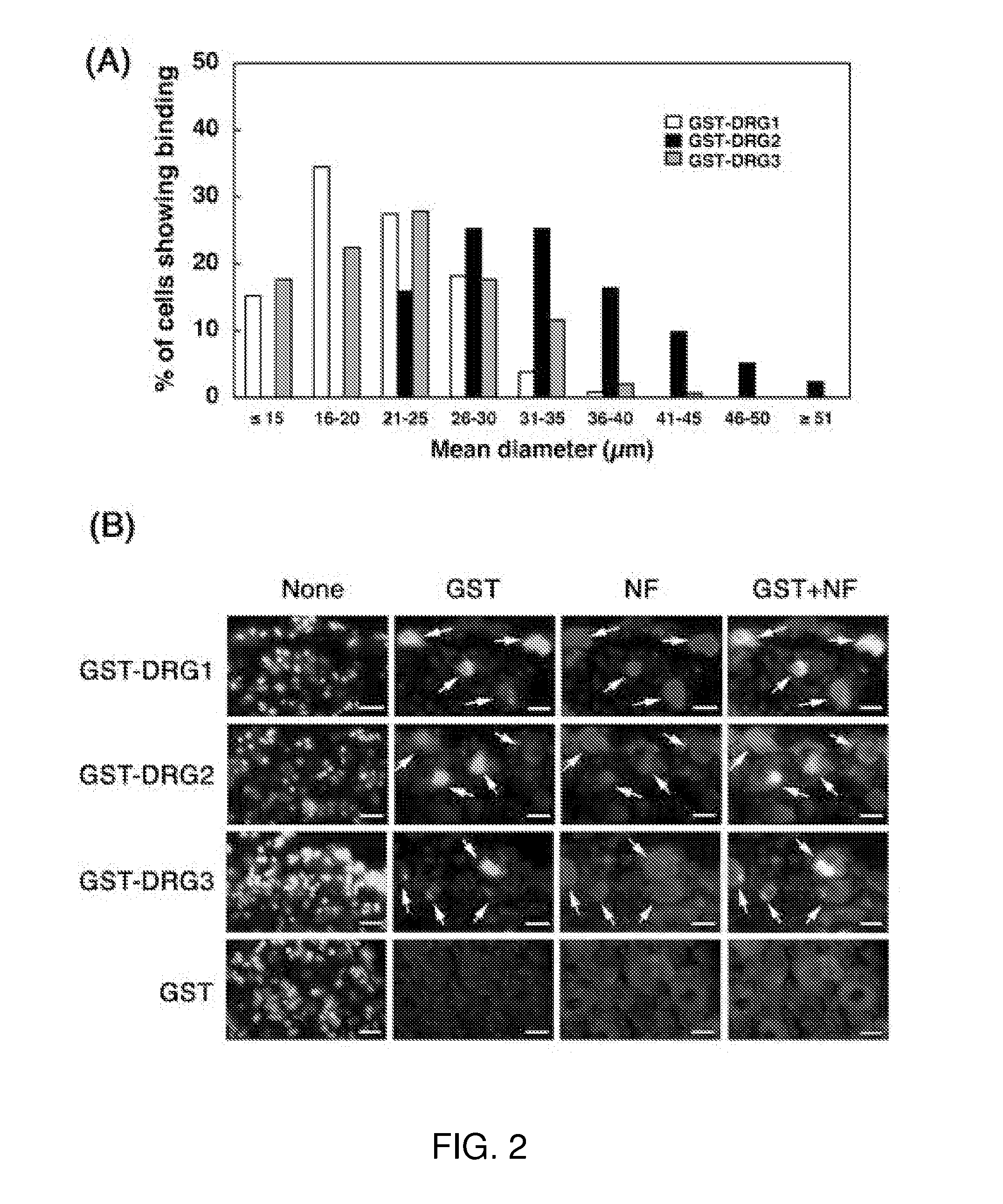
Production of Fiber-Modified Helper Virus
[0107]In a routine method for HDAd production, all necessary factors for packaging HDAd genome are supplied in trans by a helper virus (HV), a first generation Ad. In this scheme, HDAd that expresses the gene of interest (e.g., a therapeutic transgene) can be made to target specific organ or cell type simply by using an appropriate HV at the final amplification (Kim et al., 2001). There is no need to engineer a new HDAd (FIG. 3), in specific embodiments. To produce targeted HV, the inventors first attenuated the natural tropism by ablating the binding sites of Ad5 fiber to its canonical primary docking receptors, CAR and HSPG (Kritz et al., 2007). To circumvent the very poor infectivity of KO1S* mutant Ad (which lack both binding sites) toward 293 cells, 293 cells were established expressing Ad5 wild type fiber (293-fiber) to incorporate wild type fiber into the capsid of HV-KO1S* (FIG. 3a). Fiber-modified HV generated on 293-fiber had both w...
example 3
HV Containing DRG Neuron Targeting Motifs Efficiently and Specifically Transduces DRG Neurons in Vitro and in Vivo
[0108]As compared to wild-type fiber HV (HV-WF), HV-KO1S* showed 100-fold lower infectivity to 293 cells (FIG. 4b, note log scale), indicating that HV-KO1S* had lost its capacity to interact with primary docking sites on 293 cells. As the different DRG motif-containing HVs were engineered on an HV-KO1S* backbone, they also transduced 293 cells poorly (FIG. 4b, see also FIG. 6a). Transduction of isolated DRG neurons by HV-WF was about 100-fold less efficient than that of 293 cells. In contrast, HVs containing DRG targeting motifs 1-3 all displayed markedly enhanced transduction of isolated DRG neurons (FIG. 4c, note log scale). Interestingly, HV-DRG1 showed the highest transduction efficiency followed by DRG2 and DRG3. Compared to HV-WF, HV-DRG1 showed 100-fold higher efficiency of transduction of DRG neurons in vitro, while HV-KO1S* displayed minimal transduction capacit...
PUM
| Property | Measurement | Unit |
|---|---|---|
| Therapeutic | aaaaa | aaaaa |
Abstract
Description
Claims
Application Information