
Multilayer Scaffold

a multi-layer, scaffold technology, applied in the direction of prosthesis, bandages, drug compositions, etc., can solve the problems of increased morbidity and mortality of other body sites, inability to treat large lesions in this manner, and complications of elderly patients or those with complicating medical conditions (e.g. heavy smokers, diabetics) to avoid potential ethical and religious barriers to use, and avoid possible risks

Inactive Publication Date: 2011-11-24
SMITH & NEPHEW INC
View PDF2 Cites 27 Cited by
  • Summary
  • Abstract
  • Description
  • Claims
  • Application Information

AI Technical Summary

Benefits of technology

[0038]The use of synthetic materials also avoids the possible risk of disease transmission which may be associated with materials derived from animal or human sources and further avoids the potential ethical and religious barriers to the use of such materials.
[0039]It is particularly advantageous that the synthetic material used for first and second layers is biodegradable / bioresorbable. That is, the fibres transiently degrade / resorb within the physiological environment, with the hydrolysis by-products generated during resorption being excreted by normal biochemical pathways. It is particularly advantageous that the scaffold is completely resorbable as this eliminates the need for invasive and painful removal of the scaffold after wound healing is complete.
[0050]At least one of the layers of the scaffold can further comprise active agents which can promote wound healing. For example, agents which improve scar resolution and prevent scar formation, for example insulin, vitamin B, hyaluronic acid, mitomycin C, growth factors, such as TGFβ, cytokines or corticosteroids. These agents can be associated with the fibres, for example attached to the fibres or impregnated within the fibres.
[0072]Changing any of the conditions above to an intermediate situation whereby fibres retain enough solvent to allow bonding together with other fibres on the collector without substantially altering the fibrous nature of the scaffolds, to improve scaffold strength and retention of structure

Problems solved by technology

Although most small cancer lesions are sutured following excision, large lesions often cannot be treated in this manner.
This relatively expensive procedure results in a good quality repair, but causes additional morbidity to another body site.
Elderly patients or those with complicating medical conditions (e.g. heavy smokers, diabetics) can suffer complications after a graft or flap procedure.
These patients can also suffer from poor healing, resulting in repeated visits to a clinician and extended treatment times.
The graft or flap option is not always available to dermatologists, who can either attempt to close the wound by suturing, leave it to heal by secondary intention or refer it to a plastic surgeon.
Suturing may not be possible where the excised area is too large, and this upper size limit is reduced in areas of the body where the skin is tighter or scarring is more of a problem (such as the face).
Leaving the wound open to heal by secondary intention invites infection and can result in scarring.
Referral to a plastic surgeon increases the overall treatment cost and can lead to the potential problems discussed above.
There are concerns regarding the use of materials derived from natural polymers, due to the potential risk from pathogen transmission, immune reactions, poor mechanical properties and a low degree of control over the biodegradability2.
However the skin is a complex, multilayered organ, and in a number of clinical instances, full thickness wounds require repair and / or regeneration.

Method used


Image

  • Multilayer Scaffold
  • Multilayer Scaffold
  • Multilayer Scaffold

Examples


example 1

[0106]A non-woven monolayer scaffold was prepared by electrospinning a solution of poly(glycolic acid) (PGA) in 1,1,1,3,3,3-hexafluoropropan-2-ol (hexafluoroisopropanol, HFIP).

Solution Preparation.

[0107]PGA supplied by PURAC Biomaterials (with an approximate weight-average molecular weight of 130,000) was melt-extruded at 260-274° C. using a Rondol Linear 18 single screw extruder and then immediately quenched in water at 5-10° C. This extruded PGA was used to prepare a 7 w / w % solution in spectrophotometric grade HFIP supplied by Apollo Scientific Ltd (corresponding to a solution viscosity of approximately 0.35 Pa·s). This solution was left rolling overnight at 21° C. until dissolved. Prior to electrospinning, the solution of PGA in HFIP was filtered through a 10.0 μm Whatman Polydisc HD filter (polypropylene filter, 50 mm diameter) directly into a 20 mL syringe (polypropylene, lubricant-free, 20.0 mm internal diameter). The resulting polymer solution was free from visible particula...

example 2

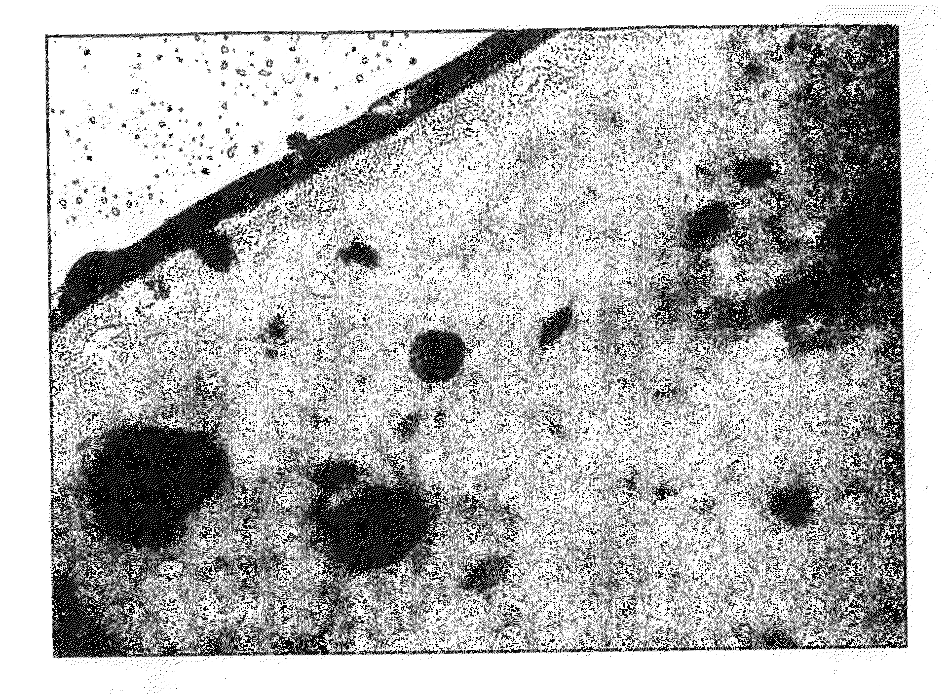
[0118]An 8 w / w % solution of PGA in HFIP was prepared and used to prepare a non-woven monolayer scaffold material using the same general method described in Example 1. This concentration of PGA in HFIP corresponds to a solution viscosity of approximately 0.55 Pa·s. FIG. 4 shows an SEM image of the scaffold acquired at a magnification of 10,000.

Results

[0119]Thickness=120-140 μm across the central 65% of the scaffold length.

Mean fibre diameter=0.51 μm±0.12 μm.

Largest Detected Pore Diameter=2.29 μm

[0120]Mean-Flow Pore Diameter (median pore diameter)=1.15 μm

Diameter at Maximum Pore Size Distribution=0.94 μm.

example 3

[0121]A 9 w / w % solution of PGA in HFIP was prepared and used to prepare a non-woven monolayer scaffold material using the same general method described in Example 1, although no aqueous sodium chloride was added to the solution of PGA in HFIP. This concentration of PGA in HFIP corresponds to a solution viscosity of approximately 0.85 Pa·s. In addition, the electrospinning duration was increased to 68 minutes. FIG. 5 shows an SEM image of the scaffold acquired at a magnification of 6,000.

Results

[0122]Thickness=100-110 μm across the central 70% of the scaffold length.

Mean fibre diameter=0.81 μm±0.38 μm.

Largest Detected Pore Diameter=3.44 μm

[0123]Mean-Flow Pore Diameter (median pore diameter)=1.87 μm

Diameter at Maximum Pore Size Distribution=1.58 μm.


PUM

PropertyMeasurementUnit
diameteraaaaaaaaaa
diameteraaaaaaaaaa
diameteraaaaaaaaaa

Abstract

The invention generally relates to biodegradable and / or bioresorbable fibrous articles and more particularly to products and methods having utility in medical applications.

Description

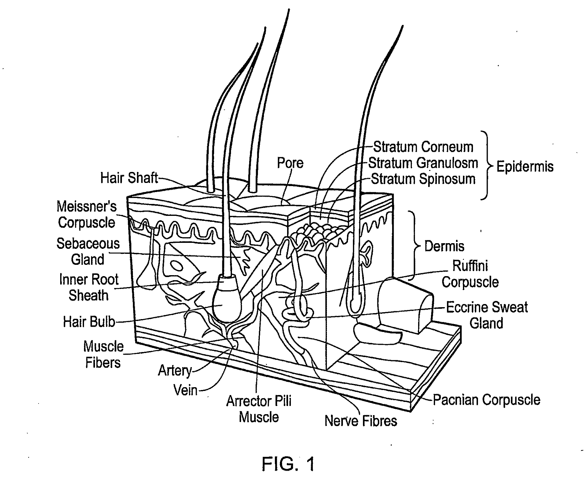
RELATED APPLICATIONS[0001]The application claims priority to UK application No. 0801405.2 entitled “Multilayer Scaffold”, filed on Jan. 25, 2007 and UK patent application No. 0802767.4 entitled “Multilayer Scaffold”, filed on Feb. 15, 2007, the entire contents of which are hereby incorporated by reference.FIELD OF THE INVENTION[0002]The invention generally relates to biodegradable and / or bioresorbable fibrous articles and more particularly to products and methods having utility in medical applications.BACKGROUND TO THE INVENTION[0003]Skin is the largest organ in the body, covering the entire external surface and forming about 8% of the total body mass1. Skin is composed of three primary layers as illustrated in FIG. 1: the epidermis, the dermis, and the hypodermis (subcutaneous adipose layer).[0004]The epidermis contains no blood vessels, and cells in the deepest layers are nourished by diffusion from blood capillaries extending to the upper layers of the dermis. The main type of ce...

Claims


Application Information

Patent Timeline
Patent Type & Authority Applications(United States)
IPC IPC(8): A61K9/70A61P17/02
CPCA61L27/3813A61L27/60A61L27/58A61L27/56A61P17/02A61F2/105A61L27/18A61L2430/34D01F6/625D10B2401/10D10B2401/12D10B2509/00
Inventor SMITH, JENNIFER MARGARETRAXWORTHY, MICHAEL JOHNIDDON, PETER DAMIEN
Owner SMITH & NEPHEW INC
Learn More