Tethered Conformation Capture

a technology of conformation capture and tethered conformation, which is applied in the field of tethered conformation capture, can solve the problems of low resolution and throughput of most methods used to address 3d organization of the genome, and the inability to analyze 3d organization of chromatin remains elusive, and achieves the effect of less noise and higher resolution

Inactive Publication Date: 2011-11-24
UNIV OF SOUTHERN CALIFORNIA
View PDF6 Cites 42 Cited by
  • Summary
  • Abstract
  • Description
  • Claims
  • Application Information

AI Technical Summary

Benefits of technology

[0010]It is one object of the present invention to provide methods and systems for the determination of 3-dimensional chromatin structure at higher resolutions and with less noise.

Problems solved by technology

While involvement of the spatial organization of loci or conformation of the chromatin fiber has been shown in some examples (Osborne et al., 2004; Lee et al., 2005; Spilianakis and Flavell, 2004; Cai et al., 2007), understanding 3D organization of chromatin at a genome level remains elusive.
The ability to analyze 3D organization of chromatin has been hampered by technical limitations.
Most of the methods that are used to address 3D organization of the genome have low resolution and throughput.
This first generation of capture methods is limited in scope in that it can only evaluate a few chosen interactions at a time.
Therefore, none of these methods provides a genomic or regional context for the interaction frequencies to reveal the more significant aspects of the 3D organization of the locus or changes therein.
The inventors have found, however, that ligation at low concentrations is disadvantageous because it extremely difficult to use the technique in conformation capture with both high resolution and low noise.
The inventors have discovered that low concentrations are not effective at eliminating the ligation between different crosslinked DNA complexes.
These intermolecular ligations, which manifest as false interactions in techniques such as Hi-C, appear to be a major source of noise in conformation capture assays.
Moreover, the theoretical resolution limit of a genome-wide conformation capture assay such as Hi-C is determined by the size of DNA fragments generated during the conformation capture method.
Smaller DNA fragments result in higher resolution conformation capture, but also result in high concentration of DNA fragments which in turn result in increased intermolecular ligation reactions and increased noise.
It is thus very difficult using known methods to achieve both high resolution and low noise in known conformation capture techniques.

Method used


Image

  • Tethered Conformation Capture
  • Tethered Conformation Capture
  • Tethered Conformation Capture

Examples


example 1

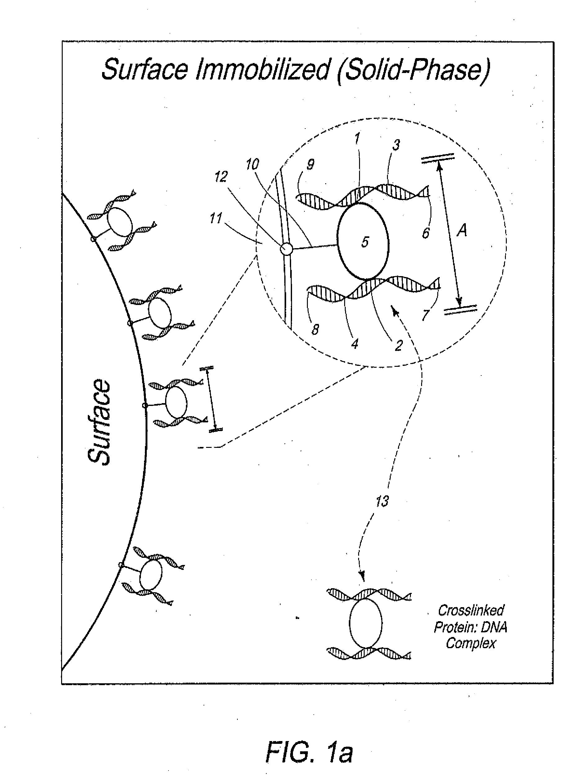
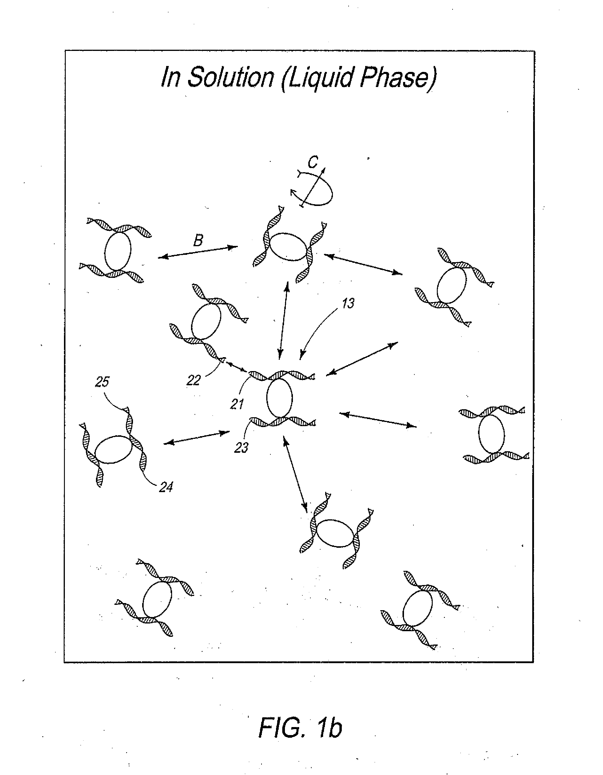
[0060]Overview. In one example of the present invention, cells were crosslinked with formaldehyde and treated with Iodoacetyl-PEG2-Biotin to biotinylate the cysteine residues of all proteins. Chromatin was then digested with a restriction enzyme, leaving 5′ overhangs. After digestion, crosslinked protein-DNA complexes were immobilized on the surface of streptavidin-coated magnetic beads through biotinylated proteins and excess streptavidin was blocked. The 5′ overhangs were filled in with an α-thio-triphosphate containing nucleotide analogue inserted before a biotinylated nucleotide. Blunt DNA ends were then ligated while immobilization prevented free diffusion of the complexes, therefore promoting intramolecular ligations. After ligation, DNA is purified, which separates it from the surface and crosslinked proteins. The biotinylated nucleotides on DNA ends that did not participated in ligation were then removed using E. coli Exonuclease III (ExoIII). ExoIII was used to catalyze rem...

example 2

[0088]We created a TCC library from 25 million GM12878 human lymphoblast cells (Coriell Institute, Camden, N.J.) using HindIII as the restriction enzyme. For comparison, a Hi-C library of 25 million GM12878 cells digested with HindIII was also created as described by Lieberman et al. The two libraries were sequenced on Illumina Genome Analyzer II platform (Illumina, San Diego, Calif.) in a paired-end format. For every dataset, the two reads of each cluster were filtered for ligation junctions and then aligned to build GRCh37 / hg19 of the human genome using Bowtie (Langmead et al., 2009).

[0089]Two types of read pairs do not contain information about the 3D organization of the genome and are a result of error in the process. The first group is the DNA molecules that do not include a ligation junction yet bind to streptavidin coated beads and appear in the final library. In sequencing, these molecules which we refer to as “dirt” result in read pairs that align only 300-700 bp apart to o...

example 3 (

TCC has Reduced Level of Noise)

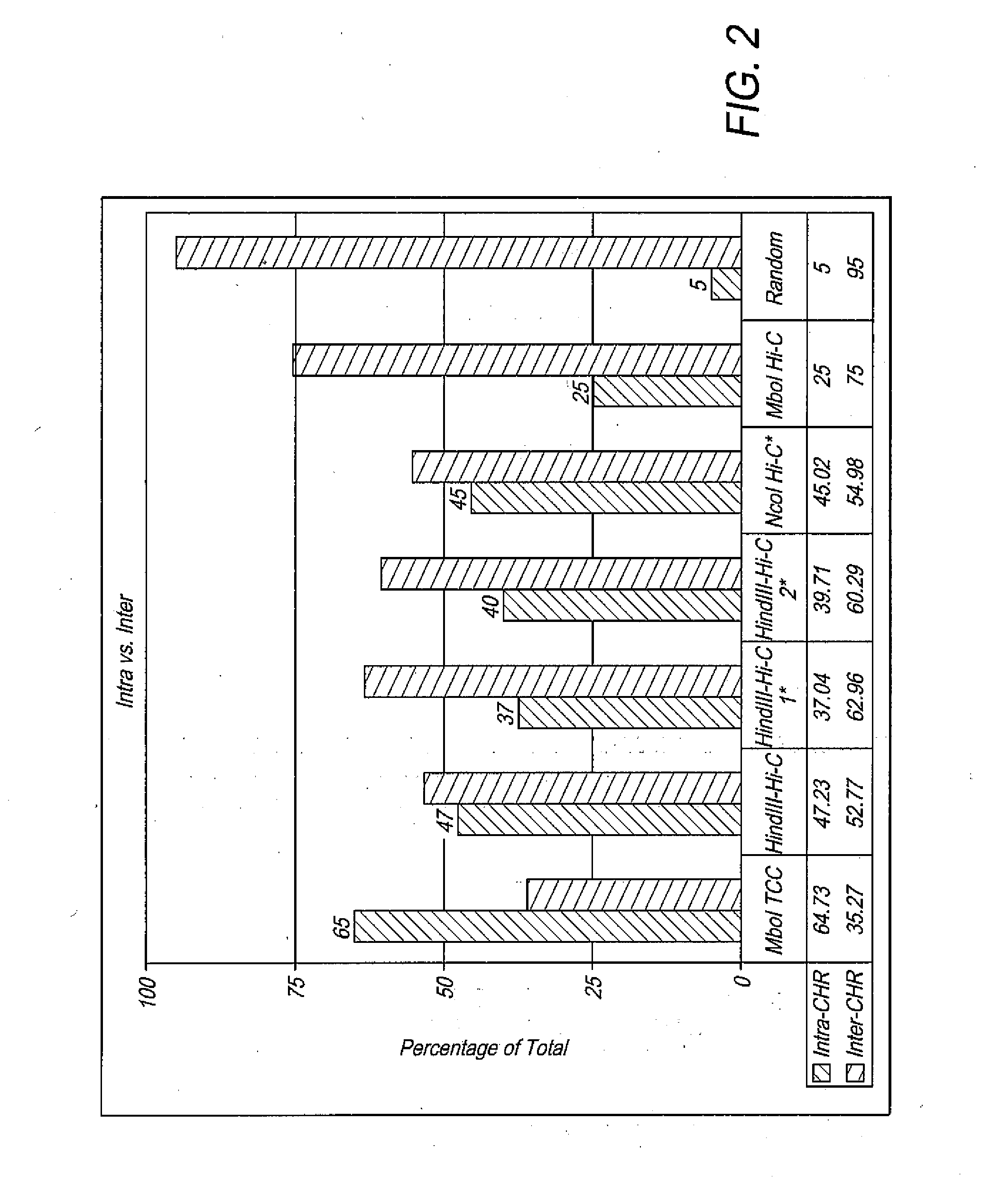
[0092]Differing intermolecular ligation levels should manifest itself in the portion of interchromosomal interactions in the data. In a random ligation of HindIII-digested human DNA, 95% of all ligation events are expected to take place between fragments that belong to different chromosomes. In other words, in a Hi-C or a TCC experiment, 95% of the noise caused by intermolecular ligations comes in the form of interchromosomal connections. These random ligations are expected to be uniformly distributed throughout the interaction matrix.

[0093]Due to existence of chromosome territories (Cremer and Cremer, 2001) and polymer-like properties of DNA (Lieberman-Aiden et al., 2009), a higher portion of contacts than expected by random that in fact are present in the cells are expected to be intrachromosomal. This is why intermolecular ligation noise appears mostly as interchromosomal interactions in the data. The portion of intrachromosomal and interchromosomal...


PUM

PropertyMeasurementUnit
temperatureaaaaaaaaaa
surface areaaaaaaaaaaa
volumeaaaaaaaaaa

Abstract

Disclosed are methods and systems for determining the three-dimensional structure of chromatin in eukaryotic cells. More specifically, disclosed are methods and systems for obtaining chromatin structural information by surface immobilization, i.e tethering crosslinked protein:DNA complexes and/or ligated DNA complexes to media such as beads, gels, and or matrices during the conformation capture assay. In general, the method includes contacting a cell with a cross-linking reagent to cross-link DNA and protein in the cell; lysing the cell, producing cross-linked protein:DNA complexes by cutting the chromatin using a chemical, physical or enzymatic method, substantially immobilizing the cross-linked protein:DNA complexes, ligating the cross-linked protein:DNA complexes intramolecularly such that the ligated protein:DNA complexes represent structural organization of the chromatin; characterizing the ligated DNA by sequencing or other methods; and identifying any structural organization of the chromatin. The structural organization preferably includes information relating to interacting loci of the chromatin.

Description

[0001]This invention was made with government support under Contract Nos. R01 GM064642, R01 HL076334 and R01 GM077320 awarded by the National Institute of Health. The government has certain rights in the invention.FIELD OF THE INVENTION[0002]The present invention, Tethered Conformation Capture (TCC) relates in general to methods and systems for determining the three-dimensional structure of chromatin in Eukaryotic Cells. More specifically, the invention provides improved methods and systems for obtaining chromatin structural information by surface immobilization, i.e tethering crosslinked protein:DNA complexes and / or ligated DNA complexes to media such as beads, gels, matrices during the conformation capture assay.BACKGROUND OF THE INVENTION[0003]The three-dimensional (3D) organization of eukaryotic genomes plays an important role in various nuclear processes such as transcription, replication, and DNA repair (Wolffe, 1998). Beyond packaging by nucleosomes, the folding and spatial a...

Claims


Application Information

Patent Timeline
Patent Type & Authority Applications(United States)
IPC IPC(8): C40B30/00C12Q1/68
CPCG01N33/5308G01N33/68C12Q1/68C12Q2523/101
Inventor CHEN, LINKALHOR, REZA
Owner UNIV OF SOUTHERN CALIFORNIA
Why Eureka
Try Eureka