
Method and kit for assessing viable cells

a cell and kit technology, applied in the field of cell analysis methods and procedures, can solve the problems of extended incubation time, time-consuming, complex methods presently employed,

Active Publication Date: 2011-12-22
CHEMOMETEC AS
View PDF2 Cites 18 Cited by
  • Summary
  • Abstract
  • Description
  • Claims
  • Application Information

AI Technical Summary

Benefits of technology

[0059]It has been found that the labelling agent according to the invention is particularly effective in staining living cells only thereby offering the opportunity of distinguishing living cells from dead cells in a sample. A great advantage of the present invention is the fast result obtained.
[0060]The term cell mobility generally refers movements of the cell, such as cell motility as well as cell differentiation and cell proliferation, wherein motility generally refers to the ability of some cells to move spontaneously and actively, such as sperm cells, propelled by the regular beat of their flagellum, or the bacterium E. coli, which swims by roting a helical prokaryotic flagellum.
[0061]As shown in the Examples the labelling agent is present in the nucleus as well as the cytosol of the cell.SAMPLES
[0062]The sample may be any sample, such as a biological sample, comprising cells for which viability should be determined. The method according to the invention applies to analysis of any type of cell or biological material or tissue, including the raw materials and processes associated with the manufacture, storage and transportation of said products, for the presence of viruses, bacteria, fungi, protozoa or components of these organisms.
[0063]In particular a biological sample may be selected from a body fluid sample, a tissue sample, a fermentation sample, a liquid cultivation sample, a cell culture sample, a water sample, such as mammalian and yeast cell cultures, a beverage sample, a pharmaceutical sample, a microelectronic product, and cells suspended in a liquid. More particular the biological sample is selected from a blood sample, a urine sample, a saliva sample, a semen sample, a solubilised tissue sample, a milk sample, or selected from a liver sample, a kidney sample, a muscle sample, a brain sample, a lung sample.
[0064]The biological sample may be selected from any species, such as a human sample, a mouse sample, a rat sample, a monkey sample, a dog sample

Problems solved by technology

However, methods presently employed are quite complex and time consuming.
This technique has however drawbacks including extended incubation times (several minutes).

Method used


Image

  • Method and kit for assessing viable cells
  • Method and kit for assessing viable cells
  • Method and kit for assessing viable cells

Examples


example 1

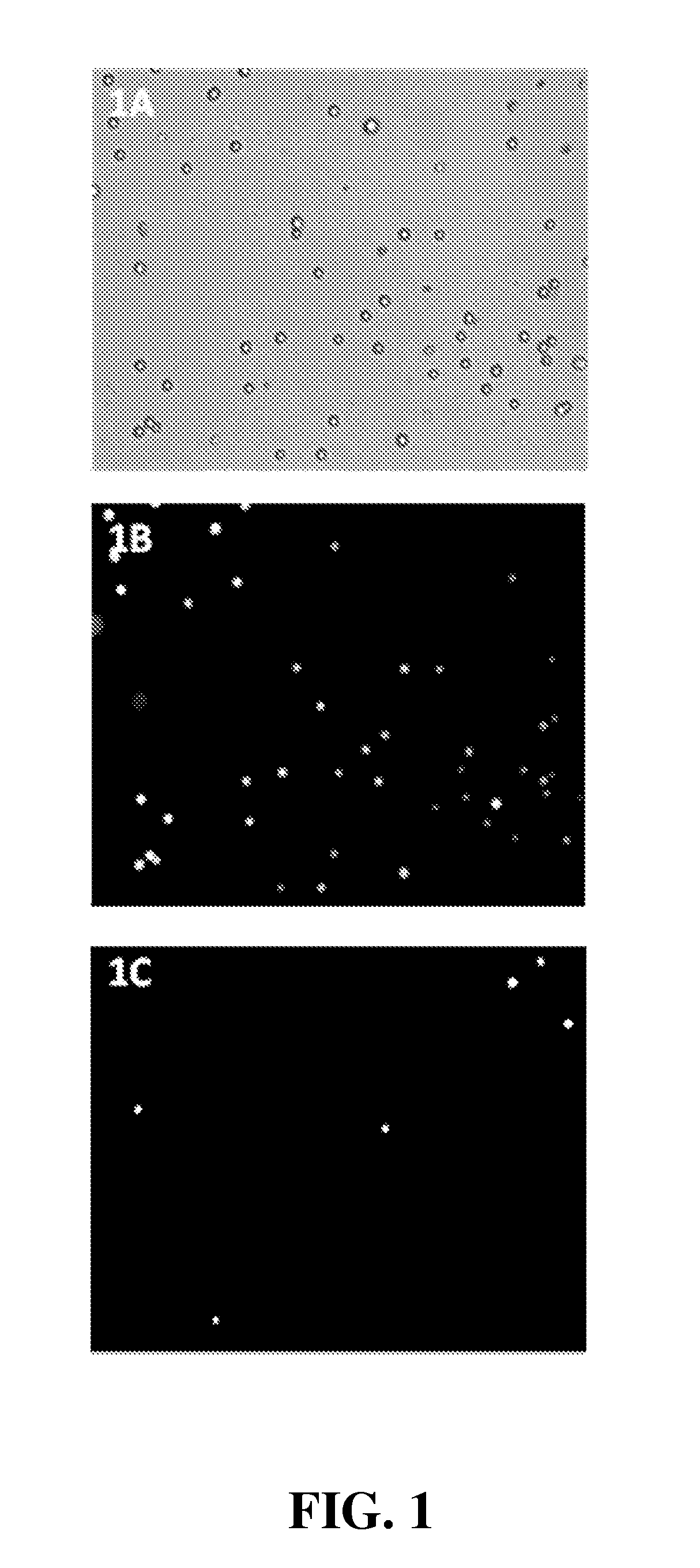
[0150]Use of NAM to determine viability of proliferating Jurkat (JM) cells (a T lymphocyte cell line)

[0151]Materials and Methods. Jurkat (JM) cells were grown at 37° C. in a humidified air atmosphere with 5% CO2 in RPMI (Invitrogen, #61870) supplemented with 10% heat-inactivated fetal bovine serum (Invitrogen, #10108-165). 90 μL proliferating Jurkat cells (cell density 1.2×10⁶, 99% viable determined using the NC-100 NucleoCounter system following the manufacturer's (ChemoMetec A / S) protocol) were added 10 μL NAM (N-(9-acridinyl)maleimide, Sigma, #01665, CAS no. 49759-20-8) dissolved in DMSO (100 μg NAM pr. mL DMSO) and mixed by pipetting. Cells were loaded into a NucleoCassette, containing the DNA stain propidium iodide (PI). The cells were investigated using an Olympus IX50 fluorescence microscope, and images were captured using a Lumenera CCD camera and in-house developed software. PI and NAM fluorescence were detected using, respectively, U-MWG2 (green long pass: 510-550 nm) and ...

example 2

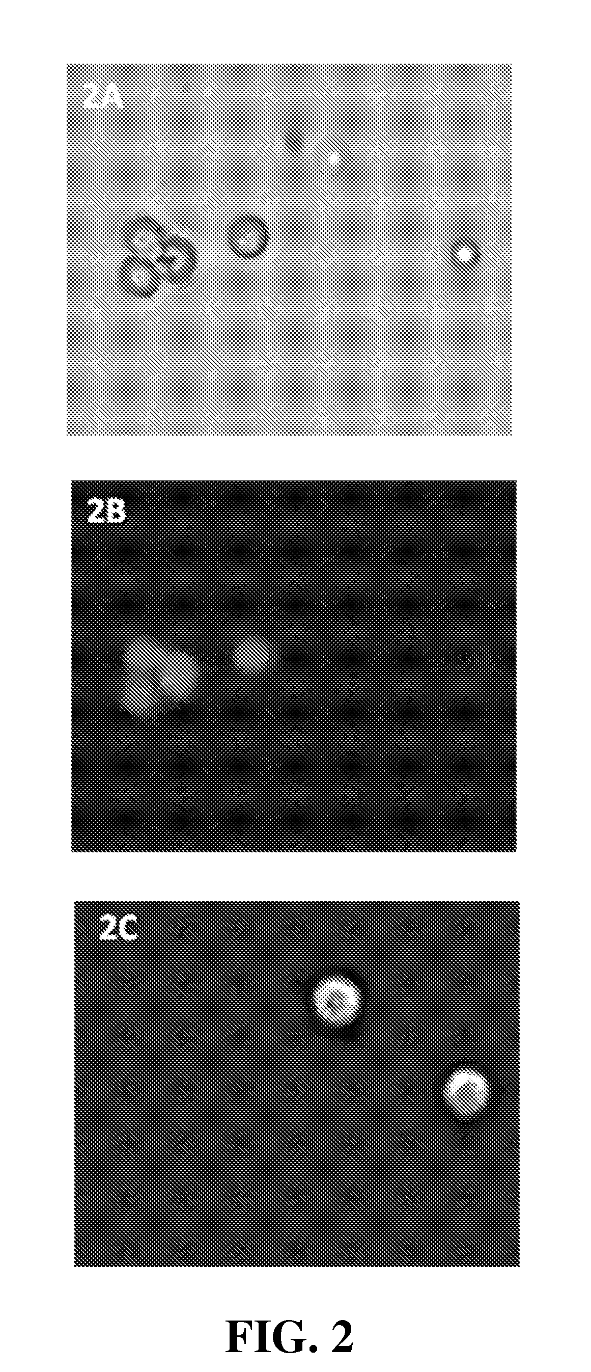
[0153]Use of NAM to determine viability of HEK293 cells (a Human Embryonic Kidney cell line)

[0154]Materials and Methods. HEK293 cells were grown at 37° C. in a humidified air atmosphere with 5% CO2 in DMEM (Invitrogen, #31966) supplemented with 10% heat-inactivated fetal bovine serum (Invitrogen, #10108-165). Cells were harvested with 0.5 mL of trypsin (Invitrogen, #25300) and neutralized with 5 mL medium (DMEM+10% FCS) two days after they had reached full confluency. These outgrown HEK293 cells (cell density 1.6×10⁶, 78% viable determined using the NC-100 NucleoCounter system following the manufacturer's (ChemoMetec) protocol) were stained with 10 μg / mL NAM (Sigma, #01665). Another cell sample were treated with 0.25% Triton X-100 (Sigma, #T9284) and hereafter added 10 μg / mL NAM. After staining, each cell sample was loaded into a NucleoCassette, containing the DNA stain propidium iodide (PI). The cells were investigated using an Olympus IX50 fluorescence microscope, and images were ...

example 3

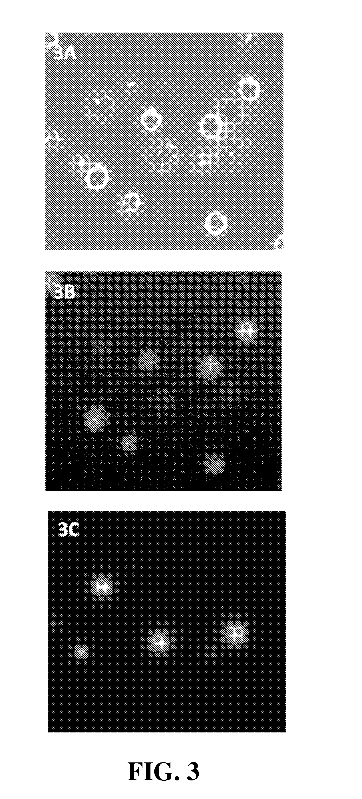
[0156]Use of NAM to determine viability of Drosophila S2 cells

[0157](Drosophila melanogaster Schneider line-2 (S2) cells were originally derived from late embryonic stage Drosophila embryos.)

[0158]Materials and Methods. Drosophila S2 cells were grown at 28° C. without shaking in Schneider's Drosophila medium (Invitrogen, #21720) supplemented with 10% heat-inactivated fetal bovine serum (Invitrogen, #10108-165). S2 cells (cell density 1.7×10⁷, 99% viable determined using the YC-100 NucleoCounter system with diploid settings following the manufacturer's (ChemoMetec) protocol) were diluted 10 times in PBS and stained with 10 μg / mL NAM (Sigma, #01665). Cells were loaded into a NucleoCassette, containing the DNA stain propidium iodide (PI). The cells were investigated using an Olympus IX50 fluorescence microscope, and images were captured using a Lumenera CCD camera and in-house developed software. PI and NAM fluorescence were detected using, respectively, U-MWG2 (green long pass: 510-55...


PUM


Abstract

The present invention provides simple, rapid methods and procedures for analyzing cells, hereunder quantitative and qualitative assessment of cells, such as viability. The present invention relates to the use of various optionally substituted reporter compounds particularly detectable upon their reaction with thiol-containing species present in higher concentrations in intact (e.g., living) cells than in non-intact (e.g., dead, stressed and apoptotic) cells. The present invention also relates to the use of various optionally substituted reporter compounds particularly detectable upon their reaction with species present in intact and / or non-intact cells. Moreover, the present invention relates to the use of measuring techniques and / or instruments coupled with the use of various optionally substituted reporter compounds. The invention further relates to compositions used in methods for analyzing cells, such as a composition comprising N-(7-dimethylamino-4-methyl-3-coumarinyl)-maleimide (DACM).

Description

[0001]All patent and non-patent references cited in the application, or in the present application, are also hereby incorporated by reference in their entirety.FIELD OF INVENTION[0002]The present invention provides simple, rapid methods and procedures for analyzing cells, hereunder quantitative and qualitative assessment of cells. The present invention relates to the use of various optionally substituted reporter compounds particularly detectable upon their reaction with species (e.g., sulphur-containing species, hereunder thiol-containing species) present in higher concentrations in intact (e.g., living) cells than in non-intact (e.g., necrotic, stressed and apoptotic) cells. The present invention also relates to the use of various optionally substituted reporter compounds particularly detectable upon their reaction with species present in intact and / or non-intact cells. Moreover, the present invention relates to the use of measuring techniques and / or instruments coupled with the u...

Claims


Application Information

Patent Timeline
IPC IPC(8): C12Q1/02
CPCC09B15/00C09B57/00G01N33/583G01N33/502C09B57/02G01N21/6428G01N33/582G01N2021/6439G01N2201/06113G01N2201/062G01N2510/00
Inventor SKINDERSO, METTE ELENASORENSEN, HELLE FROBOSEKJ.AE BUTTED.RULFF, SOREN
Owner CHEMOMETEC AS
Learn More