External magnetic force for targeted cell delivery with enhanced cell retention

Inactive Publication Date: 2012-10-04
CEDARS SINAI MEDICAL CENT
View PDF0 Cites 30 Cited by
  • Summary
  • Abstract
  • Description
  • Claims
  • Application Information

AI Technical Summary

Benefits of technology

[0015]In some embodiments, the methods provided herein lead to a functional improvement in the damaged heart which is manifest as increased cardiac output. In some embodiments, the increase in cardiac output comprises an increase in left ventricular ejection fraction. In some embodiments, delivery of the magnetized cells yields an increase of at least 5% in the left ventricular ejection fraction. In one embodiment left ventricular ejection fraction is increased by about 10%. In some embodiments, the amount of viable cardiac tissue is increased in addition to the increased cardiac output. In some embodiments, anatomical improvements in the damaged heart tissue, such as an increase in cardiac wall thickness, are detected. In some embodiments, wherein the damaged cardiac tissue is a result of a myocardial infarction the methods provided herein result in decreases in scar tissue formation. In some embodiments, the functional and/or anatomical improvements in the damaged cardiac tissue are due to proliferation of the delivered cells within or adjacent to the region of damaged cardiac tissue. In some embodiments, the functional and/or anatomical improvements in the damaged cardiac tissue is due to paracrine modulators released from the delivered cells, wherein the paracrine modulators improve the viability of cardiac tissue and/or recruit endogenous cardiac cells to the region of damaged cardiac tissue.
[0016]In several embodiments, there is provided a method for the repair or regeneration of damaged cardiac tissue, comprising delivering magnetically-labeled stem cells to a subject having a heart comprising a region of damaged cardiac tissue with compromised cardiac function, wherein the stem cells are cardiac stem cells, applying a magnetic field around or adjacent to the damaged cardiac tissue leading to enhanced delivery, retention, or engraftment of the magnetically-labeled stem cells which results in functional improvement in the region of damaged cardiac tissue. In some embodiments, the magnetically-labeled stem cells are delivered within or adjacent to the region of damaged c

Problems solved by technology

Heart disease is a leading cause of fatalities in modern societies.
However, a major challenge of delivering and localizing cells to a target tissue or organ such as the heart remains.
Additionally, cell retention rate after delivery is low, regardless of the delivery method.
It is also difficult to direct cells to a particular organ or tissue where their healing action is intended.
In some embodiments, the cardiac tissue is damaged as a result of injury or disease, and has reduced cardiac function.
These stresses may negatively impact the viability and/or functionality of the cells such that re

Method used


Image

  • External magnetic force for targeted cell delivery with enhanced cell retention
  • External magnetic force for targeted cell delivery with enhanced cell retention
  • External magnetic force for targeted cell delivery with enhanced cell retention

Examples


example 1

Isolation of Cardiac-Derived Stem Cells from Cardiac Biopsy Specimens

[0220]Pluripotent stem cells can be isolated from cardiac biopsy specimens or other cardiac tissue using any known methods, for example, the multi-step process described in U.S. Publication No. 2008 / 026792, which is incorporated herein by reference in its entirety.

[0221]Utilizing such method, cardiac tissue was first obtained via percutaneous endomyocardial biopsy or via sterile dissection of the heart. It shall be appreciated, however, that in other embodiments, the original tissue may be obtained by other means (e.g., surgical specimens, fresh cadaveric tissue, etc.). In some embodiments, fresh tissue harvesting is unnecessary, as stem cells that have previously been isolated and banked (e.g., stored frozen) are used. Once obtained, tissue specimens were stored on ice in a high-potassium cardioplegic solution (containing 5% dextrose, 68.6 mmol / L mannitol, 12.5 meq potassium chloride, and 12.5 meq sodium bicarbona...

example 2

Isolation of Porcine Cardiac-Derived Cells

[0225]Porcine CDCs were isolated and cultured according to Davis et al., (2009) PLoS One 4(9):e7195. Briefly, porcine endomyocardial specimens were sampled from the right ventricular septum using the bioptome. Biopsy specimens were stored on ice in high-potassium cardioplegic solution (5% glucose, 68.6 mM mannitol, 12.5 meq potassium chloride, 12.5 meq sodium bicarbonate, 10 units / mL heparin) to maintain tissue viability during transport. Tissue was processed within 2 hours. The myocardial specimens were then cut into fragments less than 1 mm3, washed and partially digested with collagenase. These tissue fragments were cultured as cardiac explants on fibronectin (20 μg / mL; Sigma) coated dishes in cardiac explant media (CEM; Iscove's Modified Dulbecco's Medium (GIBCO), fetal bovine serum (10% (mini swine only); HyClone, Logan, Utah), 100 U / ml penicillin G (GIBCO), 100 U / mL streptomycin (GIBCO), 2 mmol / L L-glutamine (Invitrogen, Carlsbad, Cali...

example 3

Labeling of CDCs With SPIO

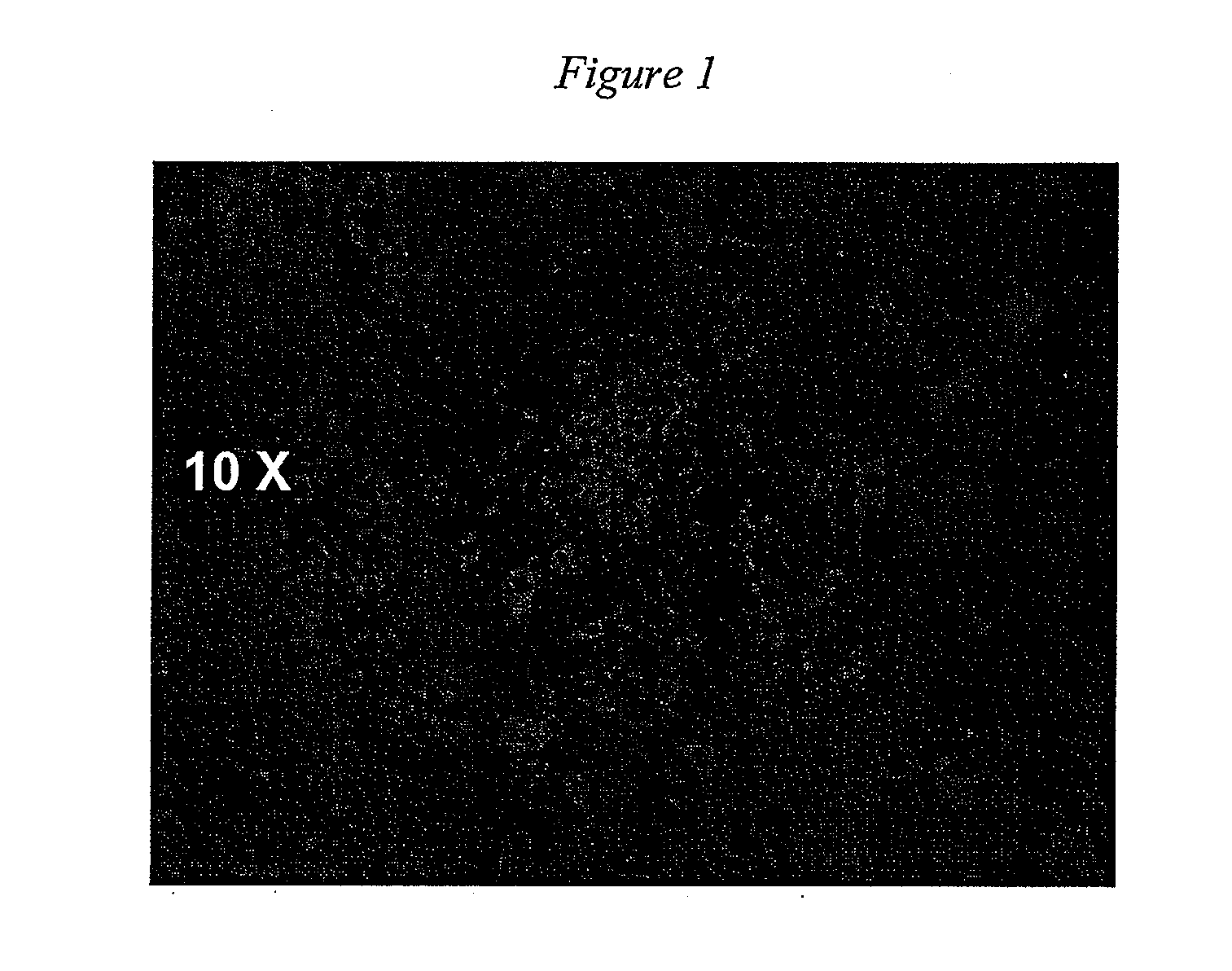
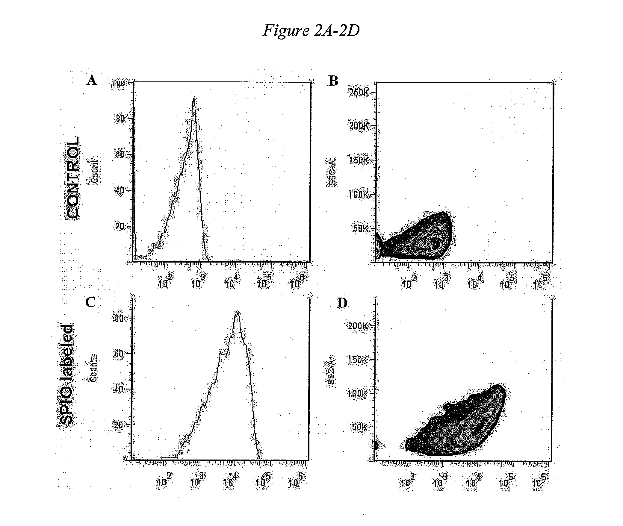
[0226]SPIO microsphere particles (0.9-μm diameter, Bangs Laboratories, IN) were incubated with CDCs (1×10⁶ cells in 15 ml medium) in a T75 flask with a ratio of microspheres to cells at 500:1. As discussed herein, additional ratios of microspheres (or other labeled particle to cells may be used in some embodiments (e.g., 100:1; 250:1; 1000:1; 2000:1, 4000:1, etc.). The cells were incubated overnight at 37° C. and 95% air / 5% CO2 to incorporate SPIO into the cells.

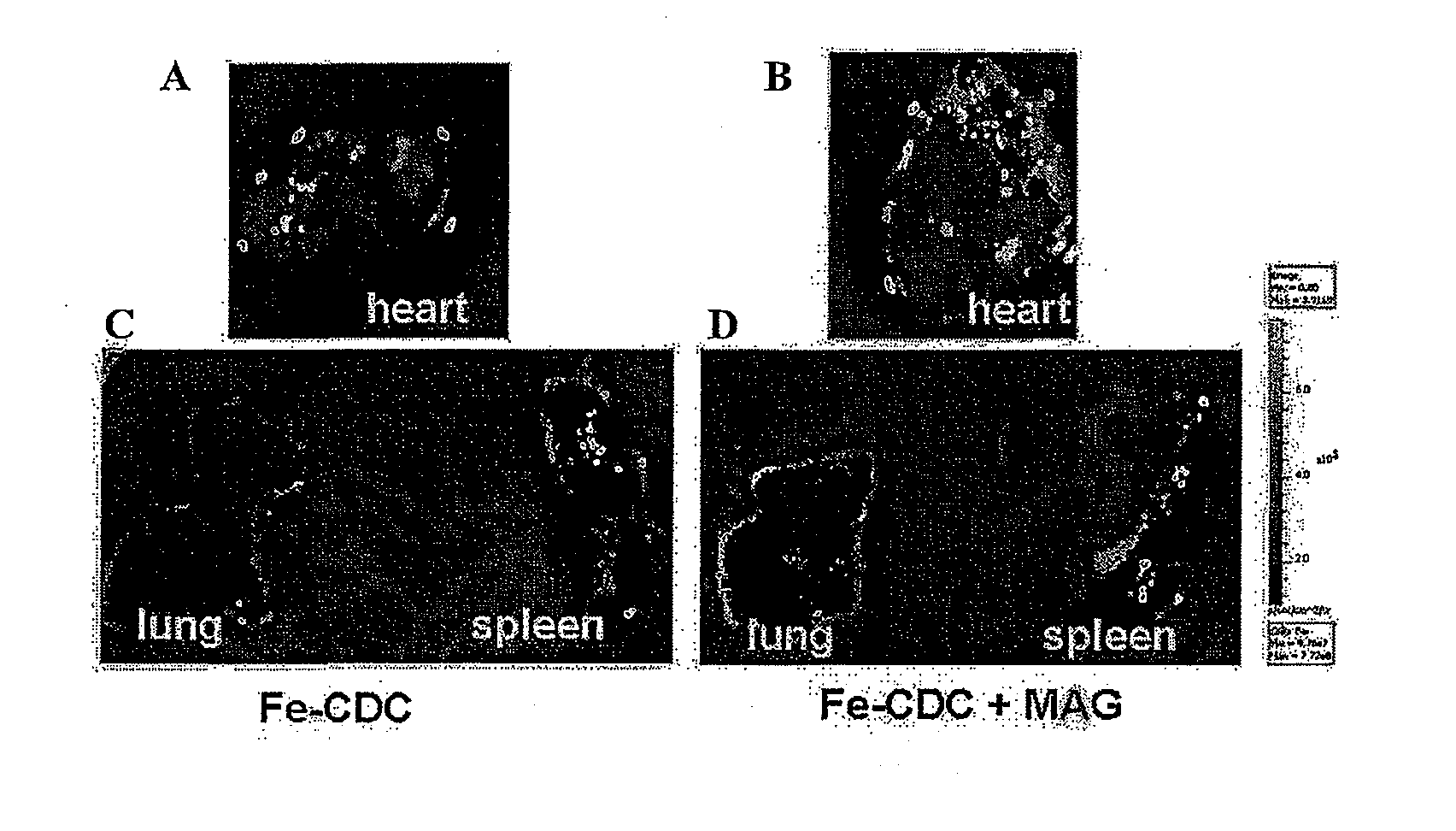
[0227]The labeling efficiency was assessed by microscopic examination and flow cytometry. CDCs labeled with SPIO (with a dragon green fluorescence tag) overnight were examined under a fluorescence microscopy (Excitation 488 nm; Emission 520 nm). A green color was observed, which indicates that the cells were successfully labeled with the SPIO (see FIG. 1). The labeling efficiency was also analyzed by flow cytometry. As shown in FIG. 2, compared to the control group (Panels A and B), the histogram sh...


PUM


Abstract

Disclosed herein are compositions and methods for the improved delivery of cells to a target tissue. In some embodiments, the compositions comprise stem cells, in particular cardiac stem cells, and the target tissue is damaged or diseased cardiac tissue. In several embodiments, the methods, in combination with the compositions, yield enhanced delivery, retention, and/or engraftment of the cells into the target tissue, thereby inducing improved functional recovery.

Description

CROSS-REFERENCE TO RELATED APPLICATIONS[0001]This application claims the benefit of U.S. Provisional Application No. 61 / 255,438, filed on Oct. 27, 2009 the disclosure of which is expressly incorporated by reference herein.STATEMENT REGARDING FEDERALLY SPONSORED R&D[0002]This invention was made with Government support under NIH Contract HL083109, an RO1 grant awarded by the National Heart, Lung, and Blood Institute. The Government has certain rights in the invention.BACKGROUND[0003]1. Field of the Invention[0004]Embodiments of the present application relate generally to compositions and methods for the enhanced delivery, retention and / or the engraftment of cells in a target tissue or organ using cell magnetization, optionally in combination with one or more vascular permeability agents. In particular, cardiac cells may be delivered to damaged cardiac tissue and the enhanced delivery, retention and / or the engraftment of the cells facilitates repair and / or regeneration of cardiac tissu...

Claims


Application Information

Patent Timeline
IPC IPC(8): A61M37/00A61N2/10C12N5/071
CPCA61M25/0068A61M25/0082A61M25/0133A61N2/06A61M25/10A61N2/002A61M25/04
Inventor MARBAN, EDUARDOCHENG, KE
Owner CEDARS SINAI MEDICAL CENT
Why Eureka
Try Eureka