
System and Method for Detecting and Quantifying Active T-cells or Natural Killer Cells

a technology of active t-cells and natural killer cells, applied in combinational chemistry, chemical libraries, instruments, etc., can solve the problems of high mortality rate, undesirable use of immuno-suppressants, loss of essential immune function, etc., and achieve no or limited adverse effects

Inactive Publication Date: 2012-11-29
ONFELT BJORN +4
View PDF4 Cites 2 Cited by
  • Summary
  • Abstract
  • Description
  • Claims
  • Application Information

AI Technical Summary

Benefits of technology

[0030]By comparing the data generated by the software for optical analyses with patient data stored in the accumulated database the system hereby disclosed provides a novel empirical approach for predicting GVHD-GVL after SCT. Since the number of SCT to patients in need thereof is limited due to the risk of GVHD, a reliable prediction method would have great advantages.

Problems solved by technology

SCT is commonly performed in order to restore essential immune function after chemotherapy and radiation since this, besides desired anti-tumor effects, often causes destruction of the patient's hematopoietic system, leading to loss of essential immune function.
However, even though stem cell donors are selected based on matching human leukocyte antigen (HLA), complications regularly occur where transplanted cells perceive the recipient's cells as foreign, leading to a patient-directed immune response and GVHD.
Thus, immuno-suppressants are undesirable to use both considering patient health and the high cost to society.
However, severe GVHD is associated with high mortality rates, large treatment costs and socioeconomic effects.
Unfortunately, there are no reliable methods for predicting the risk of GVHD or chance of GVL prior to selection of donors for SCT.
This makes the detection impossible by conventional experimental methods based on bulk populations of cells.
Nevertheless, this technique has many drawbacks, as it does not allow multiple readings of the same cell through time making it unsuitable for investigations of dynamic cell-cell interactions.
In this regard, one particular challenge has been to develop devices for studies of non-adherent cells.
Such devices are needed since it is difficult to retain such cells at a given location during periods of manipulation, incubation and read-out, for example.
The presence of electrodes enables electromagnetic manipulation of the cells and although it makes every trapping site individually addressable, it severely limits the number of cells that can be trapped on a given area.
A drawback with the hydrodynamic forces trapping methods is that the liquid pressure strain induced on the cell and the liquid flow induced surface stress is likely to affect the cells in an assay.
Another drawback with this trapping method is the limitation on the cells' natural movements, thus limiting the interaction or limiting multiple interaction events.
In the case of cell interaction studies involving imaging analysis, it has to be noted that pressure or flow fluctuations often have an impact on image quality, as these fluctuations will cause the images to be blurry or out of focus.
The problem with this patent is that there is little mobility for the tightly trapped living cell in each cell carrier and this impedes any mechanical manipulation of the cell, including its extraction.
This is not suitable for two cell interaction assays, as the wells fit only one cell.
Enlarging the well to fit two cells with different geometry will not work, as the smaller cell will escape.
The size of the wells limits the volume of culture media contained in each well and thereby the availability of nutrients.
As a consequence, cell interaction studies and long-term assays are not possible with this kind of device.
Moreover with the prior devices, even in those cases in which more than one individual cell can be held in a well or compartment, it is not possible to control exactly how many cells end up in each well.
Importantly, the latter methods assess target cell death with single cell resolution but do not provide information on the single cell level for the effector cell population.
It is also the case that high-throughput screening for analyzing significant populations of cells might not be achievable in the method described in patent WO07 / 116309.

Method used


Image

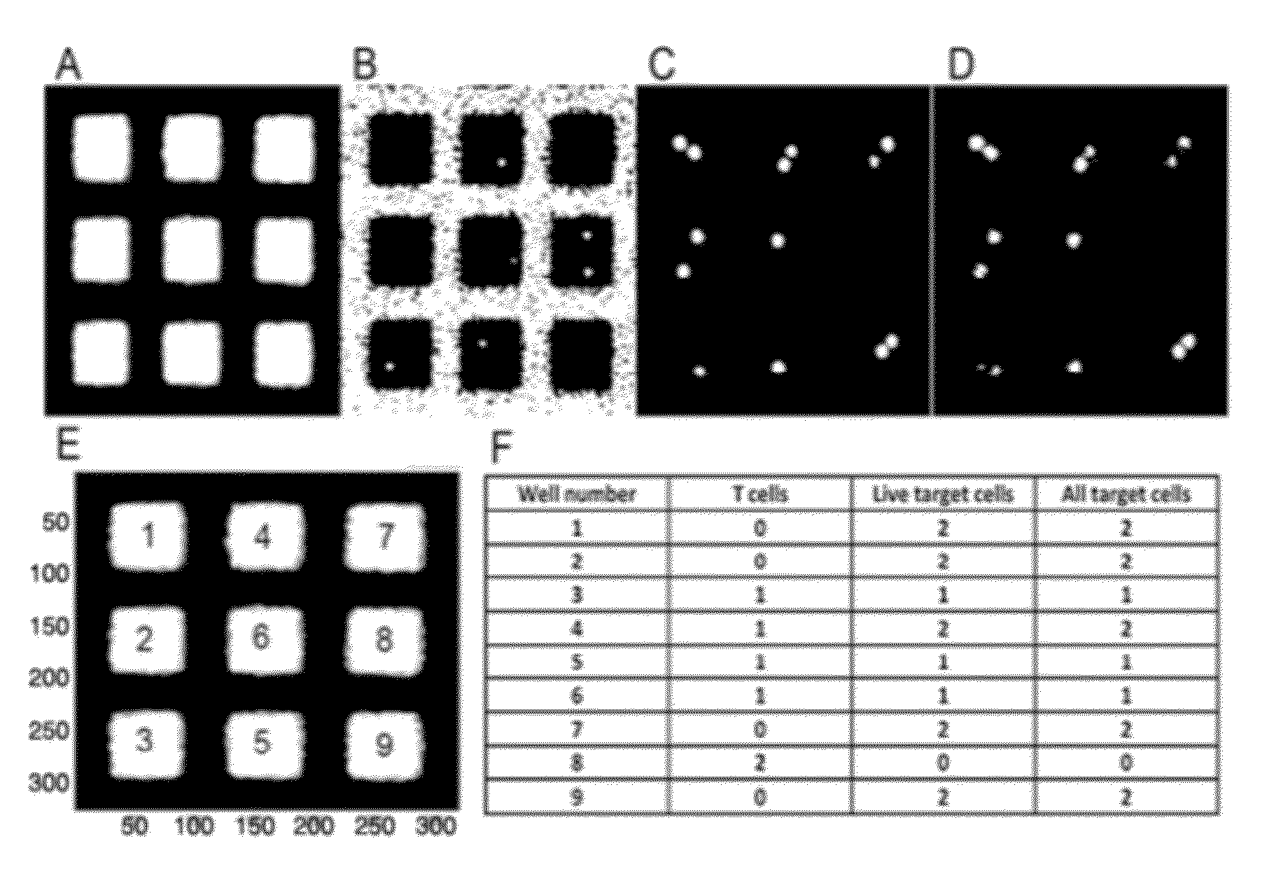
  • System and Method for Detecting and Quantifying Active T-cells or Natural Killer Cells
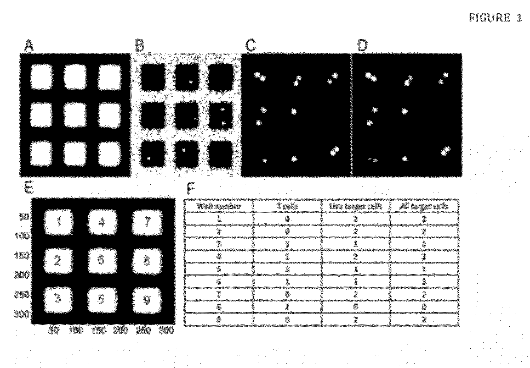
  • System and Method for Detecting and Quantifying Active T-cells or Natural Killer Cells
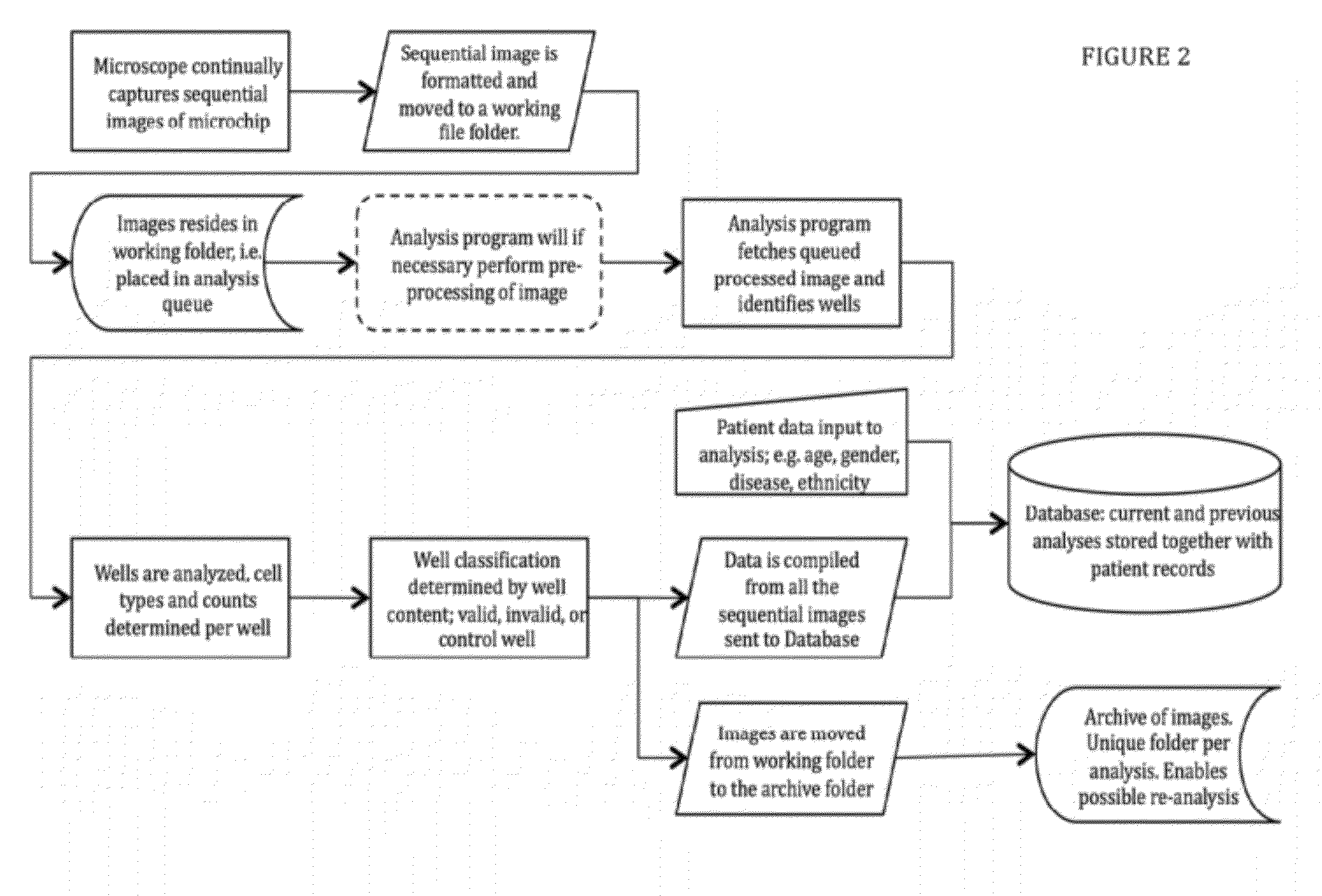
  • System and Method for Detecting and Quantifying Active T-cells or Natural Killer Cells

Examples


example 1

Screen for Alloreactive Cells

[0102]In order to obtain a proof of concept, the donor and recipient cells used were miss-matched in 3 out 6 major HLA alleles (considering HLA-A, HLA-B and DRBI). This is a higher degree of miss-match compared to the real transplantation setting where seldom more than 1 miss-matched major HLA allele is allowed. The advantage in using a higher degree of miss-match for this experimental setup is that the frequency of reactive T cells will be much higher facilitating the detection and quantification of these specific killing T cells.

1. Effector Cell Preparation.

[0103]To obtain T cells, 20 ml of peripheral blood was drawn in two 10 ml heparinized collection tubes. Blood was diluted 1:1 in phosphate-buffered saline (PBS) prior to a 20 min separation of peripheral blood mononuclear cells (PBMCs) by Ficoll density gradient centrifugation at 400 g without brakes. The PBMCs were collected and washed by diluting in PBS and centrifugation for 10 min at 400 g force...

example 2

[0113]This example illustrates the steps that follow example 1, where identified specific T-cells from analysis are desired to be isolated, expanded and injected into a patient, e.g. In the case of cell therapy.

8. T Cell Harvesting.

[0114]After analysis, wells with specific cytotoxic T-cells will have been identified. Without damaging the T-cells, these are extracted from their respective wells. For increasing chance of success and cell survival when isolating the T-cells, the cells may be left in the wells to proliferate for some time before extraction. Examples of harvesting methods are by optical or acoustic traps or by magnetic labeling and extraction as known in the art, such as a micromanipulator.

9. T Cell Expansion

[0115]The specific effector cells are transferred to culture flasks containing RPMI-medium with added L-glutamine, streptomycin, penicillin, 10% human AB sera and recombinant IL-2. In order to stimulate growth of the effector T-cells, magnetic beads coupled to stimul...

example 3

11. Microchip Design Criteria and Considerations

[0117]When designing the chip dimensions one should consider different aspects such as to maximize the cellular interactions, to be able to contain sufficient nutrients, deep enough to prevent escape or movement of the cells between wells and biocompatibility; optimization of the washing steps.

[0118]The microchip enabling the present invention should meet specific design requirements as follows:

[0119]Medical or Statistical Demands[0120]useful number of cells, range 104 to 1.5×10⁶ [0121]yield after seeding, range 25% to 85%[0122]plausible number of positive cell-cell interactions, range 2 / 10 000 to 100 / 10 000

[0123]Optical Demands[0124]transparency, range from IR over optical visibility to UV[0125]optical path, range from 150 μm to 200 μm[0126]flatness, range max + / −0.1 μm

[0127]Biological Demands[0128]biocompatibility, range max 3 weeks cell survival[0129]well depths for capturing, range 100 μm to 1 mm, resulting in sedimentation time 10...


PUM


Abstract

A system and a methodology are provided with a broad range of application in immune diagnostic screening and therapy and specifically for predicting the risk of graft versus host disease (GVHD) and / or graft versus leukemia (GVL) effects prior to transplantation. The method includes the steps of incubating single T or NK cells with a few, usually three to five target cells for extended period of times and evaluating the cell contact-dependent lytic activity or activation of the single effector cells within larger populations. The results obtained are analyzed by proprietary software for automated image analysis and compared with patient data comprised in a comprehensive database containing accumulated empirical and clinical information on donor-recipient screening results and patient information. A micro device is provided for implementing the disclosed method, wherein said micro device is a multi-well microchip, having tens of thousands wells with defined characteristics thus allowing long-term assays and quantitative cell activity inspection / evaluation by high-resolution microscopy.

Description

CROSS-REFERENCE TO RELATED APPLICATIONS[0001]This application claims priority from PCT / EP2010 / 067077 filed Nov. 9, 2010, which claims priority from U.S. Provisional Application Ser. No. 61 / 280,809 filed Nov. 9, 2009.BACKGROUND OF THE INVENTION[0002]1. Field of the Invention[0003]The present invention concerns a novel combination of unique tools for improved cellular resolution in investigating functional properties of cell populations. In particular, the invention herein is a system and a methodology for detecting, isolating and / or quantifying specific active effector cells within larger populations based on interaction with pre-determined desired target cells. In particular embodiments the reactivity on a single cell level and specifically the lytic activity of a single effector cell is used as basis for evaluating the population general specificity towards the target cells.[0004]More specifically, the present invention relates to the prediction of the risk of graft versus host dis...

Claims


Application Information

Patent Timeline
IPC IPC(8): C40B30/06C40B60/12
CPCG01N33/5047G01N2800/245G01N33/505
Inventor ONFELT, BJORNUHLIN, MICHAELKARRE, KLASVANHERBERGHEN, BRUNOFRISK, THOMAS
Owner ONFELT BJORN
Learn More