Methods of producing human rpe cells and pharmaceutical preparations of human rpe cells

Active Publication Date: 2013-06-13
ADVANCED CELL TECH INC
View PDF2 Cites 32 Cited by
  • Summary
  • Abstract
  • Description
  • Claims
  • Application Information

AI Technical Summary

Benefits of technology

[0077]In one embodiment, the RPE cells may improve visual acuity after transplantation. In an

Problems solved by technology

Mutant REP-1 proteins cannot escort Rab proteins, leading to a lack of functional Rab proteins.
This lack of Rab proteins causes a disruption in intracellular trafficking and leads to necrosis in the RPE.
Currently, no treatment is available and a need exists for a therapy for choroideremia.
Finally, signals sent by the retina for nourishment trigger the growth of new blood vessels (proliferative retinopathy) but these new blood vessels are abnormal and fragile.
As the structural integrity of the blood vessels deteriorate (in part due to changes in osmolarity due to insulin/sugar imbalance fundamental to diabetes), they leak blood, causing severe vision loss and even blindness.
Generally, diabetic retinopathy may only be controlled or slowed with surgery but not treated, and the patient usually continues to suffer from vision problems.
Atrophy of the submacular RPE and the development of choroidal neovascularizations (CNV) results secondarily in loss of central visual acuity.
Neovascular or exudative AMD (“wet AMD”) causes vision loss due to abnormal blood vessel growth (choroidal neovascularization) in the choriocapillaris, through Bruch's membrane, ultimately leading to blood and protein leakage below the macula.
Bleeding, leaking, and scarring from these blood vessels eventually cause irreversible damage to the photoreceptors and rapid vision loss if left untreated.
However, the goal of these therapies is to stem further vision loss and, unfortunately, existing damage cannot be reversed.
Patients with RP all suffer some vision loss, with night blindness as a typical early symptom followed by tunnel vision, and some may lose all sight.
Although treatment with vitamin A and lutein has shown some promise in slowing the progress of RP, no effective treatment is available.
Initial detachment may be localized, but without rapid treatment the entire retina may detach, leading to vision loss and blindness.
The current treatment is emergency eye surgery but only has an approximately 85% success rate, and even if successful, the patient may suffer a loss of visual acuity and visual artifacts.
Over time, these diseased cells cause a blackened hole to form in the central vision, and the ability to perceive colors is eventually affected.
Currently, there are no treatments available for Stargardt's Disease.
(2007) Br J Opthalmol 91: 349-353 describes the successfully transplanta

Method used


Image

  • Methods of producing human rpe cells and pharmaceutical preparations of human rpe cells
  • Methods of producing human rpe cells and pharmaceutical preparations of human rpe cells
  • Methods of producing human rpe cells and pharmaceutical preparations of human rpe cells

Examples


example 1

Method of Making Human RPE Cells Using hES Cells

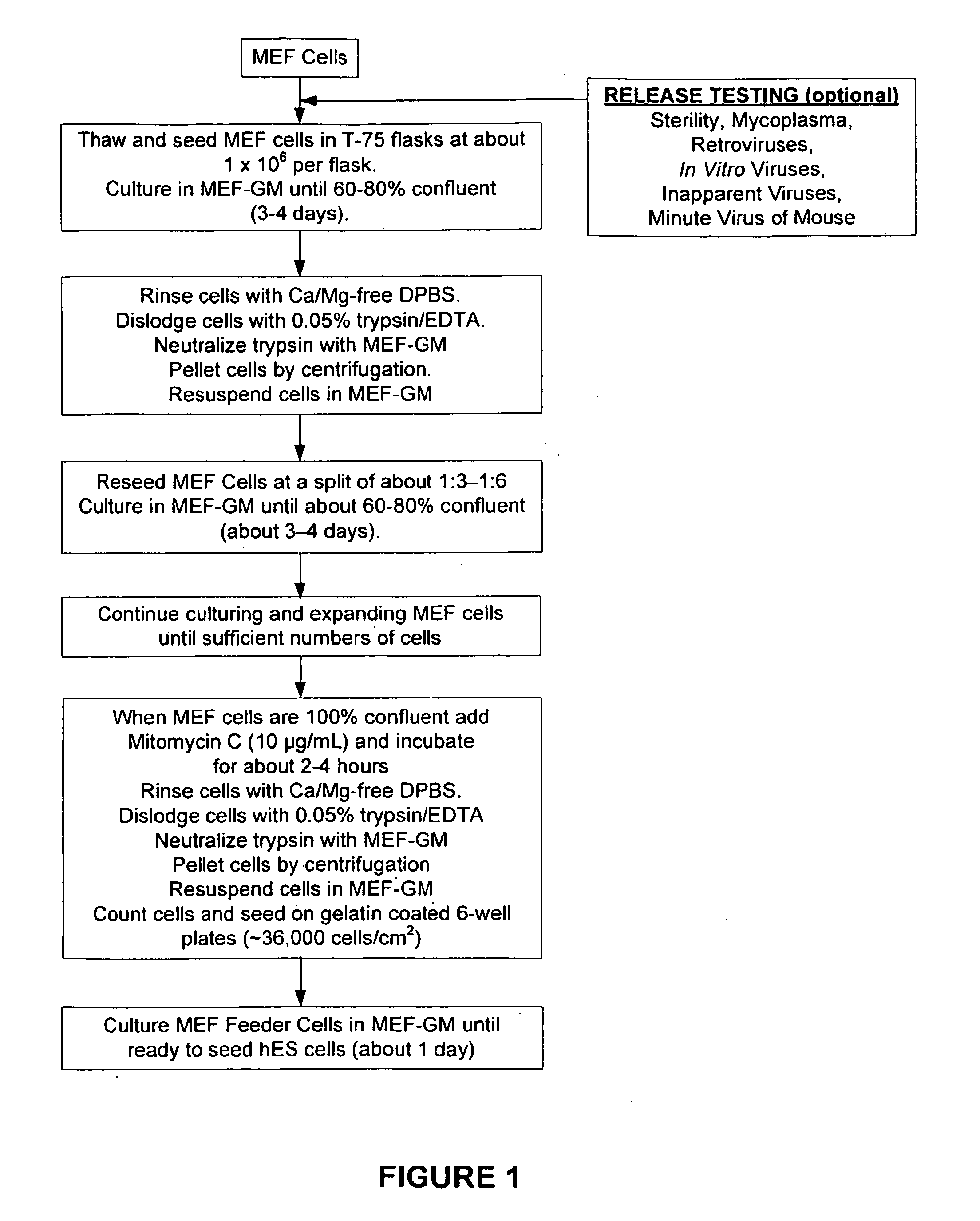
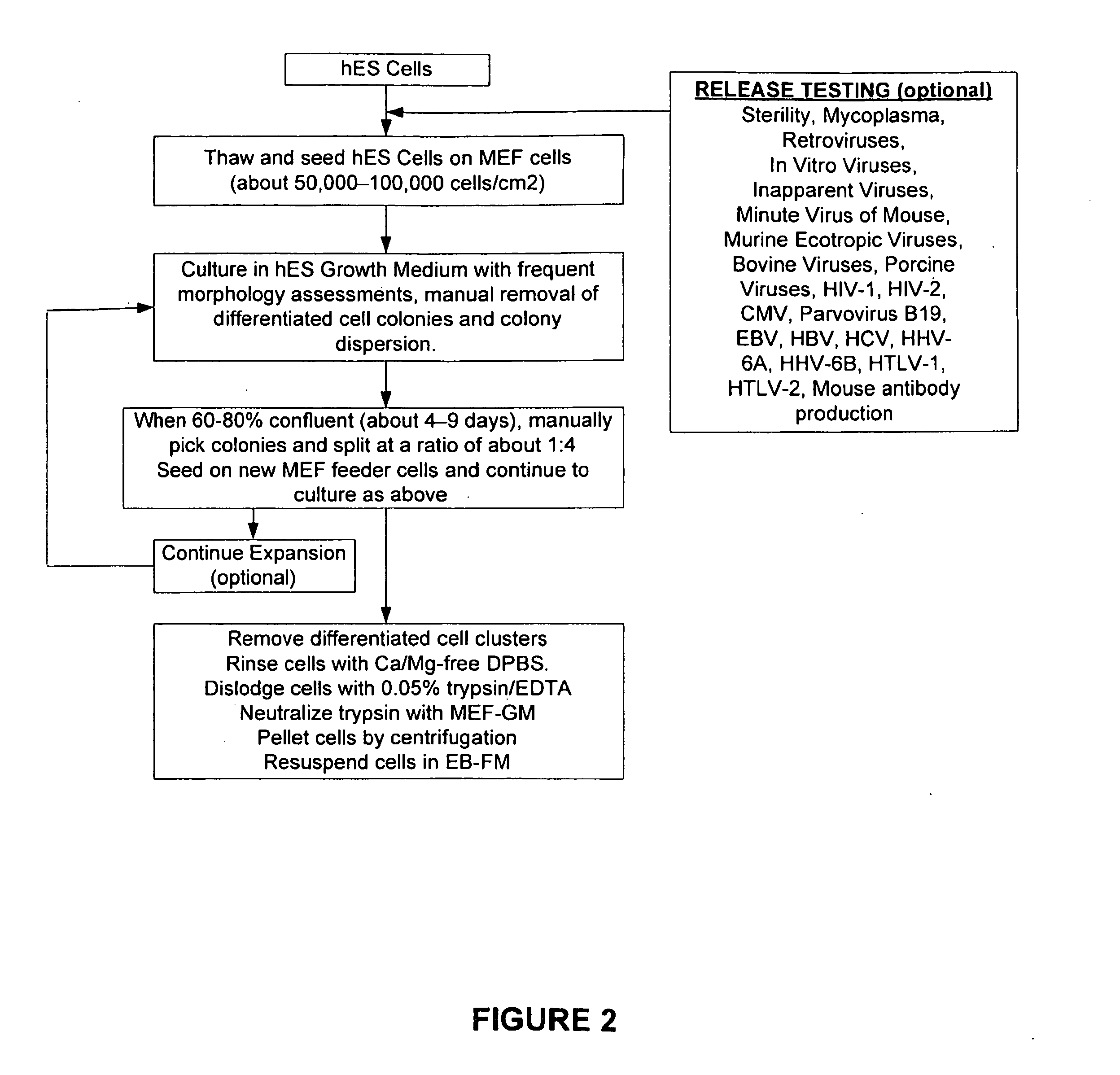
[0233]Mouse embryo fibroblasts (MEF) were grown in MEF-GM medium supplemented with about 10% fetal bovine serum (FBS). When sufficient numbers of MEFs were obtained, feeder cells were prepared by mitotically blocking the MEFs with mitomycin-C and seeding into 6-well plates coated with gelatin. See FIG. 1. Vials of hES were thawed, seeded on to the MEF feeder cells, and co-cultured in hES Growth Medium. See Table 2 and FIG. 2. The hES cells were expanded several times at a split ratio of about 1:3. When a sufficient number of hES cells were propagated, the cells were harvested and placed into suspension culture in low attachment 6-well plates in EB Formation Medium (this allows for the formation of embryoid bodies (EBs)). See Table 2 and FIG. 3.

[0234]The EBs were then be transferred to gelatin-coated 6-well plates to allow for the outgrowth of RPEs. The initial growth medium is EB Outgrowth Medium, but once the EBs were attached this wa...

example 2

Seeding and Expansion of hES Cells

[0235]Cryopreserved human embryonic stem cells (hES) cells were thawed, washed with hES-GM, and inoculated onto the mitotically inactivated mouse embryonic feeder (MEF) cells in the gelatin-coated 6-well plates. See FIG. 7. The contents of each vial (˜1 million cryopreserved cells) of hES were seeded into one well of a 6-well plate, and co-cultures of hES and MEF are incubated for about 4-9 days until about 60-80% confluent. During this time the cultures were examined microscopically: larger colonies displaying mostly hES morphology were dispersed into smaller pieces to prevent spontaneous differentiation. Mosaic colonies with large areas of undifferentiated cells were trimmed by removing those portions comprised of differentiated cells. Colonies containing predominately differentiated cells or non hES cell morphology were picked and discarded, using a stem cell cutting tool using photographs as a guide to the morphology of the colonies. See FIG. 2....

example 3

Human Embryoid Body Formation and Outgrowth

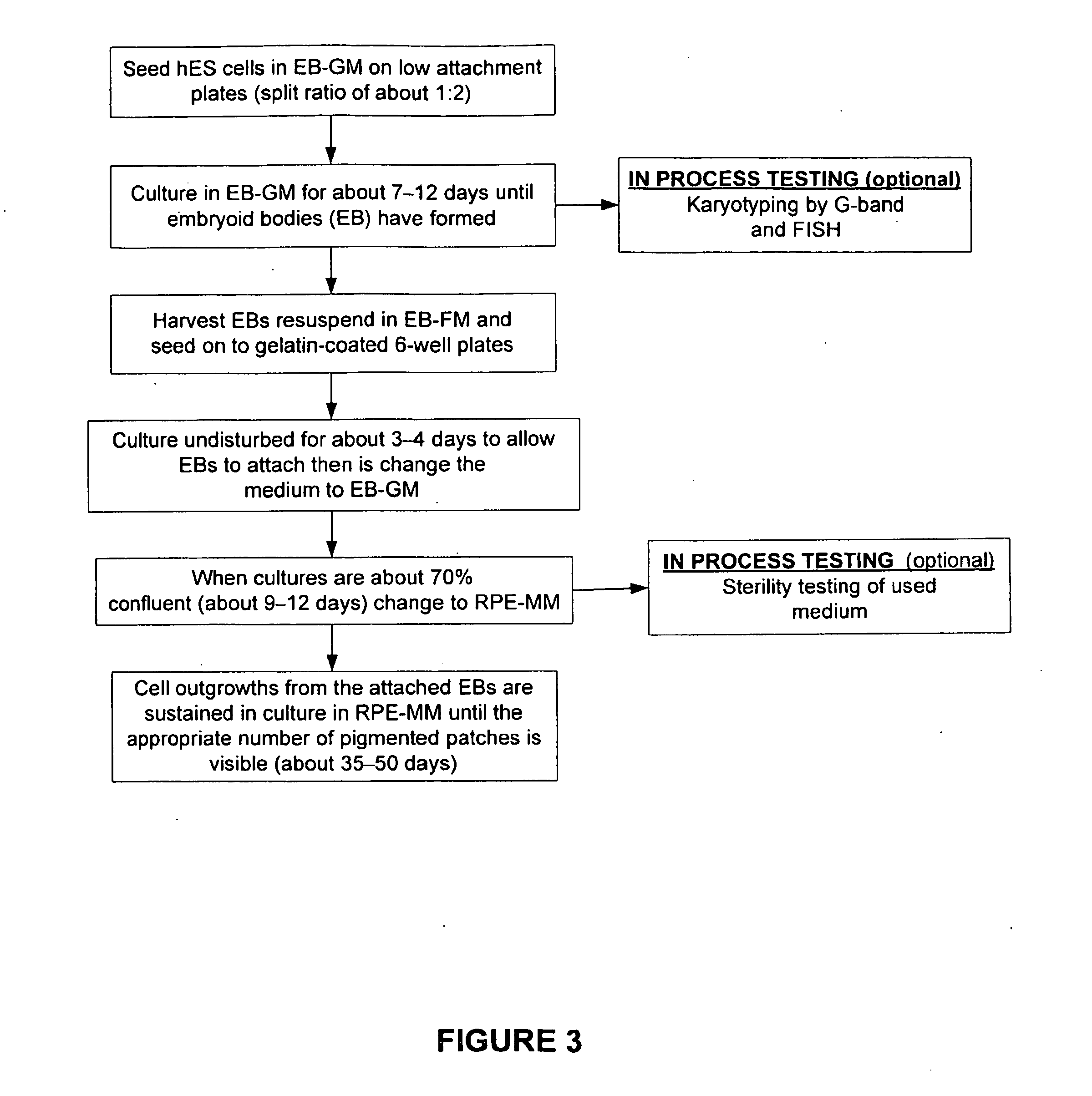
[0239]hES cells were inoculated onto low-attachment, 6-well plates (at a split ratio of 1:2) and cultured for about 7-12 days until embryoid bodies were formed and matured. Embryoid bodies in suspension were harvested from the low attachment wells, resuspended in EB-FM, and plated onto gelatin-coated 6-well culture plates. The plates were cultured undisturbed for about 3-4 days to allow the embryoid bodies to attach. At this time, medium was changed to EB Growth Medium (EB-GM). When cultures were about 70% confluent (e.g., after about 9-12 days), the medium was changed to RPE Maintenance Medium (RPE-MM). See FIG. 3. Cell outgrowths from the attached EBs were sustained in culture in RPE-MM until the appropriate number of pigmented clusters were visible (e.g., after about 35-50 days after changing to RPE-MM). Thus, embryoid bodies were formed and isolated substantially free of non-human cells and thus not “xenotransplantation” material. These...


PUM


Abstract

The present invention provides improved methods for producing retinal pigmented epithelial (RPE) cells from human embryonic stem cells, human induced pluripotent stem (iPS), human adult stem cells, human hematopoietic stem cells, human fetal stem cells, human mesenchymal stem cells, human postpartum stem cells, human multipotent stem cells, or human embryonic germ cells. The RPE cells derived from embryonic stem cells are molecularly distinct from adult and fetal-derived RPE cells, and are also distinct from embryonic stem cells. The RPE cells described herein are useful for treating retinal degenerative conditions including retinal detachment and macular degeneration.

Description

CROSS-REFERENCE TO RELATED PATENT APPLICATIONS[0001]This International Patent Application claims priority to U.S. Provisional Patent Application No. 61 / 262,002, filed Nov. 17, 2009, the disclosure of which is herein incorporated by reference.BACKGROUND OF THE INVENTIONRetinal Pigment Epithelium (RPE)[0002]The retinal pigment epithelium (RPE) is the pigmented cell layer outside the neurosensory retina between the underlying choroid (the layer of blood vessels behind the retina) and overlying retinal visual cells (e.g., photoreceptors—rods and cones). The RPE is critical to the function and health of photoreceptors and the retina. The RPE maintains photoreceptor function by recycling photopigments, delivering, metabolizing, and storing vitamin A, phagocytosing rod photoreceptor outer segments, transporting iron and small molecules between the retina and choroid, maintaining Bruch's membrane and absorbing stray light to allow better image resolution. Engelmann and Valtink (2004) “RPE C...

Claims


Application Information

Patent Timeline
IPC IPC(8): A61K35/12
CPCA61K35/12C12N2506/02C12N5/0621A61P27/02A61K35/56C12N5/00C12N5/0662
Inventor MALCUIT, CHRISTOPHERLEMIEUX, LINDAHOLMES, WILLIAMHUERTAS, PEDROVILNER, LUCY
Owner ADVANCED CELL TECH INC
Why Eureka
Try Eureka