Biopsy method and gun set devices

a biopsy method and gun set technology, applied in medical science, vaccination/ovulation diagnostics, diagnostics using spectroscopy, etc., can solve the problems of inability to prevent bleeding from the biopsy site, failure to be located exactly at the right location, and inter-contamination and the spread of sick cells, so as to minimize the damage to the various tissues that are penetrated, the effect of reducing the damage of the penetration

Inactive Publication Date: 2014-08-14
VARIABLE GAUGE CATHETER INC
View PDF14 Cites 17 Cited by
  • Summary
  • Abstract
  • Description
  • Claims
  • Application Information

AI Technical Summary

Benefits of technology

[0031]A needle that is protected by several sheaths one for each suspected unhealthy penetrated tissue in order to reduce inter-contamination and the spread of the sick cells. A sample notch is supported on an internal rod, that on the opposite side has a drug impregnated plug that is released simultaneously with the tissue harvesting process. The process is enhanced by the use of a capillary tube that brings negative pressure in the sample notch fixing the sample on the pad in order to be cut smoothly, without generating shearing in the tissue, while a positive pressure works synchronous with the cutter tube hook that detaches the drug-plug that will expand in the tissue as soon as the tube is withdrawn.
[0032]Using exchangeable cores the sample cutter may be extracted and a new tube may be introduces that delivers specialized plugs along the penetration hole, minimizing the penetration damage to the various tissues penetrated. One of the sampler tips or a specialized penetration tip, may be provided with a set of radioactive point sources operating on different energies and providing a stereoscopic view from inside the body, simultaneously with an accurate position and direction, making it redundant to the ultrasound system frequently used, and providing a coordinate localization of all operations. A set of hollow-capillary optic fiber is passing through the tube allowing a complex optical UV and IR spectroscopy in order to map the composition of the penetrated tissues, and could even resonantly damage the selected molecular bonds in the ill cells, killing the tumor.

Problems solved by technology

These ideas, which comprise the previous art, have evident difficulties with the possible misplacement of the outer cannula as it penetrates to the biopsy site, as well as failure to be located exactly at the right location to prevent bleeding.
One problem with this tip is that it is not sharp enough, so its failure to penetrate may cause more complications.
But if the gelatin is deposited prematurely before it reaches the biopsy site it cannot prevent bleeding from the biopsy site.
This device has some operational problems, as interaction with various hardness and viscosity of organs provides various volumes of biopsy tissue.
One challenge of biomedical optics is to uncouple the signals influenced by biochemistry, which themselves provide specificity for identifying diseased states, from those influenced by tissue scattering, which are typically not specific to a pathology.
In this wavelength region, silica glass fiber optics cannot be used for delivering such light.
A set of hollow-capillary optic fiber is passing through the tube allowing a complex optical UV and IR spectroscopy in order to map the composition of the penetrated tissues, and could even resonantly damage the selected molecular bonds in the ill cells, killing the tumor.
The inventors consider that most of the problems generated by the actual biopsy operations are both due to the obsolete procedure and equipment used to sample the patient's tissue and due to the penetration tool leaving a large communication hole through the internal organs where infections may propagate.
The sampling process is finished and the whole assembly is withdrawn from the body leaving a serious penetration wound behind.
The actual technique uses compression to limit the bleeding that is not fully successful for tissues placed deep in the body.
By turning the device on its axis a circular profile of the surrounding area may be obtained, and tissue fiber may be identified, but in this case the cutting edge device's profile 287 is inappropriate for this operation.
A similar technique may be used to seal military penetrations, but the plug has to be more complex introducing substitute parts for the damaged tissue.
It will be nice to place a CCD camera on the cutting tip, but this is not possible except with large needles with diameter over 4 mm, and that is what the current patent tries to avoid: making large wounds in the patient's body, for no substantial gain.

Method used


Image

  • Biopsy method and gun set devices
  • Biopsy method and gun set devices
  • Biopsy method and gun set devices

Examples


Embodiment Construction

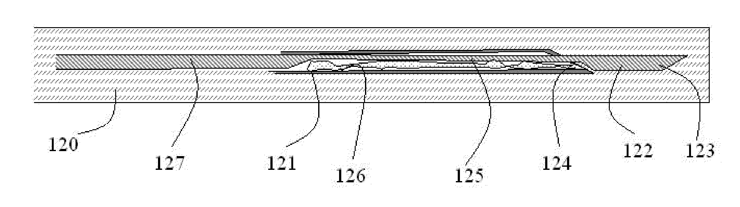
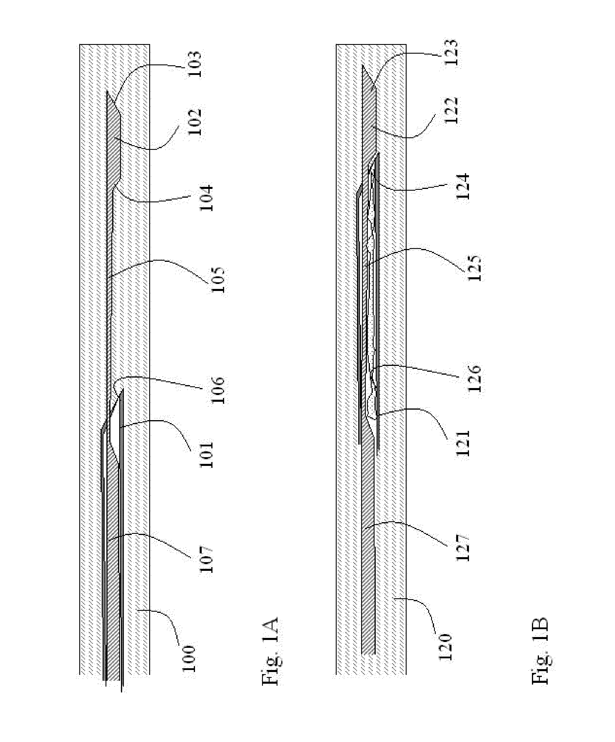
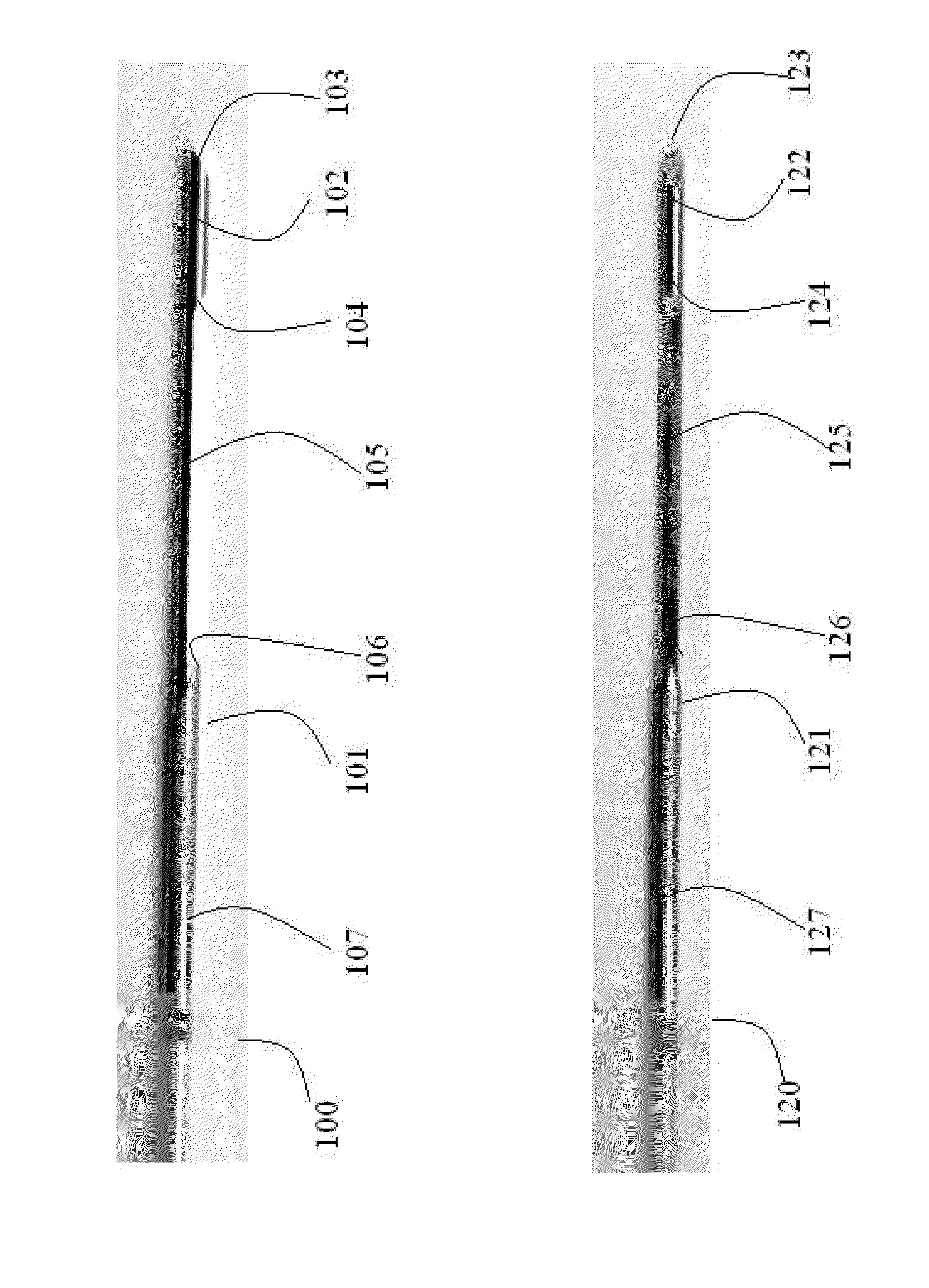
[0066]The inventors consider that most of the problems generated by the actual biopsy operations are both due to the obsolete procedure and equipment used to sample the patient's tissue and due to the penetration tool leaving a large communication hole through the internal organs where infections may propagate. Therefore we develop a new method of penetration with a more complex tool that uses a gradual approach inside sheathed tubes that first aims for very small perforation using liquid injection, in order to reduce the stress and tissue stretching as much as possible. The assembly comes as the a set of instruments to assure the gradual penetration of the ultra thin optical investigation needle with appropriate sheathing and plugging, optionally followed by thicker gauge biopsy needles with appropriate sheathing and plugging tools, with dynamic pressure compensation systems to prevent undesired pressures developing.

[0067]The method is straightforward, and starts with coordinates l...


PUM


Abstract

A novel set of biopsy-gun related tools are developed in order to make a better safer sampling and diagnosis, comprising a set of tools that takes the sample, releases a drug impregnated plug and additionally may inject drugs, measure pH and the molecular content of the penetrated tissues.
A novel technology aiming to minimize the penetrated tissue damage, using a small diameter needle with capabilities for immediate spectroscopic analysis of the tissue, and followed by sampling and plugging of the tissue when needed.
The invention describes a set of variable diameter thin tubes used to guide and insulate the penetrated organs, and a final operation of plugging the wounds with absorbable substances, impregnated with drugs.
The supplementary low intensity radiation sources could increase the processes' accuracy and safety, offering the opportunity of gathering supplementary x-densitometry information.

Description

CROSS REFERENCE TO RELATED APPLICATIONS[0001]This application claims the benefit of U.S. Provisional Application No. 61 / 762,582 from Feb. 08, 2013, and no international application.BACKGROUND[0002]A biopsy is a medical procedure to acquire cells or tissues for pathological examination. It normally involves the removal of tissue from a living subject to determine the presence or extent of disease. The tissue is generally examined under microscopy by a pathologist, and can also be analyzed chemically. When an entire lump or suspicious area is removed, the procedure is called an excisional biopsy. When only a sample of tissue is removed with preservation of the histological architecture of the tissue's cells, the procedure is called an incisional biopsy or core biopsy. When a sample of tissue or fluid is removed with a needle in such a way that cells are removed without preserving the histological architecture of the tissue cells, the procedure is called a needle aspiration biopsy.[000...

Claims


Application Information

Patent Timeline
Patent Type & Authority Applications(United States)
IPC IPC(8): A61B10/02A61B5/145A61B5/00A61B5/06
CPCA61B10/0275A61B10/0283A61B5/061A61B2562/0233A61B5/0075A61B5/6848A61B5/14539
Inventor POPA-SIMIL, LIVIUKRAMER, RADU
Owner VARIABLE GAUGE CATHETER INC
Why Eureka
Try Eureka