
Targeted Lipid-Drug Formulations for Delivery of Drugs to Myeloid and Lymphoid Immune Cells

a technology of myeloid and lymphoid immune cells and drug delivery system, applied in the field of medical arts, can solve the problems of inability to achieve human-practical strategy, inability to develop long-circulating large (>500 nm) liposomes using steric stabilization methods, and inability to achieve drug delivery. to achieve the effect of facilitating production

Inactive Publication Date: 2014-08-21
RODOS BIOTARGET
View PDF2 Cites 0 Cited by
  • Summary
  • Abstract
  • Description
  • Claims
  • Application Information

AI Technical Summary

Benefits of technology

The patent provides a system for delivering chemicals to immune cells harboring HIV. Using targeting molecules like DC-SIGN, the system can directly deliver antiviral drugs to these cells. The system can also be used for vaccination against HIV, treatment of immune cell proliferation disorders, and in the production of anti-infective vaccines and therapeutic tools. The technical effects of the patent include improved treatment and prevention options for HIV infection and related disorders.

Problems solved by technology

However, application of lipid-drug complexes to drug delivery systems was not realized until 30 years later.
Whereas RES uptake in vivo can be saturated at high doses of liposomes or by predosing with large quantities of control liposomes, this strategy may not be practical for human use because of the adverse effects related to sustained impairment of physiological functions of the RES.
Due to biological constraints, development of long circulating large (>500 nm) liposomes using steric stabilization methods has not been successful.
This process may potentially lead to a premature leakage or dissociation of drugs from liposomes.
However, therapeutic applications of systemically administered liposomes have been limited by their rapid clearance from the bloodstream and their uptake by the RES (Alving C, et al., Complement-dependent phagocytosis of liposomes: suppression by ‘stealth’ lipids, J Liposome Res 2:383-395 ).

Method used


Image

  • Targeted Lipid-Drug Formulations for Delivery of Drugs to Myeloid and Lymphoid Immune Cells
  • Targeted Lipid-Drug Formulations for Delivery of Drugs to Myeloid and Lymphoid Immune Cells
  • Targeted Lipid-Drug Formulations for Delivery of Drugs to Myeloid and Lymphoid Immune Cells

Examples


example 1

Materials and Methods

[0096]Preparation of Liposomes.

[0097]A 30 μmol lipid film composed of DOPC / Chol / DOPE-MBP (36.5:33.0:2.5 mol:mol:mol) was formed (cholesterol was purchased from Calbiochem, San Diego, Calif., USA; and DOPE and DOPE-MPB were from Avanti Polar Lipids, Alabaster, Ala., USA). Lipid films were hydrated with 1 ml 50 mM calcein (Molecular Probes, Eugene, Oreg., USA) in PBS (pH 7M), sonicated in a bath sonicator (5 min) and extruded ×5 through a 0.1 μm nucleopore filter (Avanti Polar Lipids) using a hand-held extruder. Also, freeze-thaw cycles can be employed. The mean liposome size was determined by quasielectric light scattering with a Nicomp 380 ZLS Zeta-Potential Particle Sizer (Particle Sizing Systems, Santa Barbara, Calif., USA), yielding an average diameter of 146.7±31.0 nm.

[0098]Protein A Liposomes.

[0099]To be able to test the targeting ability of different antibodies with a standardized liposome, immunoglobulin-molecules were coupled to liposomes via protein A o...

example 2

Active Targeting of Immune Cells with Monospecific or Bispecific Immunoliposomes

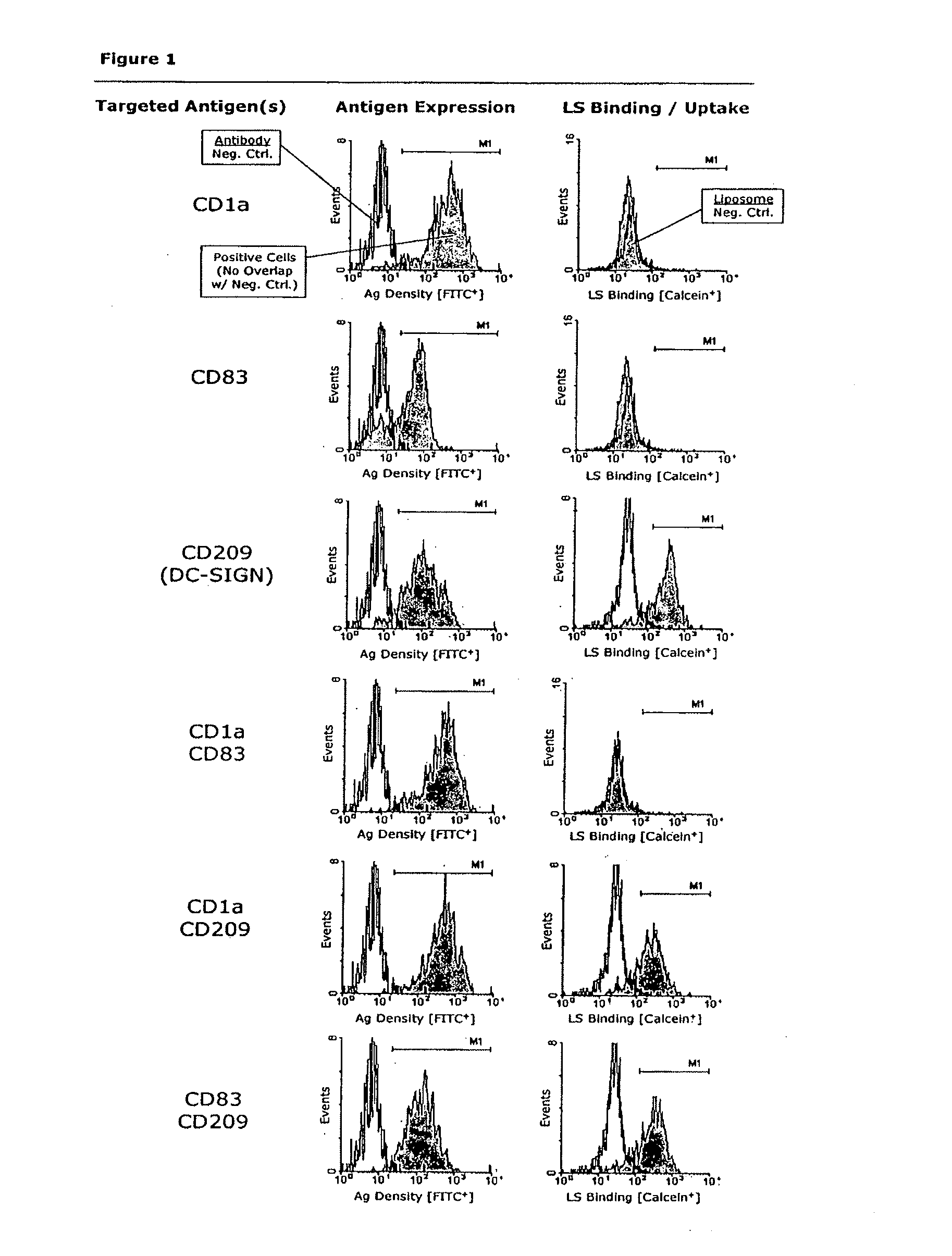
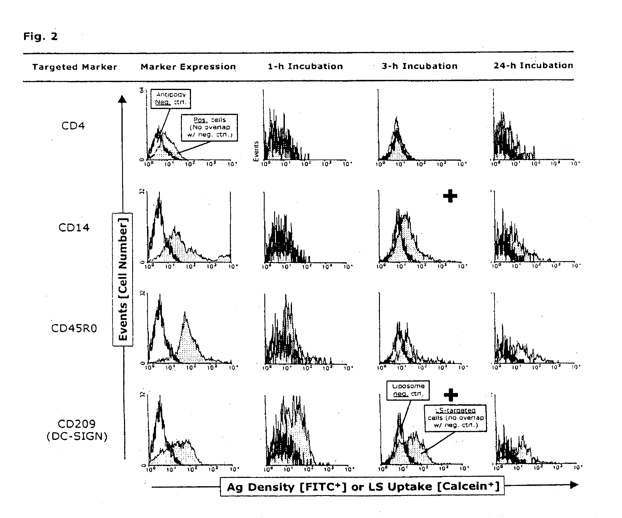
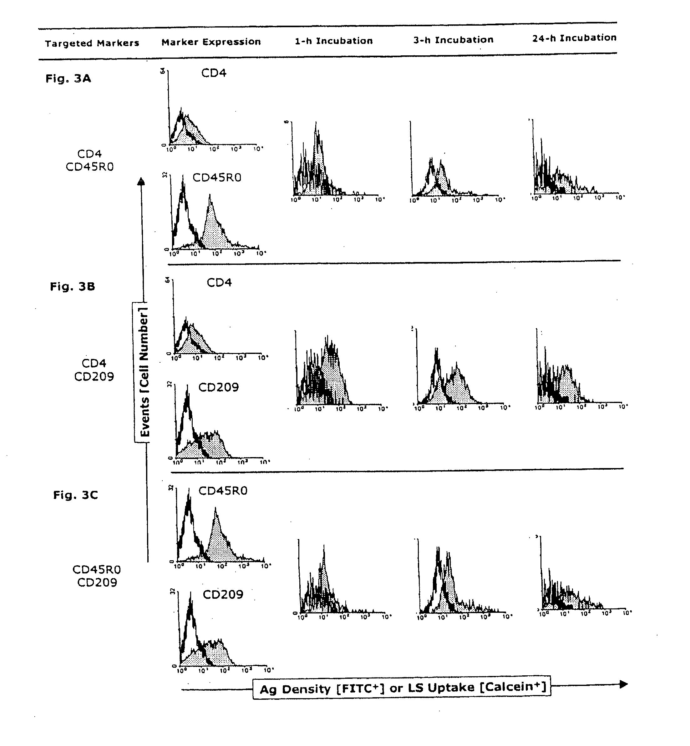
[0142]Peripheral blood mononuclear cells (PBMNCs) were evaluated according to their size (forward scatter) and granularity (side scatter) and thus were gated as naïve T and B cells; activated T-cells and B-cells; and monocytes, including a small proportion of blood dendritic cells (data not shown). Cultured monocyte-derived dendritic cells (MoDCs) were tested for expression of markers delineating their developmental stage (maturity), as well as for DC subtype markers. The DCs expressed markers typical for skin and mucosal DC phenotypes that are considered to play a key role in HIV infection. When being infected via the mucosal route, mucosal DCs are the first immune cell type to be directly infected by HIV (and integrate its genetic information as proviral DNA) and / or harvest HIV on their surface by DC-SIGN and / or take up HIV by any of various mechanisms to retain it in intracytoplasmic compartments (e.g...

example 3

Fluorescence-Microscopic Uptake Studies

[0158]After infection with HIV-1, intracytoplasmic compartments with accumulated infectious virus are demonstrable in both immature and mature MyDCs (Frank, I et al., Infectious and whole inactivated simian immunodeficiency viruses interact similarly with primate dendritic cells (DCs): differential intracellular fate of virions in mature and immature DCs, J Virol 76:2936-51 [2002]). Therefore for comparison, immature or mature MoDCs were incubated for 3, 4 or 5 h at 37° C. with anti-CD209-labeled liposomes (each at n=3). The cells were then harvested as described above and gently pipetted to dissociate homotypic clusters (as controlled by phase microscopy). Pelleted single cells were successively dissolved in 100 μl of ProLong antifade mounting medium to which was added 5 μM of the positively charged AT-binding DNA dye, 4′,6-diamidino-2-phenylindole (DAPI) (both from Molecular Probes, Eugene, Oreg., USA). Fifty μl of each preparation were trans...


PUM

PropertyMeasurementUnit
sizeaaaaaaaaaa
sizeaaaaaaaaaa
sizeaaaaaaaaaa

Abstract

A method of preferentially delivering an active agent to an immune cell, such as a myeloid progenitor cell, a dendritic cell, a monocyte, a macrophage or a T-lymphocyte, or other cell type restricted to a functional organ system or an anatomic entity, of a mammalian subject by administering a lipid-drug complex to the subject. The lipid-drug complex is comprised of an active agent, such as a drug, and an outer surface with a targeting ligand that binds a marker on the surface of the immune cell or other cell type that is infected with or susceptible to infection with an infectious agent. The other cell type that is infected with or susceptible to infection with an infectious agent may belong to a malignant tumor or a part of the immune system contributing to the development, maintenance, or exacerbation of an autoimmune disease or chronic inflammatory disease.

Description

[0001]This application is a continuation of U.S. patent application Ser. No. 10 / 943,758, filed Sep. 17, 2004, which claims priority under 35 U.S.C. 119 from U.S. provisional patent application Nos. 60 / 503,769, filed Sep. 17, 2003, and 60 / 567,376, filed Apr. 30, 2004, the entire disclosures of which are expressly incorporated by reference herein.BACKGROUND OF THE INVENTION[0002]1. Field of the Invention[0003]The present invention relates to the medical arts, and in particular, to targeted liposomal drug delivery.[0004]2. Discussion of the Related Art[0005]Myeloid dendritic cells (My-DCs) belong to the most potent group of professional antigen-presenting cells, with the unique ability to induce primary cellular and humoral immune responses (reviewed in Banchereau J, Paczesny S, Blanco P, Bennett L, Pascual V, Fay J, Palucka A K, Dendritic cells: controllers of the immune system and a new promise for immunotherapy, Ann N Y Acad Sci 987:180-7 [2003]). These cells, within the lymphoid or...

Claims


Application Information

Patent Timeline
Patent Type & Authority Applications(United States)
IPC IPC(8): A61K9/127A61K38/16A61K36/185A61K39/395A61K39/085A61K47/48A61K48/00A61P31/00C12N15/867
CPCA61K9/127A61K39/3955A61K38/168A61K36/185A61K39/395A61K39/085A61K47/6913A61P31/00A61P31/04A61P31/10A61P31/12A61P33/00A61P35/00A61P37/02
Inventor GIESELER, ROBERT K.MARQUITAN, GUIDOSCOLARO, MICHAEL J.SULLIVAN, SEAN M.
Owner RODOS BIOTARGET
Learn More