Method and apparatus for extended phase correction in phase sensitive magnetic resonance imaging

a phase correction and phase sensitive technology, applied in the field of medical imaging, can solve the problems of reduced or even reversed contrast in an ir image, almost unavoidable in an mr image, and the potential for increasing tissue contrast by ir imaging, etc., and achieves the effect of greater overall orientational smoothness

Active Publication Date: 2015-06-11
BOARD OF RGT THE UNIV OF TEXAS SYST
View PDF0 Cites 20 Cited by
  • Summary
  • Abstract
  • Description
  • Claims
  • Application Information

AI Technical Summary

Benefits of technology

[0031]In other respects, embodiments may involve a value of the vector A for an initial seed pixel being assigned as VA, and a sequenced region growing being performed to construct a vector image VA, where a value of the vector B for the same initial seed pixel is assigned as VB, and another sequenced region growing is performed to construct a vector image VB. Either vector image VA or vector image VB may be set to a final vector image V, depending on whether vector image VA or vector image VB has a greater overall orientational smoothness.

Problems solved by technology

The potential for increased tissue contrast by IR imaging, however, is not always realized because conventional MR image reconstruction preserves only the magnitude of the MR signals and may actually lead to reduced or even reversed contrast in an IR image.
One challenge in PSIR image reconstruction is a phase-correction process to separate the intrinsic signal phase in the complex image from the background or error phase, which is almost unavoidable in an MR image.
However, these approaches reduce data acquisition efficiency.
Further, spatial misregistration between the actual and calibration scans due to patient motion can also be problematic.
As such, pixels with large phase variation, such as in regions of low signal-to-noise ratio (SNR) or along tissue boundaries may corrupt the phase correction process.
The actual threshold value, however, can be critical.
If the value selected is too small, phase correction cannot reach beyond the regions defined by the threshold value and may thus be terminated prematurely.
Alternatively, if the value selected is too large, errors in phase correction may propagate and even corrupt the rest of the process.
Another limitation of the phase correction algorithms is the global polarity of a PSIR image, which cannot be unambiguously determined from the phase correction process itself.
Consequently, images from different component channels of a phased array coil cannot be readily combined and inconsistency in display may arise for different images of a multi-slice acquisition.
Presently, a commonly used method for fat suppression is chemical shift selective saturation (CHESS), which, despite its many advantages, is known to be intrinsically susceptible to both radiofrequency (RF) and magnetic field inhomogeneity.
The drawbacks of STIR include reduction in scan efficiency and signal-to-noise ratio as well as potential alteration to the image contrast.
Potential limitations of the approach include a requirement for prior image thresholding to successfully exclude low signal-to-noise regions.
The RIPE approach may also run into difficulties when two input images are substantially in-phase and substantially 180° out-of-phase, or when regions of large artifacts (e.g., near metallic implants) are present to create an incorrect initial bias for the phasor iterations.
This flexibility, however, also adds additional variables and complexity to phase error calculations that were not considered in previous algorithms.
The process may also run into difficulties when two input images are substantially in-phase and substantially 180° out-of-phase, or when regions of large artifacts (e.g., near metallic implants) are present to create an incorrect initial bias for the phasor iterations.
Furthermore, modeling of the fat signal by Eggers et al. is based on measurement using magnetic resonance spectroscopy that can only account for limited number of spectral peaks and cannot account for other confounding factors such as magnetic field strength, pulse sequence and scan parameters used, and potentially different relaxation times for the different spectral peaks.
Referenced shortcomings of some existing or traditional approaches to phase sensitive MRI are not intended to be exhaustive, but rather are among many that tend to impair the effectiveness of previously known techniques concerning image reconstruction; however, those mentioned here are sufficient to demonstrate that the methodologies appearing in the art have not been satisfactory and that a need exists for techniques described and claimed in this disclosure.

Method used


Image

  • Method and apparatus for extended phase correction in phase sensitive magnetic resonance imaging
  • Method and apparatus for extended phase correction in phase sensitive magnetic resonance imaging
  • Method and apparatus for extended phase correction in phase sensitive magnetic resonance imaging

Examples


example 1

Two-Point Dixon Example

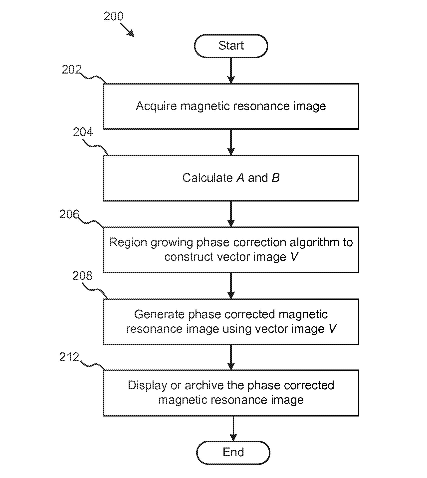
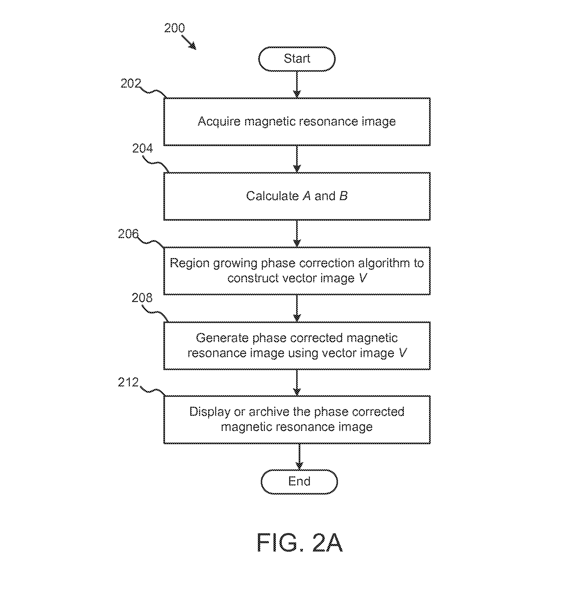
[0100]The flexible phase postprocessing strategy, as described in detail above, was implemented using MATLAB (MathWorks), and a 3D fast spoiled gradient-echo bipolar dual echo pulse sequence was used to collect raw data for a water / fat phantom (consisting of water and vegetable oil) and for the in vivo abdomen of a human subject using a 1.5T whole-body MR scanner (GE Healthcare; HDxt platform). For the in vivo imaging, an eight-channel phased array body coil was used, and the scan parameters were as follows: TR=minimum, FOV=36×27 cm, acquisition matrix=256×192, flip angle=12°, slice thickness=4 mm, total number of slices=38, and receiver bandwidth=±83.33 kHz. Minimum as well as different combinations of manually selected echo times were used for the dual-echo readout. For the phantom imaging, an eight-channel phased array head coil and scan parameters similar to those for the in vivo scanning were used except that for a fixed TE1 of 1.2 ms, TE2 varied systemat...


PUM


Abstract

Methods, apparatuses, systems, and software for extended phase correction in phase sensitive Magnetic Resonance Imaging. A magnetic resonance image or images may be loaded into a memory. Two vector images A and B associated with the loaded image or images may be calculated either explicitly or implicitly so that a vector orientation by one of the two vector images at a pixel is substantially determined by a background or error phase at the pixel, and the vector orientation at the pixel by the other vector image is substantially different from that determined by the background or error phase at the pixel. A sequenced region growing phase correction algorithm may be applied to the vector images A and B to construct a new vector image V so that a vector orientation of V at each pixel is substantially determined by the background or error phase at the pixel. A phase corrected magnetic resonance image or images may be generated using the vector image V, and the phase corrected magnetic resonance image or images may be displayed or archived.

Description

CROSS REFERENCE TO RELATED APPLICATIONS[0001]This application claims priority to U.S. Provisional patent application Ser. No. 61 / 604,413, filed Feb. 28, 2012 the entire disclosure of which is incorporated herein by reference.BACKGROUND[0002]1. Field of the Invention[0003]The present invention relates generally to the field of medical imaging. More particularly, embodiments of the invention relate to phase sensitive magnetic resonance imaging (MRI) using an extended phase correction algorithm, which among other things can process images with more flexible phases for successful water and fat imaging.[0004]2. Description of Related Art[0005]MRI has proven useful in the diagnosis of many diseases such as hepatic steatosis, cancer, multiple sclerosis, sports related injury, and bone marrow disorders. MRI provides unique imaging capabilities that are not attainable in any other imaging method. For example, MRI can provide detailed images of soft tissues, abnormal tissues such as tumors, a...

Claims


Application Information

Patent Timeline
Patent Type & Authority Applications(United States)
IPC IPC(8): G06T7/00G06K9/46G06T5/00
CPCG06T7/0012G06K9/46G06T5/00A61B5/055G01R33/565G01R33/4828G01R33/5602G01R33/5607G06F18/22G06F18/2113G06F18/24147G06T5/002G06T5/20G06T11/008G06T2207/10088G06T2207/20021
Inventor MA, JINGFEI
Owner BOARD OF RGT THE UNIV OF TEXAS SYST
Why Eureka
Try Eureka