Injectable and implantable cellular-scale electronic devices

a technology of electronic devices and cellular scales, applied in the field of biological tissue implantation, can solve problems such as heightened risk of adverse events, and achieve the effect of precise monitoring and control of biologic function

Active Publication Date: 2016-03-10
WASHINGTON UNIV IN SAINT LOUIS +1
View PDF14 Cites 122 Cited by
  • Summary
  • Abstract
  • Description
  • Claims
  • Application Information

AI Technical Summary

Benefits of technology

[0005]Provided herein are electronic devices specially configured for implantation, injection or surface mount into or onto various soft tissues, such as biological soft tissues in living animals. Ultrathin and mechanically compliant electronic device components, for example, permit access to the interior of living tissues without unduly impacting biologic function. Because the physical devices provided herein are minimally invasive, they can be used even for long-term implantation to interface with tissue that is not normally physically accessible. For ex

Problems solved by technology

For example, tissue trauma is associated with a robust immune response along with heightened risk of adverse events

Method used


Image

  • Injectable and implantable cellular-scale electronic devices
  • Injectable and implantable cellular-scale electronic devices
  • Injectable and implantable cellular-scale electronic devices

Examples


example 1

Injectable, Cellular-Scale Optoelectronics with Applications for Wireless Optogenetics

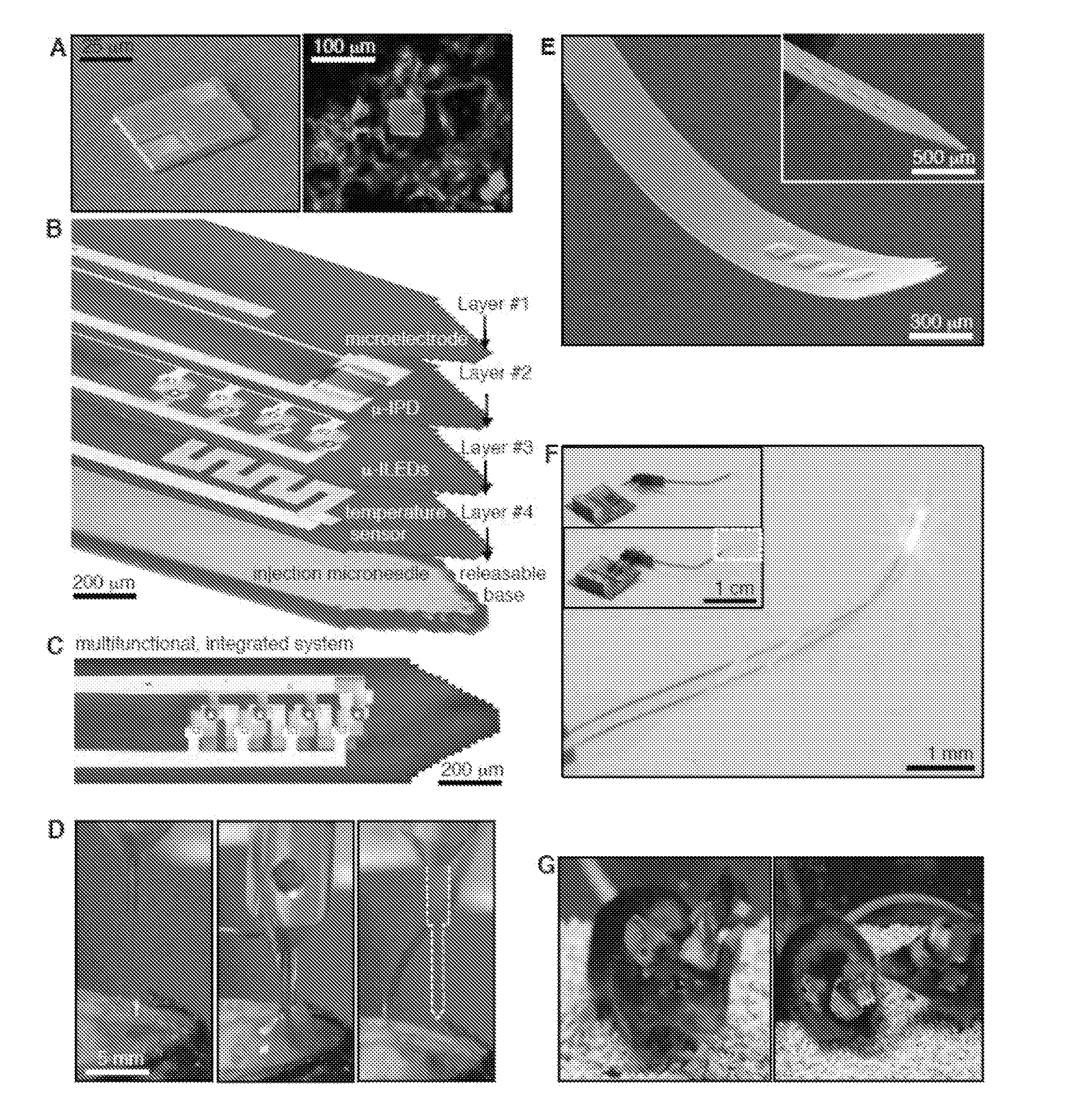
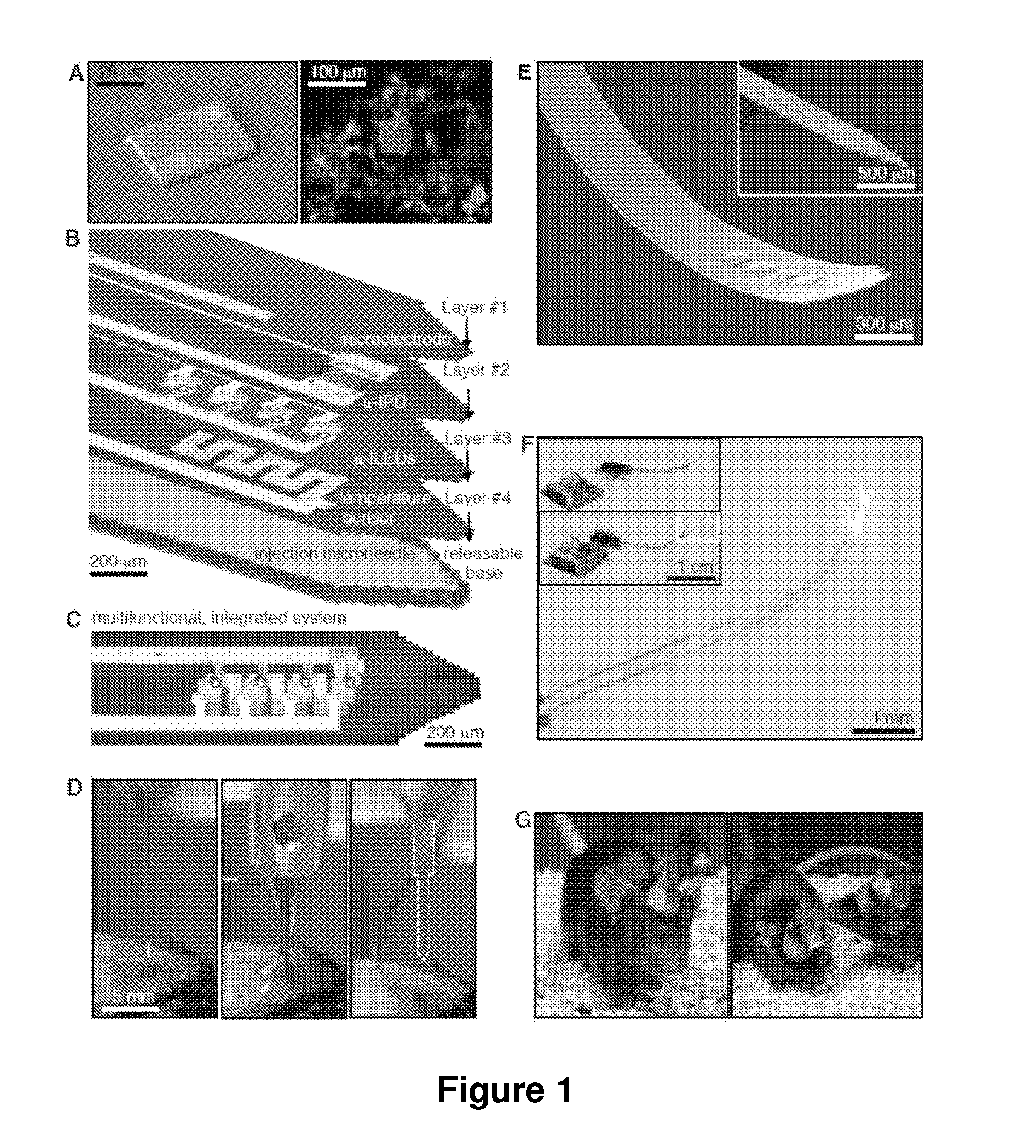
[0189]Successful integration of advanced semiconductor devices with biological systems will accelerate basic scientific discoveries and their translation into clinical technologies. In neuroscience generally, and in optogenetics in particular, an ability to insert light sources, detectors, sensors and other components into precise locations of the deep brain could yield versatile and important capabilities. Here, we introduce an injectable class of cellular-scale optoelectronics that offers such features, with examples of unmatched operational modes in optogenetics, including completely wireless and programmed complex behavioral control over freely moving animals. The ability of these ultrathin, mechanically compliant, biocompatible devices to afford minimally invasive operation in the soft tissues of the mammalian brain foreshadow applications in other organ systems, with potential for broad utili...

example 2

Methods of Making and Using Injectable Cellular-Scale Optoelectronics

[0202]Preparation of μ-ILEDs: Fabrication begins with epitaxially grown GaN on a sapphire wafer (500 μm thick double polished sapphire with 2″ diameter, Cermet Inc.). The GaN stack layers consisted of undoped GaN (3.8 μm), n-type GaN (2 μm), spacer (0.4 μm), MQW (0.14 μm), and p-type GaN (0.2 μm). Residual metal ions and GaN oxide on the surface were removed by rinsing with diluted HCl (33%). L-shaped current spreading layers of Ni (15 nm) and Au (15 nm) were formed by sputter deposition (AJA ATC 2000), followed by annealing at 500° C. for 5 min in an oxygen and nitrogen atmosphere to form an ohmic contact. After exposing n-type regions defined using photolithography (40×40 μm2 recessive square with thick AZ 2035, Microchem Inc) and etched using chorine based inductively coupled plasma reactive ion etching (ICP RIE, Plasmatherm, SLR-770), both n- and p-contact pads, each 25×25 μm2 in dimension, of 15 nm of Cr and 3...

example 3

Implanted Device for Optical Interfacing with the Sciatic Nerve

[0242]FIG. 31. Implantable or surface mounted biomedical device having printed LEDs that are useful for optogenetic applications. FIGS. 32-33 show the device implanted in a mouse for interfacing with the sciatic nerve. As desired, any other nerve location may be interfaced, such as for optogentic studies or applications. Optionally, the mouse may be transformed to express a protein, channel, or other material that is sensitive to light. A light-responsive protein channel is particularly useful in optogenic applications, such as channelrhodopsins to excite neurons, halorhodopsin for silencing and fungal opsins and enhanced bacteriorhodopsin for inhibiting neurons, including in freely moving mammals.

[0243]In FIG. 32 the devices have been implanted in male wild-type Swiss-Webster infected with a herpes simplex 1 viral construct expressing the optogenetic channel ChR2 under the control of the CMV promoter. The sciatic nerve ...


PUM


Abstract

Provided are implantable or surface mounted biomedical devices and related methods for interfacing with a target tissue. The devices have a substrate and device component supported by the substrate. The components of the device are specially configured and packaged to be ultra-thin and mechanically compliant. In particular, device thicknesses are less than 1 mm and have lateral dimensions between about 1 μm and 10 mm, depending on the application. Delivery substrates may be incorporated to assist with device implantation and handling. The devices can be shaped to provide injection in a minimally invasive manner, thereby avoiding unnecessary tissue damage and providing a platform for long-term implantation for interfacing with biological tissue.

Description

CROSS-REFERENCE TO RELATED APPLICATIONS[0001]This application claims the benefit of and priority to U.S. Provisional Patent App. Nos. 61 / 764,410 filed Feb. 13, 2013, 61 / 785,185 filed Mar. 14, 2013, and 61 / 883,524, filed Sep. 27, 2013, each of which is hereby incorporated by reference to the extent not inconsistent herewith.STATEMENT REGARDING FEDERALLY SPONSORED RESEARCH OR DEVELOPMENT[0002]This invention was made with government support under R01NS081707 and R00DA025182 awarded by the National Institutes of Health, DE-FG02-07ER46471 and DE-FG02-07ER46453 awarded by the Department of Energy and R01DA037152 awarded by the National Institute on Drug Abuse. The government has certain rights in the invention.BACKGROUND OF INVENTION[0003]The field of the invention generally relates to implantation into biological tissues. The devices and methods utilize electronic devices arranged in ultrathin functional layers, along with stacking of those functional layers in a special geometric config...

Claims


Application Information

Patent Timeline
IPC IPC(8): A61B5/00A61N5/06
CPCA61B5/0031A61N5/062A61B5/685A61M2037/0007A61M2205/04A61M2037/0061A61M2037/0053A61B2562/0285A61B2017/00004A61N2005/0612A61B2562/06A61N1/05A61B5/686A61B2562/12A61M2205/054A61M2205/05
Inventor ROGERS, JOHN A.BRUCHAS, MICHAEL RAYMONDGEREAU, ROBERTMCCALL, JORDAN GARYBRENNER, DANIELKIM, TAE-ILSHIN, GUNCHULJEONG, JAEWOONG
Owner WASHINGTON UNIV IN SAINT LOUIS
Why Eureka
Try Eureka