
Non-invasive multi-frequency oxygenation spectroscopy device using nir diffuse photon density waves for measurement and pressure gauges for prediction of pressure ulcers

a multi-frequency oxygenation and spectroscopy technology, applied in the field of frequency domain near infrared absorption devices, can solve the problems of device inability to obtain absolute values of absorption scattering coefficients, inability to contact with wounds, and inability to obtain contact probes with flexible rearranging of detector fibers, etc., to evaluate the efficacy of hyperbaric oxygen treatment and evaluate the effectiveness of wound and burn gels

Inactive Publication Date: 2016-10-13
DREXEL UNIV
View PDF4 Cites 3 Cited by
  • Summary
  • Abstract
  • Description
  • Claims
  • Application Information

AI Technical Summary

Benefits of technology

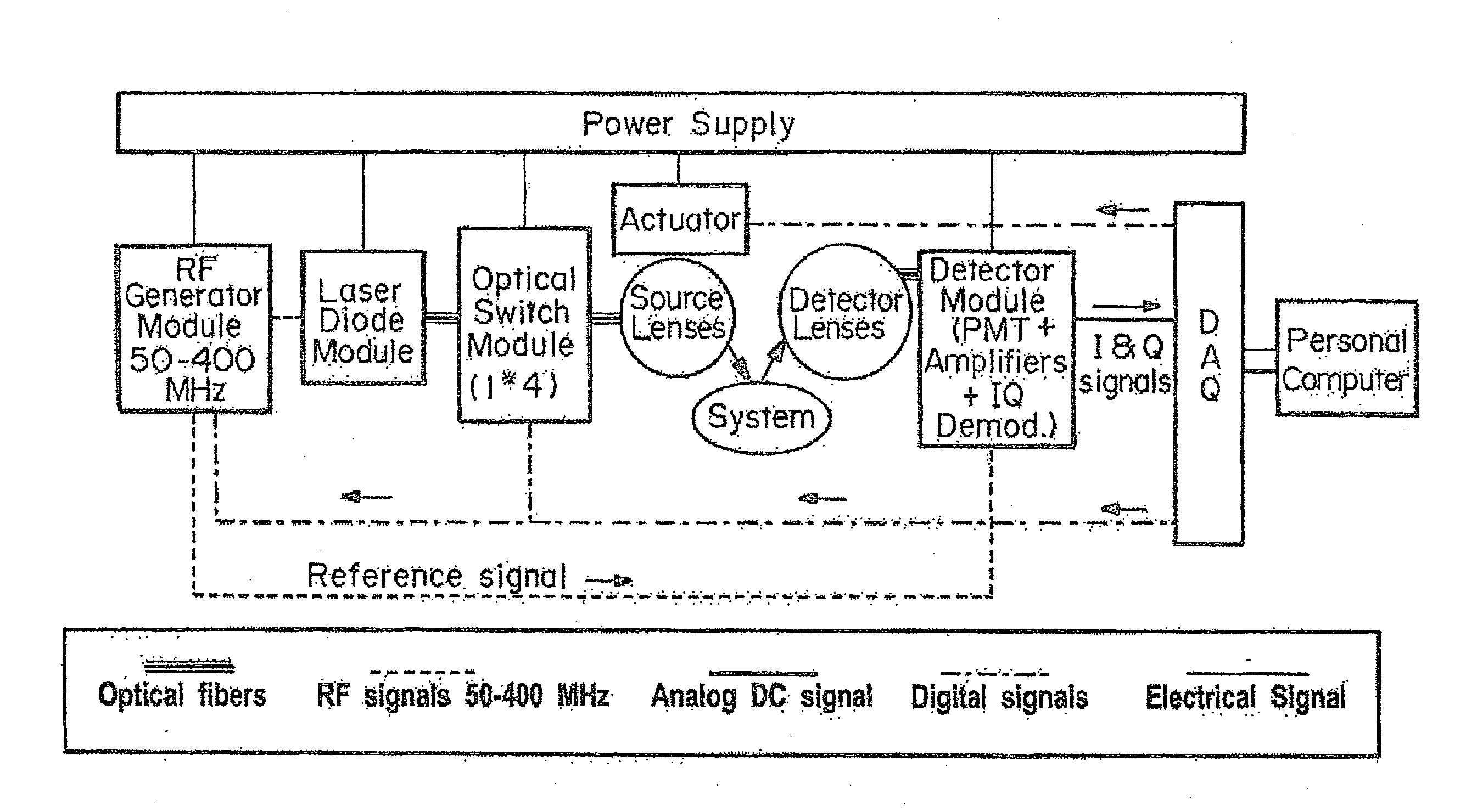
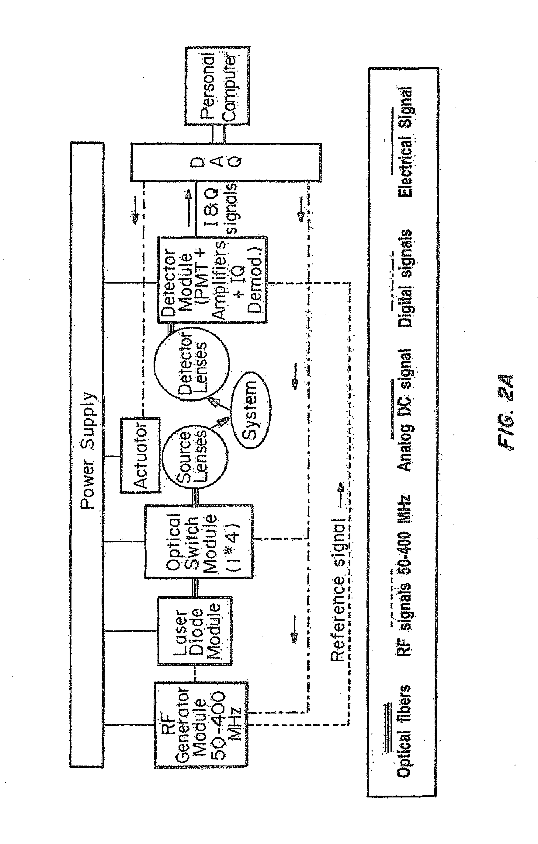
A device is described for measuring tissue damage such as pressure ulcers and other conditions characterized by decreased vascular flow. The device includes an opto-electronics module with a laser diode and an optical switch for applying modulated light to the tissue of a subject and detecting the scattered light. The device can be used to assess wound healing, pressure sores, and other diseases. The device can also be used to evaluate treatment efficiencies and the effectiveness of various therapies.

Problems solved by technology

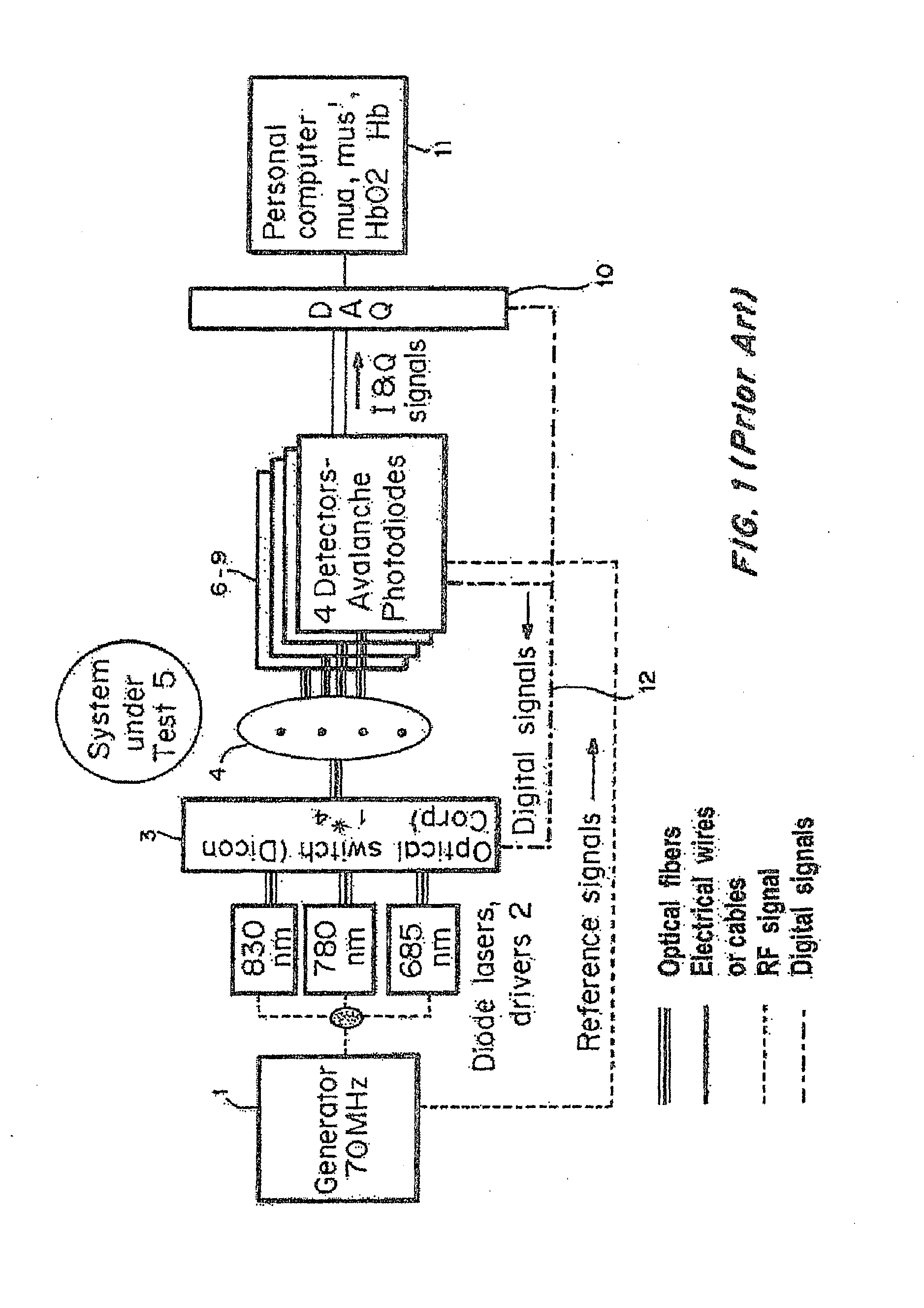
Such devices rely on optical fibers to transport the incident and scattered lights; however, the fiber optical probe is in contact with the tissue under examination.
Such contact with the wound is undesirable and the contact probe provides no flexibility in rearranging the detector fibers and optimizing the setup for wounds of different surface areas, shapes, and depths because one source and four detector fibers are permanently inserted in a rectangular piece made of plastic, Teflon, or silicon.
However, this device cannot obtain absolute values of absorption scattering coefficients but instead obtains relative changes.
Moreover, the probe also must penetrate into the burned skin, which is generally undesirable.
However, this device is very expensive and must be used on an optical table with very stable temperature and humidity conditions.
As such, it is not suitable for clinical use.
Since the power source for this method is constant, it is only possible to measure one parameter, the intensity of scattered light.
Changes in this light intensity are measured as a function of source—detector separation ρ. In the case of CW devices, however, difficulties emerge when trying to separate absorption attenuation from scattering effects.
Although a wealth of information can be obtained, this method is complex and expensive and is difficult to implement in a routine clinical setting.
Although rich in information obtained, this method is complex and expensive and difficult to implement in a routine clinical setting.

Method used


Image

  • Non-invasive multi-frequency oxygenation spectroscopy device using nir diffuse photon density waves for measurement and pressure gauges for prediction of pressure ulcers
  • Non-invasive multi-frequency oxygenation spectroscopy device using nir diffuse photon density waves for measurement and pressure gauges for prediction of pressure ulcers
  • Non-invasive multi-frequency oxygenation spectroscopy device using nir diffuse photon density waves for measurement and pressure gauges for prediction of pressure ulcers

Examples


example 1

Experimental Data and Device Calibration

Detector Linearity Testing

[0080]As with any optical detector, the APD module has a limited range where the electrical output signal is linearly proportional to the optical power of the incident light. Prior to in vitro or in vivo experiments, a calibration procedure is conducted to define the range of linearity. Any subsequent measurements which falls outside of the range of linearity is discarded and retaken after adjusting the amplitude of the signal using the optical attenuators. The results from the linearity test performed at a modulation frequency of 80 MHz are presented on FIG. 9. The linear range (slope of 0.98) observed for the APD indicates excellent detector linearity over the range of 1 to 185 mV.

Offset Testing

[0081]Offset values (i.e., voltage registered by the detector when there is no incident light) of the device were measured while changing the modulation frequency from 50 to 350 MHz. Results are shown in FIG. 10. The offset v...

example 2

Animal Study Results

[0087]An animal study for in vivo validation of the device was conducted. The purpose of the study was to differentiate superficial and deep burns using the optical data. Data analysis suggests a statistically significant difference between the optical properties of deep and superficial wounds. An in depth description of the results and study procedures follows:

Study Procedures

[0088]A Yorkshire swine weighing approximately 25 kg was purchased and transported to the DUCOM facilities. The animal was acclimated for a period of one week. Baseline measurements of healthy intact animal skin were taken following the acclimation period. Anesthesia was induced by mask prior to measurements and the animal's hair was clipped to improve the accuracy and reliability of the measurements. The animal was intubated and mechanically ventilated to control the rate of breathing and reduce motion artifacts during measurements. After hair removal, eight wound sites were symmetrically ...

example 3

Design of Probe for Non-Invasive Measurement

[0104]In exemplary embodiments, the probe described herein is used with a device the size of a small desktop computer, and the probe is gently placed on the surface of intact skin to assess tissue damage at multiple depths. Ideally, the probe could be manually held in place by a nurse at any anatomical location where DTI is suspected, and the measurement could be completed in less than one minute. The computer would immediately provide the clinician with a depth-profile of tissue oxygenation at depths ranging from 1-10 mm. The depth of bone within this range would be clearly indicated, and regions of impaired tissue oxygenation could be clearly seen.

[0105]In many cases, such a simple measurement protocol might not provide sufficient information to distinguish healthy from damage tissue with acceptable sensitivity and specificity. A more complex protocol may provide more information. When pressure is applied to the skin over bony prominence...


PUM


Abstract

Multi-frequency Diffuse Photon Density Wave (DPDW) methodology at near infrared wavelengths is used to non-invasively measure the optical absorption and reduced scattering coefficients of tissue at depths of several millimeters to quantify the depth and degree of tissue damage. A digitally-controlled stepper motor and variable RF generator enable the user to select the distance between the light source and detector and the modulation frequency through software. This allows for the collection of virtually unlimited number of data points, enabling precise selection of the volume and depth of tissue that will be characterized.

Description

CROSS-REFERENCE TO RELATED APPLICATIONS[0001]The present application is a 371 application of International Application No. PCT / US2014 / 066946, filed Nov. 24, 2014. Which claims the benefit of U.S. Provisional Application No. 61 / 906,894 filed Nov. 21, 2013, the disclosures of which are incorporated herein by reference in their entirety.FIELD OF THE INVENTION[0002]The present invention relates to a frequency domain near infrared absorption device for assessing deep tissue optical properties enabling, for example, the assessment of oxygenation of wound tissue, indicative of wound healing.BACKGROUND OF THE INVENTION[0003]A variety of instruments based on the diffuse propagation of Near Infrared (NIR) photons due to multiply scattered light have been used to obtain clinically meaningful information about living tissue, such as tissue oxygenation. Such devices rely on optical fibers to transport the incident and scattered lights; however, the fiber optical probe is in contact with the tiss...

Claims


Application Information

Patent Timeline
Patent Type & Authority Applications(United States)
IPC IPC(8): A61B5/00A61B5/1455
CPCA61B5/445A61B5/14551A61B5/0075A61B5/447A61B5/6886
Inventor PAPAZOGLOU, ELISABETHZUBKOV, LEONIDWEINGARTEN, MICHAEL S.NEIDRAUER, MICHAEL T.DIAZ, DAVID
Owner DREXEL UNIV
Learn More